New sensor chip advances rapid, cost-effective disease diagnostics

Texas A&M AgriLife Research scientists and collaborators at Iowa State University have developed a sensor chip that can detect many disease pathogens with 10 times the sensitivity of currently available methods.
The chip also eliminates the need for chemical dye reagents typically used in the diagnostic process. The new technology shows promise for rapid, low-cost point-of-care diagnostic capabilities in plants, foods, animals and humans, including detecting foodborne pathogens, bird flu and COVID-19.
Results from the new sensor are available in about 30 minutes.
In their research, published in ASC Sensors, scientists used the new sensor to detect Phytophthora infestans. The pathogen causes globally devastating late blight disease — a particular threat to potato and tomato crops.
The research was co-led by Jinping Zhao, Ph.D., AgriLife Research postdoctoral research scientist in Dallas, and Subin Mao, a Ph.D. candidate in electrical and computer engineering at Iowa State University. Serving as corresponding authors were collaborators Junqi Song, Ph.D., associate professor and plant immunity research lead with AgriLife Research in Dallas, and Long Que, Ph.D., professor of electrical engineering at Iowa State University. Seed grants from each university funded the research.
“This research advances technologies that have emerged as some of our greatest opportunities for improving agriculture, food safety and human health,” Song said. “Our publication represents a step toward realizing these powerful tools against diseases.”
Building on existing technologies

The new sensor improves upon a technique known as loop-mediated isothermal amplification, or LAMP, which is widely used to detect pathogens by amplifying their DNA.
Detection of LAMP products amplified from templates, such as pathogen DNA, often requires that the products be “labeled” by using fluorescence dyes — a costly process with low sensitivity. The new sensor diagnoses pathogens without such reagents and at high sensitivity. It also eliminates a lengthy DNA purification process that creates challenges for point-of-care use.
The new chip consists of a nanopore thin-film sensor inside a special reaction chamber. Primers are uniquely designed to be immobilized on the nanofilm, causing amplified LAMP products to become bound to the sensor, which produces signals that can be directly and easily measured with a portable spectrometer.
What’s next
The LAMP chip offers a new portable platform to detect pathogens using label-free sensors with ultrasensitivity. The research team will now work to further enhance sensitivity to a subattomolar or even lower level.
The team aims to offset current challenges to detecting and distinguishing pathogen species and strains with high-sequence similarities. They will also work to improve the specificity of detections and establish quantitative detection by integrating artificial intelligence and CRISPR gene-editing technologies.
Their goal is to achieve a viable product for broad adoption in plant, animal and human health point-of-care applications.

Read more →

Poor sense of smell linked to increased risk of depression in older adults

In a study that followed more than 2,000 community-dwelling older adults over eight years, researchers at Johns Hopkins Medicine say they have significant new evidence of a link between decreased sense of smell and risk of developing late-life depression.
Their findings, published June 26 in Journal of Gerontology: Medical Sciences, do not demonstrate that loss of smell causes depression, but suggests that it may serve as a potent indicator of overall health and well-being.
“We’ve seen repeatedly that a poor sense of smell can be an early warning sign of neurodegenerative diseases such as Alzheimer’s disease and Parkinson’s disease, as well as a mortality risk. This study underscores its association with depressive symptoms,” says Vidya Kamath, Ph.D., associate professor of psychiatry and behavioral sciences at the Johns Hopkins University School of Medicine. “Additionally, this study explores factors that might influence the relationship between olfaction and depression, including poor cognition and inflammation.”
The study used data gathered from 2,125 participants in a federal government study known as the Health, Aging and Body Composition Study (Health ABC). This cohort was composed of a group of healthy older adults ages 70-73 at the start of the eight-year study period in 1997-98. Participants showed no difficulties in walking 0.25 miles, climbing 10 steps or performing normal activities at the start of the study, and were assessed in person annually and by phone every six months. Tests included those for the ability to detect certain odors, depression and mobility assessments.
In 1999, when smell was first measured, 48% of participants displayed a normal sense of smell, 28% showed a decreased sense of smell, known as hyposmia, and 24% had a profound loss of the sense, known as anosmia. Participants with a better sense of smell tended to be younger than those reporting significant loss or hyposmia. Over follow-up, 25% of participants developed significant depressive symptoms. When analyzed further, researchers found that individuals with decreased or significant loss of smell had increased risk of developing significant depressive symptoms at longitudinal follow-up than those in the normal olfaction group. Participants with a better sense of smell tended to be younger than those reporting significant loss or hyposomia.
Researchers also identified three depressive symptom “trajectories” in the study group: stable low, stable moderate and stable high depressive symptoms. Poorer sense of smell was associated with an increased chance of a participant falling into the moderate or high depressive symptoms groups, meaning that the worse a person’s sense of smell, the higher their depressive symptoms. These findings persisted after adjusting for age, income, lifestyle, health factors and use of antidepressant medication.

“Losing your sense of smell influences many aspects of our health and behavior, such as sensing spoiled food or noxious gas, and eating enjoyment. Now we can see that it may also be an important vulnerability indicator of something in your health gone awry,” says Kamath. “Smell is an important way to engage with the world around us, and this study shows it may be a warning sign for late-life depression.”
Humans’ sense of smell is one of two chemical senses. It works through specialized sensory cells, called olfactory neurons, which are found in the nose. These neurons have one odor receptor; it picks up molecules released by substances around us, which are then relayed to the brain for interpretation. The higher the concentration of these smell molecules the stronger the smell, and different combinations of molecules result in different sensations.
Smell is processed in the brain’s olfactory bulb, which is believed to interact closely with the amygdala, hippocampus and other brain structures that regulate and enable memory, decision-making and emotional responses.
The Johns Hopkins researchers say their study suggests that olfaction and depression may be linked through both biological (e.g., altered serotonin levels, brain volume changes) and behavioral (e.g., reduced social function and appetite) mechanisms.
The researchers plan to replicate their findings from this study in more groups of older adults, and examine changes to individuals’ olfactory bulbs to determine if this system is in fact altered in those diagnosed with depression. They also plan to examine if smell can be used in intervention strategies to mitigate risk of late-life depression.
Other scientists who contributed to this research are Kening Jiang, Danielle Powell, Frank Lin and Jennifer Deal of the Johns Hopkins University School of Medicine and Bloomberg School of Public Health; Kevin Manning of the University of Connecticut; R. Scott Mackin, Willa Brenowitz and Kristine Yaffe of the University of California, San Francisco; Keenan Walker and Eleanor Simonsick of the National Institute on Aging; and Honglei Chen of Michigan State University.
No authors declared conflicts of interest related to this research under Johns Hopkins University School of Medicine policies.
This work was supported by the National Institute on Aging, the National Institute of Nursing Research and the Intramural Research Program of the National Institutes of Health: National Institute on Aging.

Read more →

Tuning T cell traits and functions with biomechanical materials

T cells experience different mechanical signals in different tissues. Researchers at Harvard’s Wyss Institute and Harvard SEAS led by David Mooney engineered a tissue-mimicking hydrogel model to show that more elastic tissues induce T cells to become effector-like T cells with strong tumor-killing potential, while more viscous tissues induce them to become memory-like T cells. This new concept could help advance adaptive T cell therapies by producing desired patient-specific T cell populations in the dish that could provide stronger effects when infused back into the same patient. The findings are reported in Nature Biomedical Engineering.
The successful campaign of adoptive T cell therapies, a type of immunotherapy in which immune T cells are collected from a patient, enhanced outside of the body, and reinfused back into the same patient, especially against blood cancers is well under way. But improving the ability to create patient-specific T cell populations with specific traits and functions could broaden clinicians’ repertoire of T cell therapies.
One way to approach this goal is to better understand how T cells’ traits and functions, including their cytotoxic effects on unwanted target cells (effector T cells) or their ability to recall and eliminate them if they show up again (memory T cells), are shaped by the mechanical resistance of the tissues they encounter while infiltrating them. The mechanical features of tissues, for example, bone, muscle, different internal organs, and blood, can vary widely, and pathological tissues such as tumor masses or fibrotic tissues are mechanically significantly different from healthy tissues.
Now, a research team at the Wyss Institute for Biologically Inspired Engineering at Harvard University and Harvard John A. Paulson School of Engineering and Applied Sciences (SEAS), led by Wyss Core Faculty member David Mooney, Ph.D., took a novel biomaterials approach to investigate the effect of tissue mechanics on the state of T cells. By engineering a 3-dimensional model of the extracellular matrix (ECM), produced by cells that are responsible for tissues’ different stiffnesses and viscoelasticities, they were able to tune both parameters independently. This enabled them to demonstrate a distinct impact of tissue viscoelasticity on T cell development and function in vitro and in vivo, and to identify a molecular pathway driving the phenomenon. The findings are reported in Nature Biomedical Engineering.
Mechanical resistance comes in the form of “stiffness,” a tissue’s (or any material’s) resistance to instantaneous deformation, and “viscoelasticity,” the type of relaxation it exhibits over time following its deformation. Explained in physical terms, a viscous (fluid) material, like honey, is more likely to flow, while an elastic (solid) material returns more rapidly to its original shape, like a rubber band after stretching — and this holds true for tissues which are composed of both solid and fluid components.
“Importantly, the phenotypes, functions, and gene expression programs of T cells trained in variations of the system correlated well with those we found in T cells in mechanically distinct tissues from patients with cancer or fibrosis,” said Mooney who also is the Robert P. Pinkas Family Professor of Bioengineering at SEAS, and leads the Wyss Institute’s Immunomaterials Initiative. “Our study provides a conceptual basis for future strategies aiming to create functionally distinct T cell populations for adoptive therapies by selectively tuning mechanical input provided by biomaterials-based engineered cell culture systems.”
Mimicking tissue mechanics in a dish

Key to their discoveries was the team’s engineering of a tunable ECM model, in which they focused on a type of collagen that they found to be key to dictating the mechanical behavior of different tissues. Collagen is a major ECM protein secreted by almost all cells in the body. Individual collagen protein molecules are naturally organized into crimped fibrils that aggregate further into fibers by chemically cross-linking themselves. Each fibril can be considered a mechanical spring, and each fiber as an assembly of springs. An ECMs stiffness depends on how densely it is packed with collagen molecules, whereas its distinct viscoelasticity depends on how densely collagen molecules are cross-linked to each other.
To mimic natural collagen-based ECM, the team fabricated hydrogels whose stiffness they could tune by varying the concentration of collagen molecules: fewer numbers of collagen molecules produced lower stiffness and higher numbers, higher stiffness. Independently, viscoelasticity became tunable by varying the amounts of a synthetic cross-linker molecule that further networked the collagen molecules. More highly cross-linked collagen molecules produced more elastic hydrogels. The resulting ECM-mimicking hydrogels equally allowed the attachment of pre-activated T cells but, importantly, enabled their stimulation with specific mechanical signals.
“To our knowledge, this is the first ECM model that allows researchers to study T cells with stiffness from viscoelasticity decoupled, and thus enables us and others in the future to investigate how immune and other cells might be mechanically regulated,” said co-first author Yutong Liu, Ph.D., who was a graduate student in Mooney’s group. “The system’s defined and uniform mechanical stimulation is vastly different from how T cells are usually cultured — cells that attach to the bottom of a culture dish encounter a highly inelastic surface, while those remaining in suspension are surrounded by the viscous medium.”
Natural consequences of mechanical action
The team performed an extensive analysis of T cells exposed to different viscoelastic conditions. “T cells that experienced a more elastic collagen matrix were more likely to develop into ‘effector-like T cells,’ whereas T cells that experienced a more viscous ECM matrix rather became ‘memory-like T cells,'” said co-first author Kwasi Adu-Berchie, Ph.D., who completed his Ph.D. in Mooney’s lab and is currently a Translational Immunotherapy Scientist at the Wyss Institute. “Importantly, we found that a T cell’s state, resulting from the viscoelasticity of a matrix, even more so from more elastic, less viscous hydrogels, becomes long-term imprinted, as the cell retains a memory of that specific matrix after being transferred to a different one. This could have broad implications for future cell manufacturing.”
Gene expression analysis led the team to the activity of a transcription factor known as AP-1 that links T cells’ reception of a more elastic, less viscous mechanical environment to a more effector-like gene expression program. The number of AP-1 complexes with specific compositions was increased, and genes depending on them for their expression were enriched, not only in T cells isolated from more elastic hydrogels, but also in T cells isolated from patients’ cancer and fibrotic tissues, which are stiffer and more elastic than neighboring healthy tissues. When they inhibited one of AP-1’s components with a drug, the effects of a more elastic collagen matrix on T cells were prevented.

To investigate how different mechanical stimulations and T cells’ predicted gene expression signatures translated into actual traits and functions, the team used therapeutic CAR-T cells engineered to bind a specific antigen of a human lymphoma cell line. CAR-T cells that were stimulated in a more elastic collagen matrix in vitro exhibited a stronger ability to kill lymphoma cells. Also in vivo, CAR-T cells stimulated in a more elastic matrix, and adoptively transferred into mice with the same type of lymphoma, were significantly more capable of reducing tumor burden in the animals and extending their lives than CAR-T cells exposed to a less elastic matrix.
“This study merges three seemingly disparate fields, biomaterials, immunotherapy, and mechanobiology, to develop an entirely new form of biomaterials-based mechanotherapeutic. It is easy to see how these findings can potentially open up new avenues to improve adoptive T cell therapies for patients in the future,” said Wyss Founding Director Donald Ingber, M.D., Ph.D., who is also the Judah Folkman Professor of Vascular Biology at Harvard Medical School and Boston Children’s Hospital, and the Hansjörg Wyss Professor of Bioinspired Engineering at SEAS.
Other authors on the study were present and former members of Mooney’s group, including David Zhang, Benjamin Freedman, Joshua Brockman, Kyle Vining, Bryan Nerger, and Andrea Garmilla. The study was funded by the National Institutes of Health (award # R01 CA276459-01), Food and Drug Administration (award # R01FD006589), and the Wellcome Leap HOPE Program.

Read more →

Researchers uncover potential biomarkers of positive response to immunotherapy

Scientists at the UCLA Jonsson Comprehensive Cancer Center have identified potential new biomarkers that could indicate how someone diagnosed with metastatic melanoma will respond to immunotherapy treatment.
The researchers found when T cells are activated, they release a protein called CXCL13, which helps attract more B cells and T cells to the tumor site. The B cells then show the T cells specific parts of the tumor, which leads to increased activation of the T cells and their ability to fight the cancer. This cooperation between T cells and B cells was associated with improved survival in patients diagnosed with metastatic melanoma who were treated with immunotherapy, but not for those who received targeted therapy (e.g., MEK inhibitors).
These findings could help guide new strategies to improve the effectiveness of melanoma cancer treatments.
“Based upon our data, increased presence of B cells and CXCL13 protein in the tumor after immunotherapy treatment may be predictive biomarkers for durable immunotherapy response in melanoma patients and may be avenues to enhance the response rate to immunotherapy in patients diagnosed with melanoma,” said co-senior author of the paper Willy Hugo, PhD, assistant professor of Medicine in the division of Dermatology at the David Geffen School of Medicine at UCLA and member of the UCLA Jonsson Comprehensive Cancer Center. “For example, combination of anti-PD1 treatments with CXCL13 or B cell-directed therapies may be strategies for patients who fail to respond to checkpoint immunotherapy alone.”
Immune checkpoint inhibitors, which harness the body’s immune system to better attack cancer cells, have revolutionized the way people with melanoma are treated. People with aggressive forms of the cancer are now living longer, healthier lives. Despite the remarkable success of using immune checkpoint inhibitors to treat people with advanced melanoma, it is still difficult to predict who will benefit from the therapy.
Identifying mechanisms that determine how tumors can become resistant to these therapies and understanding how to identify patients who will and will not respond to them is critical to developing new and improved treatments to help improve the response rate of these therapies.
To understand what may drive durable antitumor immune responses seen with checkpoint immunotherapy in some melanoma patients, and why such responses are less often seen in patients treated with other FDA-approved targeted therapies, such as mutant BRAF and MEK inhibitors, the UCLA team compared the immune responses induced by existing standard care targeted and immunotherapies for people with metastatic melanoma.
The team completed a comparative genomics analysis using published RNA-seq profiles of melanoma samples collected before and after either therapy. They found that response to immunotherapy, but not targeted therapy, is accompanied with significant infiltration of clonally diverse B cells. The increase of B cell infiltration in response to immunotherapy is accompanied by a significant upregulation of B-cell chemotactic factor, CXCL13, by T cells.
“This study suggests that CXCL13 may play an important role in bringing together T and B cells in the tumor microenvironment in patients who respond to checkpoint immunotherapy, and that this cooperation may be key to effective anti-tumor responses. Further studies are need to determine if these pathways can be boosted in non-responders to improve outcomes,” said co-senior author of the paper Melissa Lechner, MD, PhD, assistant professor of Medicine in the division of Endocrinology at the David Geffen School of Medicine at UCLA and member of the UCLA Jonsson Comprehensive Cancer Center.
These data also support a role for antigen presentation by B cells to T cells in the tumor microenvironment, and highlight the potential of using B cell-based cancer vaccines to enhance the effectiveness of immune checkpoint immunotherapies.
The team now plans to further explore these mechanisms in preclinical cancer models and test whether antigen presenting B cell and CXCL13 manipulation can improve anti-tumor immune responses in non-responders.
This work was supported in part by grants from the National Cancer Institute (1R01CA236910) and a grant from Parker Institute for Cancer Immunotherapy.

Read more →

Lean body mass, age linked with alcohol elimination rates in women

The rate at which women eliminate alcohol from their bloodstream is largely predicted by their lean body mass, although age plays a role, too, scientists found in a new study. Women with obesity — and those who are older — clear alcohol from their systems 52% faster than women of healthy weights and those who are younger, the study found.
Lean body mass is defined in the study — published in the journal Alcohol Clinical and Experimental Research — as one’s total body weight minus fat.
“We believe the strong relationship we found between participants’ lean body mass and their alcohol elimination rate is due to the association that exists between lean body mass and lean liver tissue — the part of the liver responsible for metabolizing alcohol,” said research group leader M. Yanina Pepino, a professor of food science and human nutrition at the University of Illinois Urbana-Champaign.
To explore links between body composition and alcohol elimination rates, the team conducted a secondary analysis of data from a study performed at the U. of I and another at Indiana University, Indianapolis. Both projects used similar methods to estimate the rate at which alcohol is broken down in the body.
The combined sample from the studies used in the analysis included 143 women who ranged in age from 21 to 64 and represented a wide range of body mass indices — from healthy weights to severe obesity. Among these were 19 women who had undergone different types of bariatric surgery.
In a subsample of 102 of these women, the researchers had measured the proportions of lean and fat tissue in their bodies and calculated their body mass indices. Based on their BMI, those in the subsample were divided into three groups: normal weight, which included women with BMI ranging from 18.5-24.9; overweight, those with BMI ranging from 25-29.9; and obese, participants with BMI above 30.

As the researchers expected, women with higher BMI had not only more fat mass than women of healthy weights, they also had more lean mass. On average, the group with obesity had 52.3 kg of lean mass, compared with 47.5 kg for the normal weight group.
The two studies both used an alcohol clamp technique, where participants received an intravenous infusion of alcohol at a rate controlled by a computer-assisted system. The system calculated personalized infusion rates based upon each participant’s age, height, weight and gender and was programmed so they would reach a target blood alcohol concentration of .06 percent within 15 minutes and maintain that level for about two hours
Using a breathalyzer, breath samples were collected at regular intervals throughout the experiments to estimate participants’ blood alcohol concentration and provide feedback to the system.
“We found that having a higher fat-free body mass was associated with a faster alcohol elimination rate, particularly in women in the oldest subgroups,” said Neda Seyedsadjadi, a postdoctoral fellow at the university and the first author of the study.
“The average alcohol elimination rates were 6 grams per hour for the healthy weight group, 7 grams for the overweight group, and 9 grams for the group with obesity,” she said. “To put this in perspective, one standard drink is 14 grams of pure alcohol, which is found in 12 ounces of beer, 5 ounces of table wine or 1.5 ounces shot of distilled spirits.”
The interaction between participants’ age and lean body mass accounted for 72% of the variance in the time required to eliminate the alcohol from their system, the team found.

Pepino, who also holds an appointment as a health innovation professor at Carle Illinois College of Medicine, has conducted several studies on alcohol response in bariatric surgery patients.
The findings also shed light on alcohol metabolism and body composition in women who have undergone weight loss surgery. Researchers have long known that bariatric surgery alters women’s response to alcohol but were uncertain if it affected how quickly they cleared alcohol from their systems.
Some prior studies found that these patients metabolized alcohol more slowly after they had weight loss surgery. The new study’s findings indicate that these participants’ slower alcohol elimination rates can be explained by surgery-induced reductions in their lean body mass. Weight loss surgery itself had no independent effects on patients’ alcohol elimination rates, the team found.
Additional co-authors of the current study were Dr. Blair Rowitz, associate dean for clinical affairs with the Carle Illinois College of Medicine; Vijay A. Ramchandani, a senior investigator in the section on human psychopharmocology at the National Institute on Alcohol Abuse and Alcoholism; and psychiatry professors Dr. Martin H. Plawecki and Dr. Sean J. O’Connor, and scientist in neurology Ann E.K. Kosobud, all of the Indiana University School of Medicine.

Read more →

Novel study deepens knowledge of treatment-resistant hypertension

For many patients with hypertension — an elevated blood pressure that can lead to stroke or heart attack — medication keeps the condition at bay. But what happens when medication that physicians usually prescribe doesn’t work? Known as apparent resistant hypertension (aRH), this form of high blood pressure requires more medication and medical management.
Novel research from investigators in the Smidt Heart Institute at Cedars-Sinai, published today in the peer-reviewed journal Hypertension, found that aRH prevalence was lower in a real-world sample than previously reported, but still relatively frequent — affecting nearly 1 in 10 hypertensive patients.
Through their analysis, investigators also learned that patients with well-managed aRH were more likely to be treated with a commonplace medication called mineralocorticoid receptor antagonist, or MRA. These MRA treatments were used in 34% of patients with controlled aRH, but only 11% of patients with uncontrolled aRH.
“Apparent resistant hypertension is more common than many would anticipate,” said Joseph Ebinger, MD, assistant professor of Cardiology in the Smidt Heart Institute and corresponding author of the study. “We also learned that within this high-risk population, there are large differences in how providers treat high blood pressure, exemplifying a need to standardize care.”
Study findings were based on a unique design, which used clinically generated data from the electronic health records of three large, geographically diverse healthcare organizations. Of the 2,420,468 patients analyzed in the study, 55% were hypertensive. Of these hypertension patients, 8.5%, or 113,992 individuals, met criteria for aRH.
According to Ebinger, treating aRH can be just as tricky as diagnosing it.

In fact, the “apparent” in apparent resistant hypertension stems from the fact that before diagnosis, medical professionals must first rule out other potential reasons for a patient’s blood pressure to be high.
These reasons might include medication non-adherence, inappropriate medication selection, or artificially elevated blood pressure in the doctor’s office — known as “white coat hypertension.”
“Large amounts of data tell us that patients with aRH, compared to those with non-resistant forms of hypertension, are at greatest risk for adverse cardiovascular events,” said Ebinger, director of Clinical Analytics in the Smidt Heart Institute. “Identifying these patients and possible causes for their elevated blood pressure is increasingly important.”
The takeaway, Ebinger says, is awareness — for both medical professionals and patients. He says providers should be mindful that if it’s taking four or more antihypertensive medications to control a patient’s blood pressure, they should consider evaluation for alternative causes of hypertension, or refer patients to a specialist.
Similarly, patients should lean on their medical providers to help them navigate the complex disease, including having a conversation around strategies for remembering to take their medication and addressing possible treatment side effects.
Treating patients with complex cardiac issues like aRH is at the heart of Cedars-Sinai’s expertise.
The Smidt Heart Institute was recently awarded the American Heart Association’s Comprehensive Hypertension Center Certification, recognizing the institute’s commitment to following proven, research-based treatment guidelines to care for people with complex or difficult-to-treat hypertension.
“This accreditation, coupled with our clinical and research expertise in hypertensive diseases, serves as a mark of excellence,” said Christine M. Albert, MD, MPH, chair of the Department of Cardiology and the Lee and Harold Kapelovitz Distinguished Chair in Cardiology. “These efforts signal to patients, healthcare providers, and the community that the Smidt Heart Institute is committed to delivering evidence-based, comprehensive care for hypertension.”
Funding: This work was supported in part by Cedars-Sinai Medical Center and by the National Institutes of Health grant K23-HL153888.

Read more →

A.I. May Someday Work Medical Miracles. For Now, It Helps Do Paperwork.

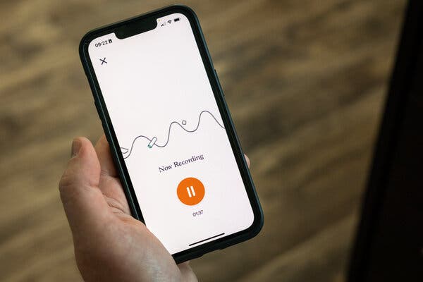
Dr. Matthew Hitchcock, a family physician in Chattanooga, Tenn., has an A.I. helper.It records patient visits on his smartphone and summarizes them for treatment plans and billing. He does some light editing of what the A.I. produces, and is done with his daily patient visit documentation in 20 minutes or so.Dr. Hitchcock used to spend up to two hours typing up these medical notes after his four children went to bed. “That’s a thing of the past,” he said. “It’s quite awesome.”ChatGPT-style artificial intelligence is coming to health care, and the grand vision of what it could bring is inspiring. Every doctor, enthusiasts predict, will have a superintelligent sidekick, dispensing suggestions to improve care.But first will come more mundane applications of artificial intelligence. A prime target will be to ease the crushing burden of digital paperwork that physicians must produce, typing lengthy notes into electronic medical records required for treatment, billing and administrative purposes.For now, the new A.I. in health care is going to be less a genius partner than a tireless scribe.Abridge, founded in 2018, provides an automated solution to a modern clerical overload in health care by using A.I. to record and generate a summary of patient visits.Audra Melton for The New York TimesFrom leaders at major medical centers to family physicians, there is optimism that health care will benefit from the latest advances in generative A.I. — technology that can produce everything from poetry to computer programs, often with human-level fluency.But medicine, doctors emphasize, is not a wide open terrain of experimentation. A.I.’s tendency to occasionally create fabrications, or so-called hallucinations, can be amusing, but not in the high-stakes realm of health care.That makes generative A.I., they say, very different from A.I. algorithms, already approved by the Food and Drug Administration, for specific applications, like scanning medical images for cell clusters or subtle patterns that suggest the presence of lung or breast cancer. Doctors are also using chatbots to communicate more effectively with some patients.Physicians and medical researchers say regulatory uncertainty, and concerns about patient safety and litigation, will slow the acceptance of generative A.I. in health care, especially its use in diagnosis and treatment plans.“At this stage, we have to pick our use cases carefully,” said Dr. John Halamka, president of Mayo Clinic Platform, who oversees the health system’s adoption of artificial intelligence. “Reducing the documentation burden would be a huge win on its own.”Recent studies show that doctors and nurses report high levels of burnout, prompting many to leave the profession. High on the list of complaints, especially for primary care physicians, is the time spent on documentation for electronic health records. That work often spills over into the evenings, after-office-hours toil that doctors refer to as “pajama time.”Generative A.I., experts say, looks like a promising weapon to combat the physician workload crisis.“This technology is rapidly improving at a time health care needs help,” said Dr. Adam Landman, chief information officer of Mass General Brigham, which includes Massachusetts General Hospital and Brigham and Women’s Hospital in Boston.For years, doctors have used various kinds of documentation assistance, including speech recognition software and human transcribers. But the latest A.I. is doing far more: summarizing, organizing and tagging the conversation between a doctor and a patient.Companies developing this kind of technology include Abridge, Ambience Healthcare, Augmedix, Nuance, which is part of Microsoft, and Suki.Ten physicians at the University of Kansas Medical Center have been using generative A.I. software for the last two months, said Dr. Gregory Ator, an ear, nose and throat specialist and the center’s chief medical informatics officer. The medical center plans to eventually make the software available to its 2,200 physicians.But the Kansas health system is steering clear of using generative A.I. in diagnosis, concerned that its recommendations may be unreliable and that its reasoning is not transparent. “In medicine, we can’t tolerate hallucinations,” Dr. Ator said. “And we don’t like black boxes.”The University of Pittsburgh Medical Center has been a test bed for Abridge, a start-up led and co-founded by Dr. Shivdev Rao, a practicing cardiologist who was also an executive at the medical center’s venture arm.Abridge was founded in 2018, when large language models, the technology engine for generative A.I., emerged. The technology, Dr. Rao said, opened a door to an automated solution to the clerical overload in health care, which he saw around him, even for his own father.“My dad retired early,” Dr. Rao said. “He just couldn’t type fast enough.”Today, the Abridge software is used by more than 1,000 physicians in the University of Pittsburgh medical system.Using A.I. software, Dr. Michelle Thompson said, has freed up two hours in her work day and has helped patients become more engaged in their care. Maddie McGarvey for The New York TimesDr. Michelle Thompson, a family physician in Hermitage, Pa., who specializes in lifestyle and integrative care, said the software had freed up nearly two hours in her day. Now, she has time to do a yoga class, or to linger over a sit-down family dinner.Another benefit has been to improve the experience of the patient visit, Dr. Thompson said. There is no longer typing, note-taking or other distractions. She simply asks patients for permission to record their conversation on her phone.“A.I. has allowed me, as a physician, to be 100 percent present for my patients,” she said.The A.I. tool, Dr. Thompson added, has also helped patients become more engaged in their own care. Immediately after a visit, the patient receives a summary, accessible through the University of Pittsburgh medical system’s online portal.The software translates any medical terminology into plain English at about a fourth-grade reading level. It also provides a recording of the visit with “medical moments” color-coded for medications, procedures and diagnoses. The patient can click on a colored tag and listen to a portion of the conversation.Studies show that patients forget up to 80 percent of what physicians and nurses say during visits. The recorded and A.I.-generated summary of the visit, Dr. Thompson said, is a resource her patients can return to for reminders to take medications, exercise or schedule follow-up visits.After the appointment, physicians receive a clinical note summary to review. There are links back to the transcript of the doctor-patient conversation, so the A.I.’s work can be checked and verified. “That has really helped me build trust in the A.I.,” Dr. Thompson said.Studies show that patients forget up to 80 percent of what physicians say during a visit, so an A.I.-generated summary is a resource for patients to return to.Maddie McGarvey for The New York TimesIn Tennessee, Dr. Hitchcock, who also uses Abridge software, has read the reports of ChatGPT scoring high marks on standard medical tests and heard the predictions that digital doctors will improve care and solve staffing shortages.Dr. Hitchcock has tried ChatGPT and is impressed. But he would never think of loading a patient record into the chatbot and asking for a diagnosis, for legal, regulatory and practical reasons. For now, he is grateful to have his evenings free, no longer mired in the tedious digital documentation required by the American health care industry.And he sees no technology cure for the health care staffing shortfall. “A.I. isn’t going to fix that anytime soon,” said Dr. Hitchcock, who is looking to hire another doctor for his four-physician practice.

Read more →

An A.I. Diagnosis Can Wait. Just Eliminate ‘Pajama Time.’

Dr. Matthew Hitchcock, a family physician in Chattanooga, Tenn., has an A.I. helper.It records patient visits on his smartphone and summarizes them for treatment plans and billing. He does some light editing of what the A.I. produces, and is done with his daily patient visit documentation in 20 minutes or so.Dr. Hitchcock used to spend up to two hours typing up these medical notes after his four children went to bed. “That’s a thing of the past,” he said. “It’s quite awesome.”ChatGPT-style artificial intelligence is coming to health care, and the grand vision of what it could bring is inspiring. Every doctor, enthusiasts predict, will have a superintelligent sidekick, dispensing suggestions to improve care.But first will come more mundane applications of artificial intelligence. A prime target will be to ease the crushing burden of digital paperwork that physicians must produce, typing lengthy notes into electronic medical records required for treatment, billing and administrative purposes.For now, the new A.I. in health care is going to be less a genius partner than a tireless scribe.Abridge, founded in 2018, provides an automated solution to a modern clerical overload in health care by using A.I. to record and generate a summary of patient visits.Audra Melton for The New York TimesFrom leaders at major medical centers to family physicians, there is optimism that health care will benefit from the latest advances in generative A.I. — technology that can produce everything from poetry to computer programs, often with human-level fluency.But medicine, doctors emphasize, is not a wide open terrain of experimentation. A.I.’s tendency to occasionally create fabrications, or so-called hallucinations, can be amusing, but not in the high-stakes realm of health care.That makes generative A.I., they say, very different from A.I. algorithms, already approved by the Food and Drug Administration, for specific applications, like scanning medical images for cell clusters or subtle patterns that suggest the presence of lung or breast cancer. Doctors are also using chatbots to communicate more effectively with some patients.Physicians and medical researchers say regulatory uncertainty, and concerns about patient safety and litigation, will slow the acceptance of generative A.I. in health care, especially its use in diagnosis and treatment plans.“At this stage, we have to pick our use cases carefully,” said Dr. John Halamka, president of Mayo Clinic Platform, who oversees the health system’s adoption of artificial intelligence. “Reducing the documentation burden would be a huge win on its own.”Recent studies show that doctors and nurses report high levels of burnout, prompting many to leave the profession. High on the list of complaints, especially for primary care physicians, is the time spent on documentation for electronic health records. That work often spills over into the evenings, after-office-hours toil that doctors refer to as “pajama time.”Generative A.I., experts say, looks like a promising weapon to combat the physician workload crisis.“This technology is rapidly improving at a time health care needs help,” said Dr. Adam Landman, chief information officer of Mass General Brigham, which includes Massachusetts General Hospital and Brigham and Women’s Hospital in Boston.For years, doctors have used various kinds of documentation assistance, including speech recognition software and human transcribers. But the latest A.I. is doing far more: summarizing, organizing and tagging the conversation between a doctor and a patient.Companies developing this kind of technology include Abridge, Ambience Healthcare, Augmedix, Nuance, which is part of Microsoft, and Suki.Ten physicians at the University of Kansas Medical Center have been using generative A.I. software for the last two months, said Dr. Gregory Ator, an ear, nose and throat specialist and the center’s chief medical informatics officer. The medical center plans to eventually make the software available to its 2,200 physicians.But the Kansas health system is steering clear of using generative A.I. in diagnosis, concerned that its recommendations may be unreliable and that its reasoning is not transparent. “In medicine, we can’t tolerate hallucinations,” Dr. Ator said. “And we don’t like black boxes.”The University of Pittsburgh Medical Center has been a test bed for Abridge, a start-up led and co-founded by Dr. Shivdev Rao, a practicing cardiologist who was also an executive at the medical center’s venture arm.Abridge was founded in 2018, when large language models, the technology engine for generative A.I., emerged. The technology, Dr. Rao said, opened a door to an automated solution to the clerical overload in health care, which he saw around him, even for his own father.“My dad retired early,” Dr. Rao said. “He just couldn’t type fast enough.”Today, the Abridge software is used by more than 1,000 physicians in the University of Pittsburgh medical system.Using A.I. software, Dr. Michelle Thompson said, has freed up two hours in her work day and has helped patients become more engaged in their care. Maddie McGarvey for The New York TimesDr. Michelle Thompson, a family physician in Hermitage, Pa., who specializes in lifestyle and integrative care, said the software had freed up nearly two hours in her day. Now, she has time to do a yoga class, or to linger over a sit-down family dinner.Another benefit has been to improve the experience of the patient visit, Dr. Thompson said. There is no longer typing, note-taking or other distractions. She simply asks patients for permission to record their conversation on her phone.“A.I. has allowed me, as a physician, to be 100 percent present for my patients,” she said.The A.I. tool, Dr. Thompson added, has also helped patients become more engaged in their own care. Immediately after a visit, the patient receives a summary, accessible through the University of Pittsburgh medical system’s online portal.The software translates any medical terminology into plain English at about a fourth-grade reading level. It also provides a recording of the visit with “medical moments” color-coded for medications, procedures and diagnoses. The patient can click on a colored tag and listen to a portion of the conversation.Studies show that patients forget up to 80 percent of what physicians and nurses say during visits. The recorded and A.I.-generated summary of the visit, Dr. Thompson said, is a resource her patients can return to for reminders to take medications, exercise or schedule follow-up visits.After the appointment, physicians receive a clinical note summary to review. There are links back to the transcript of the doctor-patient conversation, so the A.I.’s work can be checked and verified. “That has really helped me build trust in the A.I.,” Dr. Thompson said.Studies show that patients forget up to 80 percent of what physicians say during a visit, so an A.I.-generated summary is a resource for patients to return to.Maddie McGarvey for The New York TimesIn Tennessee, Dr. Hitchcock, who also uses Abridge software, has read the reports of ChatGPT scoring high marks on standard medical tests and heard the predictions that digital doctors will improve care and solve staffing shortages.Dr. Hitchcock has tried ChatGPT and is impressed. But he would never think of loading a patient record into the chatbot and asking for a diagnosis, for legal, regulatory and practical reasons. For now, he is grateful to have his evenings free, no longer mired in the tedious digital documentation required by the American health care industry.And he sees no technology cure for the health care staffing shortfall. “A.I. isn’t going to fix that anytime soon,” said Dr. Hitchcock, who is looking to hire another doctor for his four-physician practice.

Read more →

In Poorer Countries, Obesity Can Signal Financial Security

A study found that loan officers in Uganda, where information is scarce, were more likely to offer credit to heavier-looking people.In the world’s wealthiest countries, the richer people are, the thinner they tend to be.But in Uganda, one of the poorest nations, where nearly half the population eats fewer calories than they need each day, excess fat is often a sign of wealth and can help get a bank loan, according to a forthcoming article in The American Economic Review.It’s not surprising that in places where food is scarce, obesity serves as a significant marker of wealth.But what the new study points out is that in poor countries, information is also scarce. And in those situations, loan officers use whatever bits of evidence they can find to help make critical economic decisions.“Given the scarcity of readily available hard information in poor countries, wealth signals, including obesity, play a crucial role in economic interactions where individuals seek to evaluate someone’s wealth,” said Elisa Macchi, an assistant professor of economics at Brown University.As part of her research, Ms. Macchi conducted tests with 238 loan officers at 146 financial institutions in the capital city of Kampala. She asked them to review applications from fictionalized potential borrowers whose accompanying photographs were manipulated so they appeared thin or fat.It is not uncommon in Uganda for people to include a photo of themselves when submitting a loan application, and it can be one nugget of information that a loan officer uses to decide whether to even grant an applicant a first interview, Ms. Macchi said.What she discovered was that loan officers were more likely to rate the applicants as more creditworthy and more financially sound when the obese version of the photograph was attached.“The obesity premium is large, equivalent to the effect of a 60 percent increase in borrower self-reported income in the experiment,” or an additional asset like ownership of a car, the study concluded.Historically, corpulence was prized in some parts of sub-Saharan Africa. Mauritania was once notorious for the custom of brutally force-feeding young girls to make them more marriageable — a practice referred to as gavage, taken from the French term for force-feeding geese to produce foie gras. Fat was a considered both a sign of family wealth and a cultural ideal.Lately, obesity has become an increasingly worrisome health risk on the continent, a development that follows the trend in the richest nations where obesity is often correlated with poverty. The easy availability of cheap, highly processed foods that have little nutritional value allows people to satisfy hunger pangs without promoting overall health.In developing countries, changes in diets, a lack of physical activity and the use of varying modes of transportation particularly in cities are helping to drive the weight gain.“Africa is facing a growing problem of obesity and overweight, and the trends are rising,” Matshidiso Moeti, the World Health Organization’s regional director for Africa, said last year in statement. “If unchecked, millions of people, including children, risk living shorter lives under the burden of poor health.”Research has found that obesity has been associated with severe disease, and hospitalization of Covid-19 patients.The World Health Organization and other international organizations have started to work with Kenya, Tanzania and Uganda to develop programs and standards to promote healthy diets and physical activity.Cultural associations and stereotypes, though, often persist despite science-based recommendations, such as the perception that fat signals an abundance of money.But at least in the case of loan officers in Uganda, facts ultimately trumped perception. When more solid information was provided — like the loan applicant’s income, collateral and occupation — lenders used it, and the so-called obesity premium fell.“The good thing is that it’s not that entrenched,” Ms. Macchi said about preconceived notions about wealth and weight. “The moment when we give them the information, then they respond to it.”

Read more →

Sarah Ferguson, Duchess of York, Had Surgery for Breast Cancer

Ms. Ferguson, the ex-wife of Prince Andrew, had a single mastectomy after a routine mammogram. A spokesperson said her prognosis was good.Sarah Ferguson, the Duchess of York and ex-wife of Prince Andrew, the disgraced son of Queen Elizabeth II, underwent a single mastectomy after a breast cancer diagnosis, she said during an episode of her podcast that was released Monday.She received the diagnosis after a routine mammogram, she said during the episode of “Tea Talks With the Duchess & Sarah,” a new weekly podcast that she hosts with the entrepreneur Sarah Thomson. Ms. Ferguson did not say when the operation occurred but noted that the podcast was being recorded the day before it was to take place. Ms. Ferguson displayed no symptoms and did not find a lump, according to the podcast.“It’s very important that I speak about it,” said Ms. Ferguson, 63, noting that both her father and stepfather died from cancer.“I’m telling people out there because I want every person listening to this podcast to get checked,” she said.Britain’s National Health Service offers routine mammograms to women beginning at age 50. In the United States, the U.S. Preventive Services Task Force, which issues guidelines about preventive care, recommended this year that all women start routine breast cancer screening at 40. The previous recommendation had been 50.Ms. Ferguson’s surgery was successful and her prognosis is good, a spokesperson told The Associated Press. Ms. Ferguson was released from King Edward VII’s Hospital in London, which has long treated members of the British royal family.Ms. Ferguson has long been involved with the Teenage Cancer Trust, an organization in Britain that aims to help young people with cancer. Ms. Ferguson opened the trust’s first specialist cancer unit in a London hospital in 1990, according to the organization’s website.She said she was inspired to become involved with cancer charities after her stepfather died when he was 50.Ms. Ferguson married Prince Andrew in 1986 at Westminster Abbey in London. They divorced a decade later but continue to live together in Windsor. They have two daughters, Princesses Beatrice and Eugenie, and three grandchildren.Nicknamed “Fergie,” Ms. Ferguson has long been a fixture in Britain’s newspapers, which have pored over her weight, her love life, her divorce and her exclusion from the 2011 wedding of Prince William and Kate Middleton. Since then, she has made what can be described as a comeback, publishing historical novels, a children’s book and starting her podcast.She said on Monday’s episode that she was focused on getting fit and healthy after her recent diagnosis.“There’s no choice, I can’t make another excuse,” she said. “I have to go through this operation and I have to be well and strong.”

Read more →