'A new era in brain science': Researchers unveil human brain cell atlas

Salk Institute researchers, as part of a larger collaboration with research teams around the world, analyzed more than half a million brain cells from three human brains to assemble an atlas of hundreds of cell types that make up a human brain in unprecedented detail.
The research, published in a special issue of the journal Science on October 13, 2023, is the first time that techniques to identify brain cell subtypes originally developed and applied in mice have been applied to human brains.
“These papers represent the first tests of whether these approaches can work in human brain samples, and we were excited at just how well they translated,” says Professor Joseph Ecker, director of Salk’s Genomic Analysis Laboratory and a Howard Hughes Medical Institute investigator. “This is really the beginning of a new era in brain science, where we will be able to better understand how brains develop, age, and are affected by disease.”
The new work is part of the National Institute of Health’s Brain Research Through Advancing Innovative Neurotechnologies Initiative, or The BRAIN Initiative, an effort launched in 2014 to describe the full plethora of cells — as characterized by many different techniques — in mammalian brains. Salk is one of three institutions awarded grants to act as central players in generating data for the NIH BRAIN Initiative Cell Census Network, BICCN.
Every cell in a human brain contains the same sequence of DNA, but in different cell types different genes are copied onto strands of RNA for use as protein blueprints. This ultimate variation in which proteins are found in which cells — and at what levels — allows the vast diversity in types of brain cells and the complexity of the brain. Knowing which cells rely on which DNA sequences to function is critical not only to understanding how the brain works, but also how mutations in DNA can cause brain disorders and, relatedly, how to treat those disorders.
“Once we scale up our techniques to a large number of brains, we can start to tackle questions that we haven’t been able to in the past,” says Margarita Behrens, a research professor in Salk’s Computational Neurobiology Laboratory and a co-principal investigator of the new work.
In 2020, Ecker and Behrens led the Salk team that profiled 161 types of cells in the mouse brain, based on methyl chemical markers along DNA that specify when genes are turned on or off. This kind of DNA regulation, called methylation, is one level of cellular identity.

Read more →

Human Brain Cell Atlas offers unprecedented look at neuropsychiatric disorders

In a large, multi-institutional effort led by University of California San Diego, researchers have analyzed more than a million human brain cells to produce detailed maps of gene switches in brain cell types, and revealed the links between specific types of cells and various common neuropsychiatric disorders. The team also developed artificial intelligence tools to predict the influence of individual high-risk gene variants among these cells and how they may contribute to disease.
The new work, published on October 13, 2023 in a special issue of Science, is part of the National Institute of Health’s Brain Research Through Advancing Innovative Neurotechnologies Initiative, or The BRAIN Initiative, launched in 2014. The initiative aims to revolutionize understanding of the mammalian brain, in part, through the development of novel neurotechnologies for characterizing neural cell types.
Every cell in a human brain contains the same sequence of DNA, but different cell types use different genes and in different amounts. This variation produces many different types of brain cells and contributes to the complexity of neural circuits. Learning how these cell types differ on a molecular level is critical to understanding how the brain works and developing new ways to treat neuropsychiatric illnesses.
“The human brain isn’t homogenous,” said senior author Bing Ren, PhD, professor at UC San Diego School of Medicine. “It’s made up of an enormously complex network of neurons and non-neuronal cells, with each serving different functions. Mapping out the different types of cells in the brain and understanding how they work together will ultimately help us discover new therapies that can target individual cell types relevant to specific diseases.”
In the new study, the researchers analyzed more than 1.1million brain cells across 42 distinct brain regions from three human brains. They identified 107 different subtypes of brain cells and were able to correlate aspects of their molecular biology to a wide range of neuropsychiatric illnesses, including schizophrenia, bipolar disorder, Alzheimer’s disease and major depression. The researchers then use this data to create machine learning models to predict how certain sequence variations in the DNA can influence gene regulation and contribute to disease.
While these new results offer significant insights into the human brain and its pathology, scientists are still far from done with mapping the brain. In 2022, UC San Diego joined the Salk Institute and others in launching a Center for Multiomic Human Brain Cell Atlas, which aims to study cells from over a dozen human brains and ask questions about how the brain changes during development, over people’s lifespans and with disease.
“Scaling up our work to an even greater level of detail on a larger number of brains will bring us one step closer to understanding the biology of neuropsychiatric disorders and how it can be rehabilitated,” said Ren.
C-authors of the study include: Yang Eric Li, Sebastian Preissl, Michael Miller, Zihan Wang, Henry Jiao, Chenxu Zhu, Zhaoning Wang, Yang Xie, Olivier Poirion, Colin Kern, Lin Lin, Qian Yang, Quan Zhu, Nathan Zemke, Sarah Espinoza, Jingbo Shang and Allen Wang at UC San Diego, Nicholas D. Johnson Antonio Pinto-Duarte, Wei Tian Nora Emerson, Julia Osteen, Jacinta Lucero, M. Margarita Behrens and Joseph R. Ecker at the Salk Institute for Biological Studies, Kimberly Silett and Sten Linnarsson at Karolinksa Institute Anna Marie Yanny, Julie Nyhus, Nick Dee, Tamara Casper, Nadiya Shapovalova, Daniel Hirschstein, Rebecca D. Hodge Trygve Bakken, Boaz Levi and Ed Lein at Allen Institute of Brain Science and C. Dirk Keene at University of Washington Seattle.
The study was supported by the National Institutes of Health (grants UM1MH130994, U01MH114812, U54HG012510 and S10 OD026929), the National Science Foundation (grant OIA-2040727); the Nancy and Buster Alford Endowment, the Life Sciences Research Foundation, as well as gifts from Google, Adobe and Teradata.

Read more →

Researchers construct first 'multiome' atlas of cell development in the human cerebral cortex from before birth to adulthood

A team of researchers from the Icahn School of Medicine at Mount Sinai and Yale University School of Medicine has created the first “multiome” atlas of brain cell development in the human cerebral cortex across six broad developmental time points from fetal development into adulthood, shedding new light on their roles during brain development and disease.
“Multiome” refers to the simultaneous analysis of multiple types of genetic information within the same biological sample. They can include the genome, the DNA encoded in our cells; the transcriptome, the RNA copies that the cell makes from the genome; and the epigenome, chemical modifications and regulatory factors that determine the accessibility of chromatin,
As described in Science Advances, the researchers used new scientific tools to analyze and describe two kinds of information from each cell — gene expression (transcriptome) as well as DNA structure (epigenome) — enabling them to categorize cell types at different developmental stages. The data revealed specific changes in the structure of chromatin that precede gene expression. These changes are crucial for numerous processes, including the formation of neurons.
Additionally, their analysis pinpointed regions of chromatin associated with the regulation of genes known to play a pivotal role in human brain development. Notably, they revealed that these regulatory regions are often enriched for genetic signals associated with increased risk for neuropsychiatric disorders such as schizophrenia or bipolar disorder.
“Human brain development starts during embryogenesis and extends postnatally through infancy, childhood, adolescence, and young adulthood,” says Panos Roussos, M.D., Ph.D., Professor of Psychiatry, and Genetics and Genomic Sciences, Director of the Center for Disease Neurogenomics at Icahn Mount Sinai, and senior author of the paper. “Given the variable age of onset of different neurodevelopmental disorders, it is critical to examine the effect of risk factors across the full spectrum of brain development. Through the development of this atlas, we have gained a deeper understanding of the intricate regulatory mechanisms underlying brain development and disease.”
This work was supported by the National Institutes of Health’s Brain Research Through Advancing Innovative Neurotechnologies® Initiative (The BRAIN Initiative®). The comprehensive atlas is now accessible to other researchers through an online repository so others can engage with the data, visualize it effectively, and use it for their own research. The paper is included in a package of 21 research studies across Science, Science Advances and Science Translational Medicine that detail research conducted as part of the National Institutes of Health’s BRAIN Initiative Cell Census Network (BICCN), a program launched in 2017 to create an atlas of the human and non-human primate brain at the cell-type level in unprecedented detail.
The basis for neuropsychiatric disease in adulthood can often be influenced by alterations in the cellular composition of the brain that arise during development. In addition to creating the first atlas of human brain cell development in the human cerebral cortex,the research team prioritized 152 risk genes that play a causal role in a range of neuropsychiatric disorders. Their findings go beyond existing knowledge by mapping cell type and temporally specific genetic loci implicated in neuropsychiatric disorders. For example, they discovered that Tourette syndrome is associated with oligodendrocytes, while obsessive-compulsive disorder is associated with astrocytes. Both of these associations between diseases and cell types were previously unknown, and the discoveries contribute to a deeper understanding of the complex relationships between different cell types and neuropsychiatric disorders.
“It is important to recognize that the most effective therapeutic interventions should be customized to target deficiencies in gene function as specific developmental stages,” says Jaroslav Bendl, Ph.D., Assistant Professor of Psychiatry, and Genetics and Genomic Sciences, at Icahn Mount Sinai and a co-author of the work. “Only by doing so will we be able to minimize further damage and improve outcomes for individuals affected by these disorders.”
Having demonstrated that an atlas of cellular development in the human cerebral cortex was possible, the team is currently expanding their study by analyzing a larger sample cohort and including different brain regions. By so doing, they aim to achieve increased resolution and gain deeper understanding of the intricate regulatory mechanisms and further unravel the complex regulatory logic underling brain development and disease.

Read more →

Cell atlases of the human brain

In two parallel projects, researchers at Karolinska Institutet have been involved in creating the most comprehensive atlases of human brain cells to date. The two studies, which are published in Science, provide clues on different brain diseases and give hope for medical advancements in the future, such as new cancer drugs.
Knowing what cells constitute the healthy brain, where different cell types are located and how the brain develops from the embryo stage is fundamental to the ability to compare and better understand how diseases arise. There are at present advanced atlases of the mouse brain, but not for the human brain. Until now.
A brain-cell census
“We’ve created the most detailed cell atlases of the adult human brain and of brain development during the first months of pregnancy,” says Sten Linnarsson, professor of molecular system biology at the Department of Medical Biochemistry and Biophysics at Karolinska Institutet in Sweden. “You could say that we’ve taken a kind of brain-cell census.”
The first project was led by Kimberly Siletti from Linnarsson’s group. It was conducted in close collaboration with Ed Lein at the Allen Institute for Brain Science in Seattle, USA, as part of the international Human Cell Atlas initiative, and based on three donated human brains from adults. The researchers analysed more than three million individual cell nuclei using the technique of RNA sequencing, which reveals each cell’s genetic identity. All in all, the researchers studied cells from just over a hundred brain regions and found over 3,000 cell types, some 80 per cent of which were neurons, the remainder being different kinds of glial cells.
“A lot of research has focused on the cerebral cortex, but the greatest diversity of neurons we found in the brainstem,” says Professor Linnarsson. “We think that some of these cells control innate behaviours, such as pain reflexes, fear, aggression and sexuality.”
Groundwork for medical advances
The researchers could also see that the cells’ identity reflects the place in the brain where they first developed in the fetus, which links to the second project. Here, Emelie Braun and Miri Danan-Gotthold from Sten Linnarsson’s group collaborated with the Swedish consortium for the Human Developmental Cell Atlas to analyse over a million individual cell nuclei from 27 embryos at different stages of development (between 5 and 14 weeks of fertilisation). The study enabled the researchers to show how the entire brain develops and is organised over time.

Read more →

Engineered bacteria paint targets on tumors for cancer-killing T cells to see

For several years, researchers have been successfully using chimeric antigen receptor (CAR) T cells to target specific antigens found on blood cells as a cure for patients with leukemia and lymphoma. But solid tumors, like breast and colon cancers, have proven to be more difficult to home in on. Solid tumors contain a mix of cells that display different antigens on their surface-often shared with healthy cells in the body. Thus, identifying a consistent and safe target has impeded the success of most CAR-T cell therapy for solid tumors at the first phase of development.
Breakthrough approach to fighting cancer
Synthetic biologists at Columbia Engineering report today a new approach to attacking tumors. They have engineered tumor-colonizing bacteria (probiotics) to produce synthetic targets in tumors that direct CAR-T cells to destroy the newly highlighted cancer cells.
“Our probiotic platform enables CAR-T cells to attack a broad range of tumor types,” said Tal Danino, associate professor of biomedical engineering, who led the study published today by Science. “Traditional CAR-T therapies have relied on targeting natural tumor antigens. This is the first example of pairing engineered T cells with engineered bacteria to deliver synthetic antigens safely, systemically, and effectively to solid tumors. This could have a significant impact on the treatment of many cancers.”
Painting targets on solid tumors
Danino’s lab has essentially created a universal CAR-T cell that attacks a universal antigen, by programming the tumor-seeking bacteria to paint solid tumors with a synthetic marker that the CAR-T cells can recognize. The researchers expect that, with further refinements, this platform will enable the treatment of any solid tumor type without the need to identify a specific tumor antigen — thus bypassing the need to generate a custom CAR-T cell product for each cancer type and each patient.
Engineering “living medicines”
This probiotic-guided CAR-T cell (ProCAR) platform is the first time that scientists have not only successfully combined engineered probiotics with CAR-T cells, but have also demonstrated the first evidence of CARs responding to synthetic antigens produced directly within the tumor.

Read more →

The Human Brain Has a Dizzying Array of Mystery Cells

Researchers identified some 3,300 types of brain cells, an order of magnitude more than was previously known, and have only a dim notion of what most of them do.An international team of scientists has mapped the human brain in much finer resolution than ever before. The brain atlas, a $375 million effort started in 2017, has identified more than 3,300 types of brain cells, an order of magnitude more than was previously reported. The researchers have only a dim notion of what the newly discovered cells do.The results were described in 21 papers published on Thursday in Science and several other journals.Ed Lein, a neuroscientist at the Allen Institute for Brain Science in Seattle who led five of the studies, said that the findings were made possible by new technologies that allowed the researchers to probe millions of human brain cells collected from biopsied tissue or cadavers.“It really shows what can be done now,” Dr. Lein said. “It opens up a whole new era of human neuroscience.”Still, Dr. Lein said that the atlas was just a first draft. He and his colleagues have only sampled a tiny fraction of the 170 billion cells estimated to make up the human brain, and future surveys will certainly uncover more cell types, he said.Biologists first noticed in the 1800s that the brain was made up of different kinds of cells. In the 1830s, the Czech scientist Jan Purkinje discovered that some brain cells had remarkably dense explosions of branches. Purkinje cells, as they are now known, are essential for fine-tuning our muscle movements.Later generations developed techniques to make other cell types visible under a microscope. In the retina, for instance, researchers found cylindrical “cone cells” that capture light. By the early 2000s, researchers had found more than 60 types of neurons in the retina alone. They were left to wonder just how many kinds of cells were lurking in the deeper recesses of the brain, which are far harder to study.With funding from the National Institutes of Health, Dr. Lein and his colleagues set out to map the brain by inspecting how brain cells activated different genes. At least 16,000 genes are active in the brain, and they are turned on in different combinations in different types of cells.The researchers collected brain tissue from several sources, including people who had recently died and those who were undergoing brain surgery.When studying fresh brain tissue, the scientists attached glass tubes to the surface of individual cells to eavesdrop on their electrical activity, injected dye to make out their structure and finally sucked out the nuclei from the cells to inspect them more closely.Rather than carrying out these procedures by hand, the researchers designed robots to work efficiently through the samples. The robots have inspected more than 10 million human brain cells so far, Dr. Lein estimated.Bottom row: Drawings of neurons known as double bouquet cells, isolated from tissue biopsies during brain surgery. Double bouquet cells serve to prevent other neurons from sending out too many signals. Top row: Electrical activity recorded from each neuron.Allen Institute for Brain ScienceSome of the newly identified cells were found in layers of cerebral cortex on the brain’s outer surface. This region is essential for complex mental tasks such as using language and making plans for the future.But the new studies reveal that much of the brain’s diversity lies outside of the cerebral cortex. A vast number of the cell types uncovered in the project lie in the deeper regions of the brain, such as the brain stem that leads to the spinal cord.The researchers found many new types of neurons, cells that use electric signals and chemicals to process information. But neurons make up only about half the cells in the brain. The other half are far more mysterious.Astrocytes, for example, appear to nurture neurons so that they can keep working properly. Microglia serve as immune cells, attacking foreign invaders and pruning some of the branches on neurons to improve their signaling. And the researchers found many new types of these cells as well.The researchers used some of the same methods to study the brains of chimpanzees and other species. By comparing the results among species, the researchers investigated how the human brain evolved to be different from those of other primates.Previous studies had suggested that the human brain might be distinctive thanks in part to having evolved new kinds of cells. But the researchers were surprised to find that all of the cell types in human brains matched up with those found in chimpanzees and gorillas, our closest living relatives.Within those cells, researchers discovered a few hundred genes that became either more or less active in humans than in other apes. Many of those genes are close to genetic switches that turn genes on or off.Dr. Bakken and his colleagues found that a number of the genes that make humans distinct are involved in building the connections between neurons, known as synapses.“It’s really the connections — how these cells are talking to each other — that makes us different from the chimpanzees,” said Trygve Bakken, a neuroscientist at the Allen Brain Institute who worked on the primate studies.Megan Carey, a neuroscientist at the Champalimaud Center for the Unknown in Portugal who was not part of the brain atlas project, said that the research provided a staggering amount of new data for researchers to use in future studies. “I think this is a tremendous success story,” she said.Yet she also cautioned that understanding how the human brain works would not be a matter of simply cataloging each and every part down to its finest details. Neuroscientists will also have to step back and look at the brain as a self-regulating system.“There will be answers in this data set that will help us get closer to that,” Dr. Carey said. “We just don’t know which ones they are yet.”Adam Hantman, a neuroscientist at the University of North Carolina who was not involved in the study, said that the atlas would be a big help for some kinds of research, like tracing the development of the brain. But he questioned whether a catalog of cell types would elucidate complex behavior.“We want to know what the orchestra is doing,” he said. “We don’t really care what this one violinist is doing at this one moment.”

Read more →

Mary Lou Retton Crowdfunded Her Medical Debt, Like Many Thousands of Others

But unlike the Olympic gymnast, most people don’t raise enough money to cover their costs.When Mary Lou Retton, the decorated Olympic gymnast, accrued medical debt from a lengthy hospital stay, her family did what countless Americans have done before them: turned to crowdfunding to cover the bills.On Tuesday, Ms. Retton’s daughter started a fund-raising campaign on social media for her mother, who she said was hospitalized with a rare pneumonia.“We ask that if you could help in any way, that 1) you PRAY! and 2) if you could help us with finances for the hospital bill,” McKenna Kelley, Ms. Retton’s daughter, wrote in a post on Spotfund, a crowdfunding platform similar to GoFundMe.The public swiftly responded, with thousands donating $350,000 in less than two days, shattering the goal of $50,000.The United States has the highest health care prices in the world. Each year, a quarter of a million Americans start crowdfunding campaigns to pay medical bills. The Spotfund post for Ms. Retton, 55, did not share many details about her costs but noted that she did not have health insurance. (When another one of Ms. Retton’s daughters, Shayla Kelley Schrepfer, was reached by text, she did not respond to a question about why her mother was uninsured.)Unlike Ms. Retton, most patients do not meet their fund-raising goals. About 16 percent of the time, studies have found, crowdfunding campaigns generate no donations at all.About half of Americans report difficulty paying their medical bills, according to a 2022 Kaiser Family Foundation survey. The problem tends to be particularly acute among the 27.5 million Americans who do not have health insurance.Most uninsured Americans have low incomes and about two-thirds say they cannot afford to buy coverage. Some earn slightly too much for Obamacare’s subsidies or say that, even with the financial assistance, the premiums are still too expensive.Last year, Nora Kenworthy, an associate professor at the University of Washington Bothell, published the largest study to date of medical crowdfunding, which analyzed nearly a half-million GoFundMe campaigns. Her work showed that the typical fund-raiser generates about $1,970, falling far short of the $5,000 to $10,000 patients are typically seeking. The most successful campaign in her data set raised $2.4 million, but such high numbers were rare. Fewer than 12 percent of campaigns met their goals.“What is concealed in viral campaigns like this one is that the vast majority of crowdfunding efforts earn much smaller amounts of money,” Dr. Kenworthy said. “As competition in this marketplace expands, the rates of success are being driven lower.”GoFundMe offers tips on how to make campaigns successful, suggesting that campaigns include “high-quality images” of the person in need and that they share “the financial, physical, and emotional troubles” that patients are experiencing.A growing body of research, however, suggests that much of a crowdfunding campaign’s success boils down to factors outside a patient’s control, including race and income, and that crowdfunding often directs resources to those who need them the least.A 2022 study of cancer patients’ fund-raisers found that those run by patients in poor neighborhoods tended to raise the least money, leading the authors to conclude that “online crowdfunding may exacerbate socioeconomic disparities in cancer care.”Poorer patients may struggle to generate donations because of bias against them as lazy or undeserving of help, said Jeremy Snyder, a professor of health sciences at Simon Fraser University in Canada and the author of a book on the ethics of crowdfunding.And richer patients are often boosted by their social networks. “If you have a lot of wealthy friends, or live in a wealthy community, those are a lot more people who can potentially donate,” Dr. Snyder said.Racial and gender disparities also exist in crowdfunding. Dr. Kenworthy and her colleagues analyzed what makes a GoFundMe successful by looking at the 827 highest performing campaigns. She found that young white men coping with unexpected medical crises tend to attract the most support, while Black women were underrepresented among successful campaigns.Michael Levenson

Read more →

Hoosen Coovadia, 83, Medical Force in South Africa in H.I.V. Fight, Dies

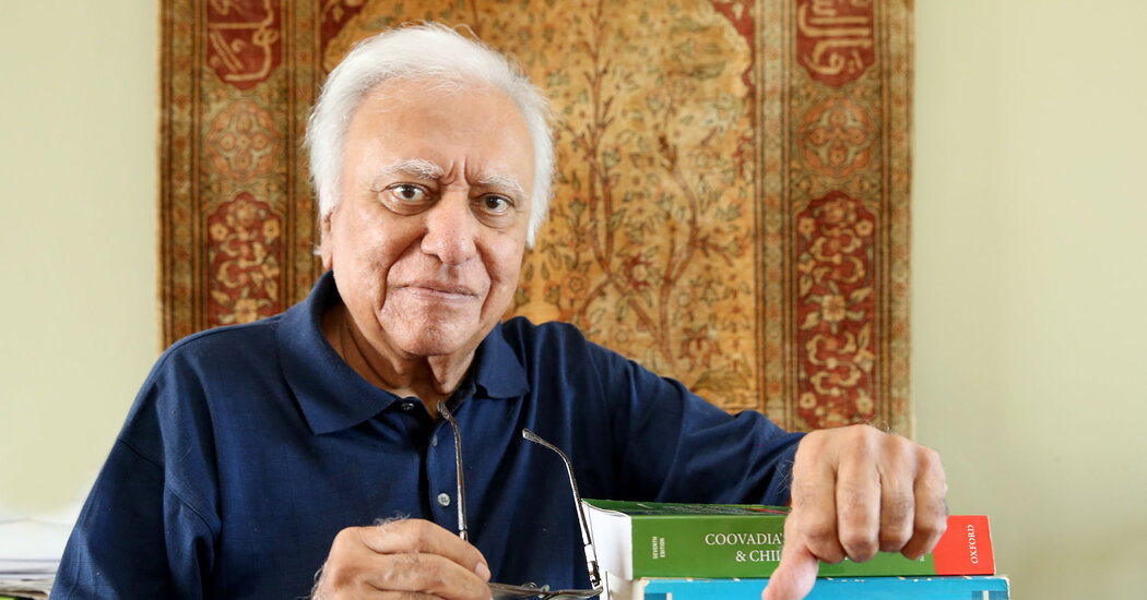
An esteemed pediatrician, he overcame apartheid’s barriers to help make his country a global leader in H.I.V. care and research.Hoosen Coovadia, a pediatrician who used science to fight for racial justice in apartheid South Africa and later transformed the approach to H.I.V. treatment for pregnant women in Africa and beyond, died on Oct. 4 at his home in Durban. He was 83.His daughter, Anuschka Coovadia, a physician in South Africa, confirmed the death, saying he had been in poor health for two years and was further debilitated by a case of Covid-19 several months ago that kept him in intensive care for weeks.Dr. Jerry Coovadia, as he was familiarly known, was a leader in the struggle against white rule in South Africa and campaigned for decades for the political transition that brought the African National Congress to power in 1994.But when, four years later, President Thabo Mbeki began to deny that the human immunodeficiency virus caused AIDS, and asserted that new treatments for H.I.V. were poisons he would not permit to be given to South Africans, Dr. Coovadia became one of the government’s fiercest critics.“Never was a doctor so vilified as Jerry Coovadia in the A.N.C. for his implacable and quietly militant stand against Thabo Mbeki,” said Zackie Achmat, the founder of the Treatment Action Campaign, a movement of people living with H.I.V. that also battled Mr. Mbeki, who was unseated in a party putsch in 2008.Dr. Coovadia and the activists he supported eventually won that fight, and he helped make South Africa a global leader in H.I.V. care and research. He also mended some relationships with the government, although he remained a vocal critic of inequity in the post-apartheid years.In a statement after Dr. Coovadia’s death, President Cyril Ramaphosa of South Africa said, “Our nation’s loss will be felt globally, but we can take pride at and comfort from the emergence of a giant of science and an icon of compassion and resilience from our country.”Hoosen Mahomed Coovadia was born on Aug. 2, 1940, in Durban. His parents, Mohamed Coovadia and Khateja Moosa, came from prosperous merchant families in Durban’s Indian community, though his father, a compulsive gambler, lost most of his wealth while Dr. Coovadia was still young.He was admitted to the University of Natal medical college, set up by the apartheid government for Black and so-called colored students like Dr. Coovadia, but after a short time he concluded that the education it offered was inferior. He applied to study in Cape Town, but the government denied him the permit to travel that he required as a nonwhite student.Instead, he traveled to India and enrolled at Grant Medical College in Bombay (now Mumbai). There he was exposed to anticolonial ideas and met other South African students, with whom he organized a political association. Prominent leaders of the anti-apartheid movement visiting India would address them.Dr. Coovadia, with a medical degree from Grant, returned to South Africa in 1966. Three years later, he married Zubeida Hamed, who had also graduated from Grant and was finishing her training in dermatology. Dr. Hamed shared her new husband’s growing interest in activism, and their home, in Durban, became a mecca for political meetings. Dr. Coovadia went to work as a pediatrician at King Edward VIII Hospital in Durban, an institution that could treat only nonwhite South Africans under apartheid, and later joined the department of pediatrics at the University of Natal Medical School (now part of the University of KwaZulu-Natal). He came under suspicion by the regime for conducting research on topics such as racial disparities in infant mortality in South Africa. He also joined the Natal Indian Congress, an anti-apartheid organization, and soon became a leader of it.In 1975, Dr. Coovadia earned a master’s degree in immunology from the University of Birmingham in Britain. Returning to South Africa, he found opposition to apartheid there swelling into open revolt. He helped found the United Democratic Front, a coalition of more than 400 trade unions, religious organizations and other civic groups opposed to white rule. In 1989, the police raided and ransacked his home in search of papers related to secret talks between the regime and the A.N.C.Dr. Coovadia in 1989 as a leader of the Natal Indian Congress, an anti-apartheid organization. He and another group member, Paddy Kearney, displayed a list of 189 people arrested for political activities. 1860 Heritage CentreA month later, South African secret police planted a bomb in front of Dr. Coovadia’s home. His son, Imraan, a novelist and professor of creative writing at the University of Cape Town, said his father had become such a prominent critic of apartheid abroad, speaking at scientific meetings, that the regime had decided to eliminate him. The bomb destroyed the second floor of the house, but the family survived.“It took weeks to get the walls rebuilt,” his daughter, Anuschka, said, “and during that time, my father’s medical students came on a schedule, protecting the house with broom poles and sticks, sitting out all night. There was so much love from his community of students.”In addition to his son and daughter, he is survived by his wife and five grandchildren.Dr. Coovadia wrote a textbook on child health now in its seventh edition, mentored dozens of students and researchers, many of whom became health ministers and key figures in global health, and conducted pioneering work on measles and pediatric kidney disorders. He advised successive South African governments from various positions, including a seat on the powerful National Planning Commission; led international research projects; published widely in scientific journals; and received awards, including the Star of South Africa, the country’s highest honor, presented by President Nelson Mandela.But it was his work on H.I.V. that had perhaps the greatest impact on global policy, and which drew him into an unexpectedly vicious political battle.In the late 1980s, he started to see babies with H.I.V. arriving at the hospital, prompting him to begin researching ways to stop the transmission of the virus from mothers to their children. “He considered it another form of oppression for these women, who were Black, who were poor, who were often rural — and on top of all of that, had H.I.V.,” said Salim Abdool Karim, a leading authority on H.I.V. globally and a former student of Dr. Coovadia’s.By the 1990s, the World Health Organization was recommending that women with H.I.V. feed their children with baby formula rather than breast milk, which could transmit the virus. But Dr. Coovadia suspected — and then proved in a series of studies — that the risk was minimal in exclusively breastfed infants, and that the health benefits for infants whose mothers did not have access to clean water with which to mix formula far outweighed the risk from H.I.V.Dr. Coovadia battled the W.H.O. and succeeded in having the policy reversed. He also helped demonstrate that giving antiretroviral drugs to pregnant women could prevent them from transmitting the virus to babies at birth.South Africa had the world’s largest number of people living with the virus by the late 1990s, but when President Mbeki, to whom Dr. Coovadia had earlier been close, created a commission to govern the AIDS response, he stacked it with rogue researchers and self-proclaimed experts known as “AIDS denialists.” Manto Tshabalala-Msimang, a health minister appointed by Mr. Mbeki, told people living with H.I.V. that they could stave off AIDS by eating garlic and beets. Dr. Coovadia was incensed.In 2000, as he prepared to become a co-chair of a major global AIDS conference in Durban, the A.N.C. government put immense pressure on him to present the denialist view.“He was such a courageous and principled person: He would not give in,” said Dr. Peter Piot, who was then the head of the United Nations AIDS agency and who joined Dr. Coovadia in “turbulent” meetings with government officials ahead of the event.President Mbeki, as head of state, opened the conference and, to the horror of much of the audience, reiterated his denialist view. The government then demanded that Dr. Coovadia allow Dr. Tshabalala-Msimang to close the event.Dr. Coovadia’s daughter recalled that 10 “thuggish” men came to the family’s hotel room the night before the conference ended and ordered him to a meeting with the minister. “When he came back, he was utterly shaken,” she said. “He didn’t sleep that night.”Knowing it might mean both the end of his professional life and ostracism from the party for which he had fought for decades, Dr. Coovadia nevertheless refused the minister the platform. Instead, the keynote speaker at the final session was Mr. Mandela, who had stepped down as president a few years earlier. He exhorted the world to bring AIDS treatment to his country and the rest of Africa.After Mr. Mbeki left office, Dr. Coovida helped South Africa roll out what became the world’s largest H.I.V.- treatment programs.

Read more →

Brain tumor treatment by targeting TUG1, a gene that controls replication stress

A new study has unravelled a crucial link between how cancer cells cope with replication stress and the role of Taurine Upregulated Gene 1 (TUG1). By targeting TUG1 with a drug, the researchers were able to control brain tumor growth in mice, suggesting a potential strategy to combat aggressive brain tumors such as glioblastomas.
“These findings have the potential to be translated into therapeutic applications, as TUG1 is highly expressed in glioblastoma,” said lead researcher Professor Yutaka Suzuki. “In this study, we successfully developed a therapeutic drug named TUG1-DDS, which selectively targets TUG1. It significantly suppressed tumor growth and improved survival, especially when administered in combination with the standard treatment of temozolomide. Therefore, it is a potentially effective therapeutic agent for treating glioblastoma.”
To understand how TUG1 could potentially treat the most dangerous forms of brain cancer, it is important to understand how cancer turns the usual processes of host cells against themselves to create an environment favorable to cancer cell growth. Even essential cell processes, such as replication, are used to the cancer’s advantage.
When a cell divides, it replicates its DNA, so that both cells have a full complement of hereditary information. The double-stranded DNA is unwound and separated into two single strands that each serve as a template for creating two identical copies by combining with RNA. A DNA:RNA hybrid structure called an R-loop helps unwind the DNA and stabilizes it as it is unwound.
To improve the conditions for cancer cells, the invasive cells hinder the natural process of DNA replication. The cells induce replication stress (RS), which results in the DNA strands breaking and unpaired single strands of DNA increasing. The result is an instability in the genome that favors tumor growth.
Cancer cells have a tricky balancing act because the increased activity can potentially backfire. RS and R-loop accumulation can also cause cancer cell death. To regulate the genome, cancer cells turn to long noncoding RNAs (lncRNAs), which allow them to repair their own DNA damage and remove unwanted R-loops.
In a new study, led by Yutaka Kondo and Miho Suzuki at Nagoya University Graduate School of Medicine, the investigators identified the role of the lncRNA TUG1. They found that TUG1 suppresses the potentially harmful R-loops together with two proteins, DHX9 and RPA32. Taken together, the TUG1-RPA-DHX9 interaction is an indispensable mechanism for regulating R-loops in regions that are known to be susceptible to DNA damage and mutations. Their findings were published in Nature Communications.
Kondo and Suzuki also found that TUG1 was rapidly up-regulated in response to RS. When they reduced TUG1 expression in cancer cells, they found severe DNA damage and cell death. “It was exciting to see the rapid increase in expression of TUG1 in response to replication stress,” said Dr. Suzuki. She continues: “Normally, it takes several hours or more for proteins to increase in response to stimuli, but RNA can be synthesized rapidly. That TUG1, an RNA molecule, increases immediately in response to replication stress indicates that it is necessary to respond quickly to critical situations.”
These findings offer hope for the development of treatment for other cancers. As Dr. Kondo explains: “TUG1 inhibitors have also been found to be effective in other types of cancer, such as pancreatic cancer and ovarian cancer. Therefore, our novel treatment, TUG1-DDS, could also be effective in other cancer types with expression of TUG1.”

Read more →

Win-win in muscle research: Faster results and fewer laboratory animals thanks to new method

To study muscle diseases, scientists rely on the mouse as a model organism. Researchers at the University of Basel have now developed a new method that is not only faster and more efficient than conventional ones but also greatly reduces the number of experimental animals needed for studying the function of genes in muscle fibers.
Researchers use the mouse as a model organism to study the structure and function of skeletal muscle, neuromuscular diseases and aging processes in muscle. The scientists are aware of their responsibility in the use of animals and have committed themselves at the University of Basel to rigorously implement the so-called 3R principles — Replacement, Reduction, Refinement — in animal-assisted research and animal husbandry.
The new method developed by Professor Markus Rüegg’s research group at the Biozentrum, University of Basel, is a further step towards reducing the number of laboratory animals. This method also opens new ways to investigate several genes simultaneously or even entire signaling pathways in muscle fibers quickly, cost-effectively and efficiently. The results of the study have now been published in Nature Communications.
The difficulty of studying genes in muscle fibers
Studying gene function in muscle is challenging. On the one hand, muscle fibers are very large and very fragile when isolated. On the other hand, in humans, they are up to half a meter long and contain thousands of nuclei. In order to change and study gene function in muscle fibers, all of the muscle fiber nuclei must be changed, which is difficult to achieve.
For some years now, scientists have been using the CRISPR/Cas9 method to study gene function. This method uses a virus to introduce the so-called Cas9 protein and a specifically designed guide RNA into the organism and thus into the nuclei. The Cas9 protein cuts the genomic DNA at the site recognized by the guide RNA. This combination of Cas9 protein and guide RNA allows altering gene function in the cell.
The CRISPR-Cas9 method can be split up
However, to ensure that the virus only alters the gene expression of muscle fibers and not those of other organs at the same time, the research team combined the CRISPR/Cas9 method with another method: First, the researchers succeeded in breeding mice with the Cas9 protein already present in their muscle fibers — but only there. They then introduced the desired guide RNA into the organism with a so-called adeno-associated virus, which infects muscle.

Read more →