Twins peak with more born than ever before

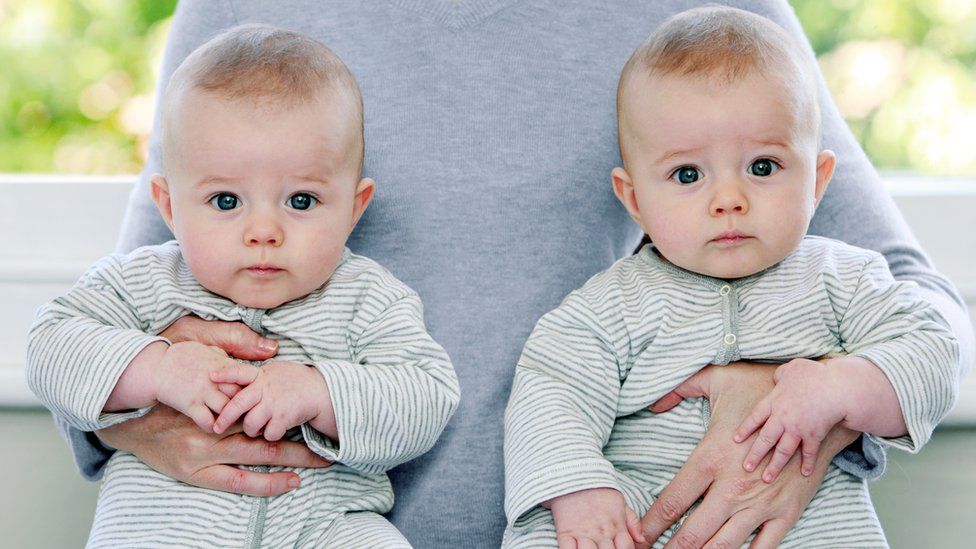
SharecloseShare pageCopy linkAbout sharingimage copyrightGetty ImagesMore twins are being born than ever before but the world may now have reached peak twin, researchers say.About 1.6 million twins are born each year worldwide, with one in every 42 children born a twin.Delayed childbearing and medical techniques such as IVF have seen the rate of twin births rise by a third since the 1980s.But it could be all downhill from here as the focus shifts to one baby per pregnancy, which is less risky.According to a global overview in the journal Human Reproduction, the peak was reached because of large increases in twinning rates in all regions over 30 years – from a 32% rise in Asia to a 71% rise in North America.The researchers collected information on twinning rates from 165 countries for 2010 to 2015, and compared them with rates for 1980 to 1985. The number of twins born per thousand deliveries is now particularly high in Europe and North America – and worldwide it’s gone from nine per 1,000 deliveries to 12.But twin rates in Africa have always been high and haven’t changed much over the past 30 years, which could be due to population growth.Helping handAfrica and Asia make up about 80% of all twin deliveries in the world at present.Prof Christiaan Monden, the study’s author from the University of Oxford, said there was a reason for that.”The twinning rate in Africa is so high because of the high number of dizygotic twins – twins born from two separate eggs – born there,” he said.”This is most likely to be due to genetic differences between the African population and other populations.”Twinning rates in Europe, North America and Oceanic countries have been catching up – and the increasing use of medically assisted reproduction since the 1970s – for example IVF, ICSI, artificial insemination and ovarian stimulation – have been the main reason.image copyrightGetty ImagesThese techniques all increase the likelihood of a multiple birth.Women choosing to start families later in life, increased use of contraception and lower fertility overall also play a role, the review says.But the emphasis is now on singleton pregnancies, which are safer, says Prof Monden.”This is important as twin deliveries are associated with higher death rates among babies and children, and more complications for mothers and children during pregnancy, and during and after delivery,” he says. Twins have more complications at birth, are more often born premature and have lower birth weights and higher still birth rates.Survival chancesThe review found that the fate of twins in low and middle-income countries was more of a concern.In sub-Saharan Africa, in particular, many twins will lose their co-twin in their first year of life – more than 200,000 each year.”While twinning rates in many rich Western countries are now getting close to those in sub-Saharan Africa, there is a huge difference in the survival chances,” said Prof Jeroen Smits, a study author.Looking ahead, the researchers say India and China will play a major role in future twinning rates.Declining fertility, older mothers at birth and techniques like IVF will all have a bearing on the numbers of twins in years to come.

Read more →

Stress reduction as a path to eating less fast food

Overweight low-income mothers of young kids ate fewer fast-food meals and high-fat snacks after participating in a study — not because researchers told them what not to eat, but because the lifestyle intervention being evaluated helped lower the moms’ stress, research suggests.
The 16-week program was aimed at preventing weight gain by promoting stress management, healthy eating and physical activity. The methods to get there were simple steps tucked into lessons on time management and prioritizing, many demonstrated in a series of videos featuring mothers like those participating in the study.
“We used the women’s testimonies in the videos and showed their interactions with their families to raise awareness about stressors. After watching the videos, a lot of intervention participants said, ‘This is the first time I’ve realized I am so stressed out’ — because they’ve lived a stressful life,” said Mei-Wei Chang, lead author of the study and associate professor of nursing at The Ohio State University.
“Many of these women are aware of feeling impatient, and having head and neck pain and trouble sleeping — but they don’t know those are signs of stress.”
An analysis of the study data showed that the women’s lowered perceived stress after participating in the intervention was the key factor influencing their eventual decrease in consumption of high-fat and fast foods.
“It’s not that these women didn’t want to eat healthier,” Chang said. “If you don’t know how to manage stress, then when you are so stressed out, why would you care about what you eat?”
The research is published in a recent issue of the journal Nutrients.

advertisement

The 338 participants, overweight or obese moms between the ages of 18 and 39, were recruited from the Special Supplemental Nutrition Program for Women, Infants, and Children (WIC), which serves low-income mothers and children up to age 5. Those eligible for the program must have an annual household income no higher than 185 percent of the federal poverty line.
Chang said these women are likely to face a number of challenges that could cause them stress: financial difficulties, living in run-down neighborhoods, frequent moves, unstable romantic relationships and households bustling with little kids. It’s also common for this population to retain 10 or more pounds of pregnancy weight after childbirth and risk life-long obesity and potential problems for themselves and new babies if they become pregnant again.
During the trial, the 212 participants randomized into the intervention group watched a total of 10 videos in which women like them gave unscripted testimonials about healthy eating and food preparation, managing their stress and being physically active. Participants also dialed in to 10 peer support group teleconferences over the course of the study.
Chang and colleagues previously reported that as a group, the women in the intervention arm of the study were more likely to have reduced their fat consumption than women in a comparison group who were given print materials about lifestyle change.
This newer analysis showed that the intervention’s lessons alone did not directly affect that change in diet. When the researchers assessed the potential role of stress as a mediator, the indirect effect of the intervention — reducing participants’ perceived stress — was associated with less consumption of high-fat foods, including fast food. A 1-point reduction in the scale measuring stress was linked to a nearly 7% reduction in how frequently the women ate high-fat foods.

advertisement

The intervention focused on showing the women examples of how they could achieve a healthier and less stressful lifestyle rather than telling them what they had to change.
“I learned a lot from those women,” Chang said. “Everything needs to be practical and applicable to daily life — anytime, anywhere.”
Some examples: Comparing a bag of chips to a bag of apples — the chips might be half the price, but they supply far fewer family snacks. Or using a household responsibility chart to assign tasks to young children, and encouraging moms to reward kids with a hug or individual attention when they follow the instructions. And taking deep breaths to counter the feeling of being overwhelmed.
When it came to stress management, the researchers focused on advising the women to shift their thinking, and not to blame themselves when things go wrong, rather than to take on solving the problems that caused them stress.
“We raised their awareness about stressors in their lives, and unfortunately a lot of these problems are not within their control,” Chang said. “So we teach them ways to control their negative emotions — remember that this is temporary, and you can get through it. And give them confidence to look to the future.”
The videos from the intervention are now part of WIC’s continuing education series for mothers. This work was supported by the National Institutes of Health. Co-authors included Duane Wegener from Ohio State and Roger Brown from the University of Wisconsin-Madison.

Read more →

Preterm birth, prolonged labor influenced by progesterone balance

New research by the National Institutes of Health found that unbalanced progesterone signals may cause some pregnant women to experience preterm labor or prolonged labor. The study in mice — published online in the Proceedings of the National Academy of Sciences — provides novel insights for developing treatments.
During pregnancy, the hormone progesterone helps to prevent the uterus from contracting and going into labor prematurely. This occurs through molecular signaling involving progesterone receptor types A and B, referred to as PGR-A and PGR-B. In this first-of-its-kind study, the scientists showed how unbalanced PGR-A and PGR-B signaling can affect pregnancy duration.
“We used genetically engineered mouse models to alter the ratio of PGR-A and PGR-B in the muscle compartment of the uterus, called the myometrium,” said senior author Francesco DeMayo, Ph.D., head of the National Institute of Environmental Health Sciences Reproductive and Developmental Biology Laboratory. “Our team found that PGR-A promotes muscle contraction and PGR-B prevents such contraction, and we identified the biological pathways influenced by both forms.”
Previous research showed that PGR-A regulates processes involved in initiating childbirth and that PGR-B affects molecular pathways related to maintaining the normal course of pregnancy. This study builds on those findings, revealing that the relative abundance of PGR-A and PGR-B may be critical in promoting healthy pregnancy. The public health implications are significant.
Preterm birth affects 10% of all pregnancies and is the primary cause of neonatal morbidity and mortality worldwide, while prolonged labor increases the risks of infection, uterine rupture, and neonatal distress, according to the researchers.
The scientists pointed out that care for preterm deliveries can result in high social and economic costs, with infants born preterm at greater risk for experiencing disorders ranging from blindness to cerebral palsy. Prolonged labor can harm both mother and infant and lead to cesarean delivery.
Progesterone treatment aimed at preventing premature labor can help a subset of patients, but for other individuals, confounding factors may reduce effectiveness, noted Steve Wu, Ph.D., first author on the study and a staff scientist in DeMayo’s lab. Wu said that the research team found novel molecules that control uterine muscle contraction, and they could serve as future therapeutic targets. He added that the current study also may help to advance treatment for labor dystocia — the clinical name for abnormally slow or protracted labor.
“Although labor stimulation by oxytocin infusion is an approved measure to mitigate labor dystocia, serious side effects have been associated with this treatment,” said Wu. “Novel proteins that we identified as being part of progesterone signaling could serve as a key molecular switch of uterine contraction, through drug-dependent regulation of their activities,” he explained.
“Hormone signaling in pregnancy is complicated and involves both the hormone levels and the types of receptors in the uterus that sense the hormones,” said co-first author Mary Peavey, M.D., from the department of obstetrics and gynecology at the University of North Carolina at Chapel Hill. “This publication sheds light on how hormones influence labor and can thus be used to help women when the uterus goes into labor too soon or for a prolonged period.”

Read more →

Unique Ag-hydrogel composite for soft bioelectronics created

In the field of robotics, metals offer advantages like strength, durability, and electrical conductivity. But, they are heavy and rigid — properties that are undesirable in soft and flexible systems for wearable computing and human-machine interfaces.
Hydrogels, on the other hand, are lightweight, stretchable, and biocompatible, making them excellent materials for contact lenses and tissue engineering scaffolding. They are, however, poor at conducting electricity, which is needed for digital circuits and bioelectronics applications.
Researchers in Carnegie Mellon University’s Soft Machines Lab have developed a unique silver-hydrogel composite that has high electrical conductivity and is capable of delivering direct current while maintaining soft compliance and deformability. The findings were published in Nature Electronics.
The team suspended micrometer-sized silver flakes in a polyacrylamide-alginate hydrogel matrix. After going through a partial dehydration process, the flakes formed percolating networks that were electrically conductive and robust to mechanical deformations. By manipulating this dehydration and hydration process, the flakes can be made to stick together or break apart, forming reversible electrical connections.
Previous attempts to combine metals and hydrogels revealed a trade-off between improved electrical conductivity and lowered compliance and deformability. Majidi and his team sought to tackle this challenge, building on their expertise in developing stretchable, conductive elastomers with liquid metal.
“With its high electrical conductivity and high compliance or ‘squishiness,’ this new composite can have many applications in bioelectronics and beyond,” explained Carmel Majidi, professor of mechanical engineering. “Examples include a sticker for the brain that has sensors for signal processing, a wearable energy generation device to power electronics, and stretchable displays.”
The silver-hydrogel composite can be printed by standard methods like stencil lithography, similar to screen printing. The researchers used this technique to develop skin-mounted electrodes for neuromuscular electrical stimulation. According to Majidi, the composite could cover a large area of the human body, “like a second layer of nervous tissue over your skin.”
Future applications could include treating muscular disorders and motor disabilities, such as assisting someone with tremors from Parkinson’s disease or difficulty grasping something with their fingers after a stroke.

Story Source:
Materials provided by College of Engineering, Carnegie Mellon University. Original written by Lisa Kulick. Note: Content may be edited for style and length.

Read more →

The U.S. Is Sitting on Tens of Millions of Vaccine Doses the World Needs

#masthead-section-label, #masthead-bar-one { display: none }The Coronavirus OutbreakliveLatest UpdatesMaps and CasesRisk Near YouVaccine RolloutGuidelines After VaccinationAdvertisementContinue reading the main storySupported byContinue reading the main storyThe U.S. Is Sitting on Tens of Millions of Vaccine Doses the World NeedsDoses from the drug maker AstraZeneca sit in U.S. manufacturing plants, awaiting regulatory authorization as the world goes begging.An Emergent BioSolutions laboratory in Baltimore. The company has already produced tens of millions of doses of the AstraZeneca vaccine, which cannot be used in the United States yet.Credit…Michael Robinson Chavez/The Washington Post, via Getty ImagesNoah Weiland and March 11, 2021, 6:25 p.m. ETWASHINGTON — Tens of millions of doses of the coronavirus vaccine made by the British-Swedish company AstraZeneca are sitting idly in American manufacturing facilities, awaiting results from its U.S. clinical trial while countries that have authorized its use beg for access.The fate of those doses of AstraZeneca’s vaccine is the subject of an intense debate among White House and federal health officials, with some arguing the administration should let them go abroad where they are desperately needed while others are not ready to relinquish them, according to senior administration officials.AstraZeneca is involved in those conversations.“We understand other governments may have reached out to the U.S. government about donation of AstraZeneca doses, and we’ve asked the U.S. government to give thoughtful consideration to these requests,” said Gonzalo Viña, a spokesman for AstraZeneca.About 30 million doses are currently bottled at AstraZeneca’s facility in West Chester, Ohio, which handles “fill-finish,” the final phase of the manufacturing process during which the vaccine is placed in vials, one official with knowledge of the stockpile said.Emergent BioSolutions, a company in Maryland that AstraZeneca has contracted to manufacture its vaccine in the United States, has also produced enough vaccine in Baltimore for tens of millions more doses once it is filled into vials and packaged, the official said.But although AstraZeneca’s vaccine is already authorized in more than 70 countries, according to a company spokesman, its U.S. clinical trial has not yet reported results, and the company has not applied to the Food and Drug Administration for emergency use authorization. AstraZeneca has asked the Biden administration to let it loan American doses to the European Union, where it has fallen short of its original supply commitments and where the vaccination campaign has stumbled badly.The administration, for now, has denied the request, one official said.Some federal officials have pushed the White House to make a decision in the next few weeks. Officials have discussed sending doses to Brazil, hard hit by a worsening coronavirus crisis.“If those donation actions were to proceed, we would seek guidance from the U.S. government on replacement of doses for use in the U.S.,” Mr. Viña said.The White House did not respond to a request for comment.The administration’s hesitation is at least partly related to uncertainties with vaccine supply before a benchmark of late May laid down by President Biden when he promised enough vaccine doses to cover every adult in the United States. Vaccine production is notoriously complex and delicate, and problems like mold growth can interrupt a plant’s progress.Last May, the Trump administration pledged up to $1.2 billion to AstraZeneca to finance the development and manufacturing of its vaccine, which it developed with the University of Oxford, and to supply the United States with 300 million doses if it proved effective. Federal officials and public health experts last year viewed the vaccine, which is less expensive and easier to store for long periods than some other vaccines, as most likely to be among the first to receive authorization.That never happened, in part because of a pattern of communication blunders by AstraZeneca that weakened the company’s relationship with American regulators and slowed the vaccine’s development. Last fall, AstraZeneca’s trial in the United States — the same one that will soon report results — was grounded for nearly seven weeks because the company was slow to provide the F.D.A. with evidence that the vaccine had not caused serious neurological side effects in two volunteers.The company is now grappling with another safety scare. Acting out of precaution, health authorities in Denmark, Norway and Iceland suspended use of the AstraZeneca’s vaccine on Thursday after several reports across the continent of severe blood clots.European official and the company said there was not evidence of any causal link. In the vast majority of cases, the emergence of such medical conditions has nothing to do with the vaccine. Some percentage of people are expected to fall ill by chance after getting vaccinated, as would happen in any group of people.The Coronavirus Outbreak

Read more →

Researchers reveal 3D structure responsible for gene expression

For the first time ever, a Northwestern University-led research team has peered inside a human cell to view a multi-subunit machine responsible for regulating gene expression.
Called the Mediator-bound pre-initiation complex (Med-PIC), the structure is a key player in determining which genes are activated and which are suppressed. Mediator helps position the rest of the complex — RNA polymerase II and the general transcription factors — at the beginning of genes that the cell wants to transcribe.
The researchers visualized the complex in high resolution using cryogenic electron microscopy (cryo-EM), enabling them to better understand how it works. Because this complex plays a role in many diseases, including cancer, neurodegenerative diseases, HIV and metabolic disorders, researchers’ new understanding of its structure could potentially be leveraged to treat disease.
“This machine is so basic to every branch of modern molecular biology in the context of gene expression,” said Northwestern’s Yuan He, senior author of the study. “Visualizing the structure in 3D will help us answer basic biological questions, such as how DNA is copied to RNA.”
“Seeing this structure allows us to understand how it works,” added Ryan Abdella, the paper’s co-first author. “It’s like taking apart a common household appliance to see how everything fits together. Now we can understand how the proteins in the complex come together to perform their function.”
The study will be published March 11 in the journal Science. This marks the first time the human Mediator complex has been visualized in 3D in the human cell.

advertisement

He is an assistant professor of molecular biosciences in Northwestern’s Weinberg College of Arts and Sciences. Abdella and Anna Talyzina, both graduate students in the He lab, are co-first authors of the paper.
Famed biochemist Roger Kornberg discovered the Mediator complex in yeast in 1990, a project for which he won the 2006 Nobel Prize in Chemistry. But Mediator comprises a daunting 26 subunits — 56 total when combined with the pre-initiation complex — it’s taken researchers until now to obtain high-resolution images of the human version.
“It’s a technically quite challenging project,” He said. “These complexes are scarce. It takes hundreds of liters of human cells, which are very hard to grow, to obtain small amounts of the protein complexes.”
A breakthrough came when He’s team put the sample on a single layer of graphene oxide. By providing this support, the graphene sheet minimized the amount of sample needed for imaging. And compared to the typical support used — amorphous carbon — graphene improved the signal-to-noise ratio for higher-resolution imaging.
After preparing the sample, the team used cryo-EM, a relatively new technique that won the 2017 Nobel Prize in Chemistry, to determine the 3D shape of proteins, which are often thousands of times smaller than the width of a human hair. The technique works by blasting a stream of electrons at a flash-frozen sample to take many 2D images.

advertisement

For this study, He’s team captured hundreds of thousands of images of the Med-PIC complex. They then used computational methods to reconstruct a 3D image.
“Solving this complex was like assembling a puzzle,” Talyzina said. “Some of those subunits were already known from other experiments, but we had no idea how the pieces assembled together or interacted with each other. With our final structure, we were finally able to see this whole complex and understand its organization.”
The resulting image shows the Med-PIC complex as a flat, elongated structure, measuring 45 nanometers in length. The researchers also were surprised to discover that the Mediator moves relative to the rest of the complex, binding to RNA polymerase II at a hinge point.
“Mediator moves like a pendulum,” Abdella said. “Next, we want to understand what this flexibility means. We think it might have an impact on the activity of a key enzyme within the complex.”
The paper was supported by Northwestern’s Chemistry of Life Sciences Institute, the Chicago Biomedical Consortium with support from the Searle Funds at The Chicago Community Trust, the American Cancer Society (award number IRG-15-173-21), the H Foundation (award number U54-CA193419) and the National Institutes of Health (award numbers R01-GM135651 and P01-CA092584).

Read more →

New insight into how cancer spreads

Breast cancer is harmful enough on its own, but when cancer cells start to metastasize — or spread into the body from their original location — the disease becomes even more fatal and difficult to treat.
Thanks to new research published in Oncogene from the lab of University of Colorado Cancer Center associate director of basic research Heide Ford, PhD, in collaboration with Michael Lewis, PhD, from Baylor College of Medicine, doctors may soon have a better understanding of one mechanism by which metastasis happens, and of potential ways to slow it down.
“Metastasis is a huge problem nobody’s tackled very well,” says Ford, who holds the Grohne Endowed Chair in Cancer Research at the University of Colorado School of Medicine. “People don’t know how to inhibit the process of metastasis, nor how to inhibit the growth of metastatic cells at secondary sites. And that’s what kills most cancer patients. A lot of common drugs, whether they’re targeted drugs or chemotherapies that are less targeted, do pretty well at inhibiting the primary tumor, but by the time cells metastasize, they’ve changed enough that they don’t get inhibited by those drugs.”
The transformation Ford and her team are studying happens when cells called epithelial cells, which are more adherent to one another and less likely to spread to other parts of the body, start to take on the characteristics of mesenchymal cells, which are more migratory and more likely to invade other parts of the body. This transformation is referred to as the epithelial-to-mesenchymal transition.
“When the epithelial cancer cells take on these characteristics of mesenchymal cells, they become less attached to their neighbor and they become more able to degrade membranes, so they can get into the bloodstream more easily,” Ford says.
In 2017, Ford published a paper showing that the metastasis process is helped along when cells that have undergone the epithelial-to-mesenchymal transition start “talking” to cells that haven’t, making those cells more likely to gain metastatic properties.
In a new paper published in December, Ford and her researchers, in a collaborative study done with Lewis and colleagues at Baylor College of Medicine, posit that the crosstalk is facilitated by a naturally occurring protein called VEGF-C.
“VEGF-C is secreted by the cells. It binds to receptors on these neighboring cells and then activates a pathway called the hedgehog signaling pathway, though it bypasses the traditional way of activating this pathway,” Ford says. “That turns on a signaling mechanism that ultimately results in activation of a protein called GLI that makes these cells more invasive and more migratory.”
In their new paper, Ford, Lewis and their researchers show that if you can inhibit production of VEGF-C, you can significantly slow metastasis.
“If you take out the receptor that receives the signal from the cells that have not undergone a transition, or if you take VEGF-C out of the mix, you can’t stimulate metastasis to the same degree,” she says. “If you remove that ability for these different cell types to crosstalk, now these cells that never underwent a transition can’t move as well anymore. They can’t metastasize as efficiently.”
The researchers are now in the early stages of animal trials to find out the best way to target that signaling pathway in order to better inhibit metastasis. They want to find out if they can stop metastasis from happening at all, and if they can slow its progression in patients in whom the metastatic process has already begun — and to see if they can inhibit tumor growth at the secondary site.
“For many years, people said there was no point in finding inhibitors to metastasis because by the time someone comes into the clinic, the horse is out of the barn, so to speak. The cells have already gotten out of the primary tumor and you can’t do anything about it,” Ford says. “But that’s not necessarily true. Now, data show that if you have cells that have metastasized to a second site — say you have breast cancer and the cells went into the lungs — those cells that are in the lungs could in fact start metastasizing to other sites. You want to stop that process no matter where you are in this progression.”

Read more →

Standard vital signs could help estimate people's pain levels

A new study demonstrates that machine-learning strategies can be applied to routinely collected physiological data, such as heart rate and blood pressure, to provide clues about pain levels in people with sickle cell disease. Mark Panaggio of Johns Hopkins University Applied Physics Laboratory and colleagues present these findings in the open-access journal PLOS Computational Biology.
Pain is subjective, and monitoring pain can be intrusive and time-consuming. Pain medication can help, but accurate knowledge of a patient’s pain is necessary to balance relief against risk of addiction or other unwanted effects. Machine-learning strategies have shown promise in predicting pain from objective physiological measurements, such as muscle activity or facial expressions, but few studies have applied machine learning to routinely collected data.
Now, Panaggio and colleagues have developed and applied machine-learning models to data from people with sickle cell disease who were hospitalized due to debilitating pain. These statistical models classify whether a patient’s pain was low, moderate, or high at each point during their stay based on routinely collected measurements of their blood pressure, heart rate, temperature, respiratory rate, and oxygen levels.
The researchers found that these vital signs indeed gave clues into the patients’ reported pain levels. By taking physiological data into account, their models outperformed baseline models in estimating subjective pain levels, detecting changes in pain, and identifying atypical pain levels. Pain predictions were most accurate when they accounted for changes in patients’ vital signs over time.
“Studies like ours show the potential that data-driven models based on machine learning have to enhance our ability to monitor patients in less invasive ways and ultimately, be able to provide more timely and targeted treatments,” Panaggio says.
Looking ahead, the researchers hope to leverage more comprehensive data sources and real-time monitoring tools, such as fitness trackers, to build better models for inferring and forecasting pain.

Story Source:
Materials provided by PLOS. Note: Content may be edited for style and length.

Read more →

AI analysis of how bacteria attack could help predict infection outcomes

Insights into how bacterial proteins work as a network to take control of our cells could help predict infection outcomes and develop new treatments.
Much like a hacker seizes control of a company’s software to cause chaos, disease-causing bacteria, such as E. coli and Salmonella, use miniature molecular syringes to inject their own chaos-inducing agents (called effectors) into the cells that keep our guts healthy.
These effectors take control of our cells, overwhelming their defences and blocking key immune responses, allowing the infection to take hold.
Previously, studies have investigated single effectors. Now a team led by scientists at Imperial College London and The Institute of Cancer Research, London, and including researchers from the UK, Spain and Israel, has studied whole sets of effectors in different combinations.
The study, published today in Science, investigated data from experiments in mice infected with the mouse version of E. coli, called Citrobacter rodentium, which injects 31 effectors.
The results show how effectors work together as a network, allowing them to colonise their hosts even if some effectors are removed. The investigation also revealed how the host’s immune system can bypass the obstacles the effectors create, triggering complementary immune responses.

advertisement

The researchers suggest that knowing how the makeup of effector networks influences the ability of infections to take hold could help design interventions that disrupt their effects.
Study lead Professor Gad Frankel, from the Department of Life Sciences at Imperial, said: “The data represent a breakthrough in our understanding of the mechanisms of bacterial infections and host responses. Our results show that the injected effectors are not working individually, but instead as a pack.
“We found there is an inherent strength and flexibility to the network, which ensures that if one or several components don’t work, the infection can go on. Importantly, this work has also revealed that our cells have a built-in firewall, which means that we can deal with the hacker’s corruptive networks and mount effective immune responses that can clear the infection.”
Study co-lead Professor Jyoti Choudhary, from the Functional Proteomics Lab at The Institute of Cancer Research, London, said: “Our study shows that we can predict how a cell will respond when attacked by different combinations of bacterial effector proteins. The research will help us to better understand how cells, the immune system and bacteria interact, and we can apply this knowledge to diseases like cancer and inflammatory bowel disease where bacteria in the gut play an important role.
“We hope, through further study, to build on this knowledge and work out exactly how these effector proteins function, and how they work together to disrupt host cells. In future, this enhanced understanding could lead to the development of new treatments.”
During their experiments, the team were able to remove different effectors when infecting mice with the pathogen, tracking how successful each infection was. This showed that the effector network produced by the pathogen could be reduced by up to 60 percent and still produce a successful infection.

advertisement

The team collected data on more than 100 different synthetic combinations of the 31 effectors, which Professor Alfonso Rodríguez-Patón and Elena Núñez-Berrueco at the Universidad Politécnica de Madrid used to build an artificial intelligence (AI) algorithm.
The AI model was able to predict the outcomes of infection with Citrobacter rodentium expressing different effector networks, which were tested with experiments in mice. As it is impossible to test in the lab all the possible networks that 31 effectors can form, employing an AI model is the only practical approach to studying biological systems of this complexity.
Co-first author Dr David Ruano-Gallego from the Department of Life Sciences at Imperial, said: “The AI allows us to focus on creating the most relevant combinations of effectors and learn from them how bacteria are counteracted by our immune system. These combinations would not be obvious from our experimental results alone, opening up the possibility of using AI to predict infection outcomes.”
Co-first author Dr Julia Sánchez-Garrido, from the Department of Life Sciences at Imperial, added: “Our results also mean that in the future, using AI and synthetic biology, we should be able to work out which cell functions are essential during infection, enabling us to find ways to fight the infection not by killing the pathogen with antibiotics, but instead by changing and improving our natural defence responses to infection.”
This project was supported by The Wellcome Trust.

Read more →

Scientists discover cellular stress enzyme that might play key role in neurodegenerative diseases such as ALS

An enzyme called MARK2 has been identified as a key stress-response switch in cells in a study by researchers at Johns Hopkins Bloomberg School of Public Health. Overactivation of this type of stress response is a possible cause of injury to brain cells in neurodegenerative diseases such as Alzheimer’s, Parkinson’s, and Amyotrophic Lateral Sclerosis. The discovery will make MARK2 a focus of investigation for its possible role in these diseases, and may ultimately be a target for neurodegenerative disease treatments.
In addition to its potential relevance to neurodegenerative diseases, the finding is an advance in understanding basic cell biology.
The paper describing the discovery appears online March 11 in PLoS Biology.
The study focused on the cellular response to “proteotoxic” stress — a buildup of damaged or aggregated proteins within the main part of the cell, which is a central feature of neurodegenerative diseases. It has been known that cells respond to this type of stress by reducing their production of new proteins, and that a signaling enzyme likely mediates this response. The researchers, after ruling out other signaling enzymes, were able to show that the signaling enzyme MARK2 has this role.
“Further studies of this previously unrecognized signaling pathway should expand our understanding of protein regulation in cells and the role of this process in the development of human diseases,” says Jiou Wang, PhD, a professor in the Department of Biochemistry and Molecular Biology at the Bloomberg School.
Together, Alzheimer’s, ALS, and other neurodegenerative disorders afflict well over 50 million people worldwide. To date there is no disease-slowing treatment, let alone a cure, for any of them — primarily because their causes are not well understood.

advertisement

One possible set of causes of neurodegenerative disorders relates to the proteotoxic stress and the response in brain cells. When this response is activated, reducing protein synthesis, it ideally minimizes the protein burden of the cell under proteotoxic stress, thereby allowing it to recover from the stress. But the long-term reduction of protein synthesis could end up starving the cell of needed proteins, injuring it, and potentially triggering cell death. In other cases, the failure of the proteotoxic stress response, rather than its overactivation, may be the problem, so that protein overload leads to cell injury or death.
To fully understand either scenario, scientists need to understand the signaling pathway that senses proteotoxic stress and switches on the proteotoxic stress response. Wang and colleagues in their new study set out to discover it.
Like others in this field, the research team already knew that the molecule at the end of this pathway that switches off protein production is a member of a broad class of signaling enzymes called kinases. They also knew in advance that there are several specific kinases that switch off protein production in the same way, but in response to other types of cellular stress, such as viral infection. The challenge in this study was to find the specific kinase that throws this switch in response to proteotoxic stress in the main part of the cell.
The researchers first identified the kinase MARK2 as one of several candidates for their inquiry by sifting through a large database, produced with prior research, of various kinases and the proteins they potentially act upon. Following up their leads with various cell-free and cell culture experiments, they were able to show that MARK2, and no other candidate kinase, can switch off the protein-making machinery in cells in response to proteotoxic stress, even when the other four known protein-shutdown kinases are absent.
Looking upstream in the signaling pathway, the team found that MARK2 is activated by another signaling kinase, PKCδ, which becomes available for its MARK2-activating role under conditions of proteotoxic stress, thus effectively acting as a proteotoxic stress sensor.
As a preliminary check on the clinical relevance of these findings, the researchers examined a mouse model of familial ALS and samples of spinal cord tissue from human ALS patients. They found evidence that this PKCδ-MARK2 pathway is highly active in these cases compared to non-ALS mice and humans.
“These findings are consistent with the idea that in ALS, for example, this PKCδ-MARK2 pathway is highly active and reducing protein production, which over the long term contributes to the disease process,” Wang says.
Having clarified the basics of how this pathway works, Wang and colleagues are now planning to study it in different neurodegenerative disease models to determine whether the pathway could be targeted to treat such diseases.
“I suspect that this PKCδ-MARK2 pathway will ultimately be shown to be relevant not only in neurodegenerative disorders but in many other diseases including cancers,” Wang says.

Read more →