Genetic base editing treats sickle cell disease in mice

Sickle cell disease (SCD) is the most common deadly genetic disorder, affecting more than 300,000 newborns worldwide each year. It leads to chronic pain, organ failure, and early death in patients. A team led by researchers at the Broad Institute of MIT and Harvard and St. Jude Children’s Research Hospital has now demonstrated a base editing approach that efficiently corrects the mutation underlying SCD in patient blood stem cells and in mice. This gene editing treatment rescued the disease symptoms in animal models, enabling the long-lasting production of healthy blood cells.
The root of SCD is two mutated copies of the hemoglobin gene, HBB, which cause red blood cells to transform from a circular disc into a sickle shape — setting off a chain of events leading to organ damage, recurrent pain, and early mortality. In this study, the researchers used a molecular technology called base editing to directly convert a single letter of pathogenic DNA into a harmless genetic variant of HBB in human blood-producing cells and in a mouse model of SCD.
“We were able to correct the disease-causing variant in both cell and animal models using a customized base editor, without requiring double-stranded DNA breaks or inserting new segments of DNA into the genome,” says co-senior author David Liu, Richard Merkin Professor and director of the Merkin Institute of Transformative Technologies in Healthcare at the Broad Institute, professor at Harvard University, and Howard Hughes Medical Institute investigator. “This was a major team effort, and our hope is that base editing will provide a promising basis for a therapeutic strategy down the road for sickle cell disease.”
“Our study illustrates the power and excitement of multidisciplinary collaborations for creating novel mechanism-based cures for genetic diseases,” says co-senior author Mitchell Weiss, chair of the St. Jude Department of Hematology. “In particular, we combined expertise in protein engineering, base editing, and red blood cell biology to create a novel approach for treating and possibly curing sickle cell disease.”
The work appeared in Nature, led by co-first authors Gregory Newby at the Broad Institute and Jonathan Yen, Kaitly Woodard, and Thiyagaraj Mayuranathan at St. Jude Children’s Research Hospital.
An improved approach
Currently, the only established method to cure SCD is a bone marrow transplant — but finding an appropriate bone marrow donor for a patient is difficult, and patients who undergo a transplant can suffer dangerous side effects. While there are a number of gene editing treatments under development that avoid these risks by modifying a patient’s own bone marrow directly, these experimental therapies rely on introducing new DNA or cleaving genomic DNA in cells, which can also cause adverse effects.

Read more →

No evidence that the shift to farming made males and females more physically similar

Scientists have proposed that males and females started to become more similar in size and shape after the origin of farming due to natural selection. However, a new evolutionary and genomic analysis by George Perry of Pennsylvania State University and colleagues, published in the journal PLOS Genetics, finds no evidence that this occurred.
The males and females of many species often have slightly different sizes and shapes — think lions and lionesses, for example. The same is also true for humans, with adult males being slightly taller and heavier on average than females. Some scientists think that the differences between the sexes used to be greater, but that the shift to farming and a more equal division of labor about 10,000 years ago created evolutionary pressure that pushed males and females to become more similar. Others, however, think that any changes that have occurred in that time are just due to chance.
In the new study, the researchers tested this hypothesis by seeing if genetic variations linked to certain physical traits to a greater degree in either males or females have become more or less common during the last 3,000 years. Specifically, they looked at height, body mass, hip circumference, body fat percentage and waist circumference, using genomic data from about 194,000 females and 167,000 males from the UK Biobank. They found more than 3,000 variations in the human genome linked to those traits to a greater degree in either in females or males. However, only variations associated with one of the traits had become significantly more common — those associated with higher body fat in females.
Overall, the findings contradict the longstanding idea that sex differences have become less pronounced due to natural selection since humans transitioned from hunting and gathering to agriculture, at least for the UK population. Additionally, the researchers point out that their study demonstrates the value of using genomic approaches to test anthropological hypotheses.
“In this study we analyzed genome-wide association study data from the UK Biobank to identify thousands of genetic variants that are differentially associated with trait variation between females and males for five body size and shape phenotypes,” the authors state. “We then studied the evolutionary history of these loci, finding no support for the longstanding hypothesis that sex differences adaptively decreased following subsistence transitions from hunting and gathering to agriculture.”
Story Source:
Materials provided by PLOS. Note: Content may be edited for style and length.

Read more →

Biocompatible hydrogel materials can rapidly recover from mechanical stress

Hydrogels are polymer materials made mostly from water. They can be used in a wide range of medical and other applications. However, previous incarnations of the materials suffered from repeated mechanical stress and would easily become deformed. A novel crystal that can reversibly form and deform, allows hydrogels to rapidly recover from mechanical stress. This opens up the use of such biocompatible materials in the field of artificial joints and ligaments.
Many of us suffer the occasional sports injury or experience some kind of pain relating to joints and ligaments at some point in our lives. For serious injuries of this nature, there is often little that can be done to repair the damage. But a new development in the field of water-rich polymer materials known as hydrogels could find its way to the operating room in around 10 years or so. And they should stand up to the same mechanical stresses our natural joint and ligament tissues experience too. They’re called self-reinforced gels.
“The problem with existing hydrogels is that they can be mechanically weak and so need strengthening,” said Associate Professor Koichi Mayumi from the Institute for Solid State Physics at the University of Tokyo. “However, previous methods to toughen them up only work a limited number of times, or sometimes just once. Those gels do not recover rapidly from stresses such as impacts well at all. So we looked at other materials which do show strong recoverability, such as natural rubber. Taking inspiration from these, we created a hydrogel that exhibits rubberlike toughness and recoverability whilst maintaining flexibility.”
Previous examples of toughened hydrogels use so-called sacrificial bonds which break when deformed. The destruction of the sacrificial bonds would dissipate mechanical energy giving the material strength, but the sacrificial bonds would take time, sometimes minutes, to recover. And sometimes they would not recover at all.
In contrast, Mayumi and his team introduced crystals which assemble into rigid shapes under strain, but very quickly revert back to a gel state when the strain is released. In other words, the overall hydrogel is extremely flexible at rest but firms up on impact, much like natural rubbers do. The crystalline structures are composed of polyethylene glycol (PEG) chains bound by hydroxypropyl-α-cyclodextrin (HPαCD) rings in a water-based hydrogel.
“As hydrogels are over 50% water, they are considered highly biocompatible, essential for medical applications,” said Mayumi. “Our next stage of research is to try different arrangements of molecules. If we simplify our structures, then we can reduce the cost of materials which will also help accelerate adoption of them by the medical industry.”
Story Source:
Materials provided by University of Tokyo. Note: Content may be edited for style and length.

Read more →

Studies reveal skull as unexpected source of brain immunity

The immune system is the brain’s best frenemy. It protects the brain from infection and helps injured tissues heal, but it also causes autoimmune diseases and creates inflammation that drives neurodegeneration.
Two new studies in mice suggest that the double-edged nature of the relationship between the immune system and the brain may come down to the origins of the immune cells that patrol the meninges, the tissues that surround the brain and spinal cord. In complementary studies published June 3 in the journal Science, two teams of researchers at Washington University School of Medicine in St. Louis unexpectedly found that many of the immune cells in the meninges come from bone marrow in the skull and migrate to the brain through special channels without passing through the blood.
These skull-derived immune cells are peacekeepers, dedicated to maintaining a healthy status quo. It’s the other immune cells, the ones that arrive from the bloodstream, that seem to be the troublemakers. They carry genetic signatures that mark them as likely to promote autoimmunity and inflammation, and they become more abundant with aging or under conditions of disease or injury. Taken together, the findings reveal a key aspect of the connection between the brain and the immune system that could inform our understanding of a wide range of brain disorders.
“There has been this gap in our knowledge that applies to almost every neurological disease: neuro-COVID, Alzheimer’s disease, multiple sclerosis, brain injury, you name it,” said Jonathan Kipnis, PhD, the Alan A. and Edith L. Wolff Distinguished Professor of Pathology & Immunology and a BJC Investigator. Kipnis is the senior author on one of the papers. “We knew immune cells were involved in neurological conditions, but where were they coming from? What we’ve found is that there’s a new source that hasn’t been described before for these cells.”
Earlier this year, Kipnis showed that immune cells stationed in the meninges keep tabs on the brain. As part of these new studies, Kipnis and Marco Colonna, MD, the Robert Rock Belliveau, MD, Professor of Pathology and the senior author on the other paper, independently launched projects to find where such cells come from. Kipnis focused on the innate arm of the immune system and Colonna on the adaptive arm. Innate immune cells are responsible for inflammation, which helps defend against infection and heal injuries, but also can damage tissues and contribute to degenerative conditions such as Alzheimer’s and Parkinson’s disease. Adaptive immune cells are capable of specifically targeting undesirables such as viruses and tumors, but they also can mistakenly home in on the body’s own healthy tissues, resulting in autoimmune diseases such as multiple sclerosis.
Colonna and colleagues — including co-first authors Simone Brioschi, PhD, a postdoctoral researcher, Wei-Le Wang, PhD, a postdoctoral researcher, and Vincent Peng, a graduate student — traced the development of B cells, antibody-producing members of the adaptive immune system. They found that most B cells in the meninges arose and matured in the skull bone marrow. As B cells mature, they must be taught to distinguish between normal proteins from the body, which pose no threat, and foreign proteins that signal infection or disease and require a response. For B cells destined for a life patrolling the boundaries of the central nervous system, the skull is a convenient site for this education.
“B cells in the bone marrow of the skull come into contact with the central nervous system and are educated by the central nervous system,” said Colonna, who is also a professor of medicine. “That would not happen if they were released into the blood. Because they are directly in contact with the brain, they learn to be tolerant of brain proteins.”
Along with the tolerant B cells derived from the skull, the researchers also discovered a population of B cells that come into the meninges from the blood. These blood-derived B cells are not trained to ignore normal central nervous system proteins. Some of these cells may wrongly recognize harmless central nervous system proteins as foreign and produce antibodies against them, Colonna said. Moreover, the number of these blood-derived B cells increases with age, providing a clue to why the risk of neuro-immune conditions is higher in older people.
Meanwhile, Kipnis’ team — led by co-first authors Andrea Cugurra, a graduate student, Tornike Mamuladze, MD, a visiting researcher, and Justin Rustenhoven, PhD, a postdoctoral researcher — was searching for the source of meningeal myeloid cells, a group of innate immune cells. They found that myeloid cells arose in the bone marrow of the skull and spinal vertebrae and entered the meninges via direct channels through the bone.
Using mouse models of multiple sclerosis and of brain and spinal cord injuries, Kipnis and colleagues found that myeloid cells swarm into the brain and spinal cord in response to injury or disease. Most of the entering cells are drawn from the resident population of myeloid cells in the meninges. These are biased toward regulating and modulating the immune response. But some myeloid cells come in from the blood, and these are more inflammatory, capable of causing damage if not properly controlled.
“Understanding where these cells come from and how they behave is a critical part of understanding the basic mechanisms of neuro-immune interactions, so we can design new therapeutic approaches for neurological conditions associated with inflammation,” said Kipnis, who is also a professor of neurosurgery, of neurology and of neuroscience. “The location of these cells in the skull makes them relatively accessible, and opens up the possibility of designing therapies to alter the behavior of these cells and treat neuro-immune conditions.”

Read more →

Researchers identify potential new antiviral drug for COVID-19

The experimental drug TEMPOL may be a promising oral antiviral treatment for COVID-19, suggests a study of cell cultures by researchers at the National Institutes of Health. TEMPOL can limit SARS-CoV-2 infection by impairing the activity of a viral enzyme called RNA replicase. The work was led by researchers at NIH’s Eunice Kennedy Shriver National Institute of Child Health and Human Development (NICHD). The study appears in Science.
“We urgently need additional effective, accessible treatments for COVID-19,” said Diana W. Bianchi, M.D., NICHD Director. “An oral drug that prevents SARS-CoV-2 from replicating would be an important tool for reducing the severity of the disease.”
The study team was led by Tracey A. Rouault, M.D., head of the NICHD Section on Human Iron Metabolism. It discovered TEMPOL’s effectiveness by evaluating a more basic question on how the virus uses its RNA replicase, an enzyme that allows SARS-CoV-2 to replicate its genome and make copies of itself once inside a cell.
Researchers tested whether the RNA replicase (specifically the enzyme’s nsp12 subunit) requires iron-sulfur clusters for structural support. Their findings indicate that the SARS-CoV-2 RNA replicase requires two iron-sulfur clusters to function optimally. Earlier studies had mistakenly identified these iron-sulfur cluster binding sites for zinc-binding sites, likely because iron-sulfur clusters degrade easily under standard experimental conditions.
Identifying this characteristic of the RNA replicase also enables researchers to exploit a weakness in the virus. TEMPOL can degrade iron-sulfur clusters, and previous research from the Rouault Lab has shown the drug may be effective in other diseases that involve iron-sulfur clusters. In cell culture experiments with live SARS-CoV-2 virus, the study team found that the drug can inhibit viral replication.
Based on previous animal studies of TEMPOL in other diseases, the study authors noted that the TEMPOL doses used in their antiviral experiments could likely be achieved in tissues that are primary targets for the virus, such as the salivary glands and the lungs.
“Given TEMPOL’s safety profile and the dosage considered therapeutic in our study, we are hopeful,” said Dr. Rouault. “However, clinical studies are needed to determine if the drug is effective in patients, particularly early in the disease course when the virus begins to replicate.”
The study team plans on conducting additional animal studies and will seek opportunities to evaluate TEMPOL in a clinical study of COVID-19.
NIH authors on the study include researchers from the National Cancer Institute, the National Institute of Allergy and Infectious Diseases, and the National Institute of Neurological Disorders and Stroke. Authors from the Pennsylvania State University are funded by NIH’s National Institute of General Medical Sciences.

Read more →

Study confirms invasive lionfish now threaten species along Brazilian coast

Since arriving to the northern Atlantic Ocean less than 30 years ago, lionfish have quickly become one of the most widespread and voracious invasive species, negatively impacting marine ecosystems — particularly coral reefs — from the northeast coast of the United States to the Caribbean Islands. In a new study, an international research team including the California Academy of Sciences presents four new records of lionfish off the coast of Brazil, confirming the invasion of the predatory fish into the South Atlantic for the first time. Their findings, published today in Biological Invasions, discuss how the lionfish may have arrived in the region, and hold important insights on how Brazil’s diving and fishing communities can help manage the invasion before it potentially devastates local ecosystems.
“For a while it was uncertain whether or not lionfish would extend into the South Atlantic,” says Academy Curator of Ichthyology and study co-author Luiz Rocha. “Now that we know they are here, it is imperative that we uncover how they arrived and work with local communities to keep the population under control. If left unchecked, lionfish could have a huge impact on local species, particularly those that exist solely in the reefs surrounding Brazil’s oceanic islands.”
Sporting maroon stripes and more than a dozen venomous spines, lionfish have long been a staple in the hobbyist aquarium trade. Like other popular aquarium fish, however, they are sometimes irresponsibly released into the wild. Indeed, it is likely that the invasion of lionfish in the Atlantic Ocean began that way.
Once they enter new waters, lionfish can quickly disrupt local ecosystems and disperse to other locations. Due to their broad diet, lack of natural predators, unique hunting style, and year-round reproduction of buoyant eggs that can travel long distances on ocean currents, lionfish have expanded faster than any other invasive marine species.
Despite those traits, lionfish have been noticeably absent in the South Atlantic — a phenomenon that the researchers attribute to the northerly flowing currents at the oceanic boundary between Brazilian and Caribbean waters. But in 2015, a local diver photographed a lionfish swimming off the southern coast of Brazil and alerted the researchers, who 11 months later found and collected the specimen confirming the species’ expansion into Brazil.
After that initial discovery, the researchers — with help from local fisherman and divers — were able to track down three additional lionfish in Brazil’s waters: two from deep coral reefs known as mesophotic reefs and one from reefs surrounding the Fernando de Noronha Archipelago around 200 miles off the country’s northeastern coast.

Read more →

AI outperforms humans in creating cancer treatments, but do doctors trust it?

The impact of deploying Artificial Intelligence (AI) for radiation cancer therapy in a real-world clinical setting has been tested by Princess Margaret researchers in a unique study involving physicians and their patients.
A team of researchers directly compared physician evaluations of radiation treatments generated by an AI machine learning (ML) algorithm to conventional radiation treatments generated by humans.
They found that in the majority of the 100 patients studied, treatments generated using ML were deemed to be clinically acceptable for patient treatments by physicians.
Overall, 89% of ML-generated treatments were considered clinically acceptable for treatments, and 72% were selected over human-generated treatments in head-to-head comparisons to conventional human-generated treatments.
Moreover, the ML radiation treatment process was faster than the conventional human-driven process by 60%, reducing the overall time from 118 hours to 47 hours. In the long term this could represent a substantial cost savings through improved efficiency, while at the same time improving quality of clinical care, a rare win-win.
The study also has broader implications for AI in medicine.

Read more →

A Surgeon Becomes a Covid Patient: 'I Had Never Faced the Reality of Death'

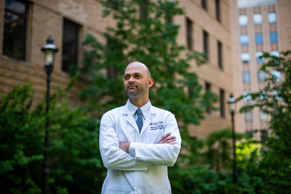
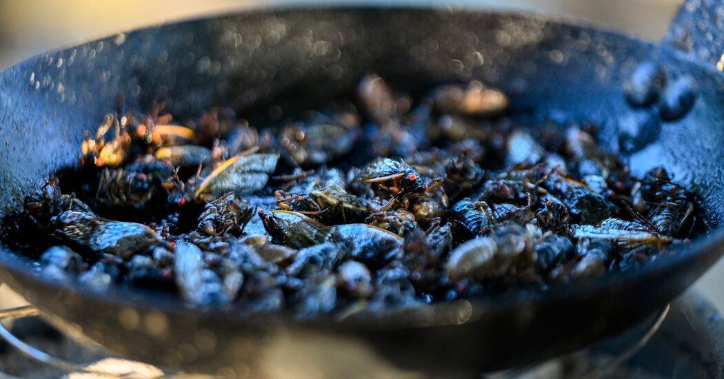
Infected early in the pandemic, Dr. Tomoaki Kato, a renowned transplant surgeon, was soon on life support, and one of the sickest patients in his own hospital.Early in the pandemic, as hospitals in New York began postponing operations to make way for the flood of Covid-19 cases, Dr. Tomoaki Kato continued to perform surgery. Patients still needed liver transplants, and some were too sick to wait.At 56, Dr. Kato was healthy and exceptionally fit. He had run the New York City Marathon seven times, and he specialized in operations that were also marathons, lasting 12 or 16 or 20 hours. He was renowned for surgical innovations, deft hands and sheer stamina. At NewYork-Presbyterian/Columbia University Irving Medical Center, where he was the surgical director of adult and pediatric liver and intestinal transplantation, his boss has called him “our Michael Jordan.”Dr. Kato became ill with Covid-19 in March 2020.“I was in a denial situation,” he said. “I thought I was going to be fine.”But he soon became one of the sickest patients in his own hospital, dependent on a ventilator and other machines to pump oxygen into his bloodstream and do the work of his failing kidneys. He came close to death “many, many times,” according to Dr. Marcus R. Pereira, who oversaw Dr. Kato’s care and is the medical director of the center’s infectious disease program for transplant recipients.Colleagues feared at first that he would not survive and then, when the worst had passed, that he might never be able to perform surgery again. But after two months in the hospital, Dr. Kato emerged with a determination to get back to work and a new sense of urgency about the need to teach other surgeons the innovative operations he had developed. His own illness also enabled him to connect with patients in ways that had not been possible before.“I really never understood well enough how patients feel,” he said. “Even though I’m convincing patients to take a feeding tube, and encouraging them, saying, ‘Even though it looks like hell now, it will get better and you’ll get through it,’ I really never understood what that hell means.”He approaches those moments differently now: “‘I was there’ are very powerful words for patients.”Dr. Kato was infected before most doctors in New York understood the siege that lay ahead.“When we really realized something serious was coming, I think it was already there,” Dr. Kato said. “No one realized how much the virus had spread through the city. The virus was everywhere.”Dr. Marcus Pereira, who oversaw Dr. Kato’s care. “He looked very sick from the moment he got here, and you realize, this potentially might not end up well,” he said. “It was a very shocking moment.”Joshua Bright for The New York TimesHis illness began with a bad backache, and then fevers that went up and down for a few days. He stayed home, periodically checking his oxygen level, and getting readings of 93 and 94 percent — results now recognized as a possible sign of Covid pneumonia.But at that early point in the pandemic, he said, “Nobody knows what Covid pneumonia is.” And he did not feel very sick, he told colleagues who kept in touch by phone.Dr. Pereira said: “I think that fooled us for a few days while he was at home. His oxygen levels were a little low, but he said, ‘I feel fine,’ and his heart rate was not that fast. He was one of the first-wave patients, and we were still learning about Covid.”‘An eye-opening moment’One morning in the shower Dr. Kato suddenly could not breathe, and he began coughing violently. He tested his oxygen again: It was dangerously low, below 90 percent. He had resisted being hospitalized, as doctors often do, but now he had no choice.“That’s when I decided to check into the hospital,” he said.Dr. Pereira, a friend as well as a colleague, was stunned by Dr. Kato’s condition.“When we actually saw him in the hospital, it was an eye-opening moment,” Dr. Pereira said. “He looked very sick from the moment he got here, and you realize, this potentially might not end up well. It was a very shocking moment. His oxygen levels were very low, he was breathing very rapidly, his heart rate was going very fast, his chest X-ray looked like he had severe Covid.”By the next day, Dr. Kato was on a ventilator.“From there,” he said, “I have no consciousness for about four weeks.”His condition worsened. Bacterial infections set in, followed by sepsis. His kidneys began to fail, and he needed dialysis. His lungs could not work well enough to use the oxygen from the ventilator, and in the middle of one desperate night a surgeon was called in to connect him to a machine that would take over for his lungs by pumping oxygen directly into his blood and taking carbon dioxide out.The machine — called ECMO, for extracorporeal membrane oxygenation — is a last resort.“When someone is on ECMO, you’re suddenly into the absolute highest-mortality group,” Dr. Pereira said. “Your chances of coming back from that are in the single digits. When he went on that, it was sort of a moment. We all felt we were about to lose him.”Dr. Kato was a star, a towering figure in his field, and to see him struck down shook the hospital staff.“It was horrific,” said Dr. Jean C. Emond, the chief of transplant services, and Dr. Kato’s boss. “It was a terrible thing touching a friend and colleague. There was this fear like, Was the world going to end — this global sense of doom. A fear of surfaces. Would you bring it home on your shoes? That deep emotional context of both the global and the personal was happening at once.”People in the highest levels of leadership at the hospital kept asking how Dr. Kato was doing.“His survival represented the fate of all, in a funny way,” Dr. Emond said.A Covid patient in Billings, Mont., hooked up to extracorporeal membrane oxygenation. “When someone is on ECMO, you’re suddenly into the absolute highest-mortality group,” Dr. Pereira said.Larry Mayer/The Billings Gazette, via Associated PressPushing the limits in surgeryDr. Emond in 2008 had lured Dr. Kato away from the University of Miami, for his rare expertise in intestinal transplants and so-called ex vivo operations for cancer, in which the surgeon cuts out abdominal organs to get at hard-to-reach tumors, and then sews the organs back in. Most important, Dr. Emond saw in Dr. Kato a willingness to push the limits of what could be done surgically to help patients.“He brought his culture of innovation,” Dr. Emond said. “And his personal capability, his ability to work for long hours, never quitting, never giving up, no matter how difficult the situation, carrying out operations that many would deem impossible.”In his first year at Columbia, Dr. Kato and his team operated successfully on a 7-year-old girl, Heather McNamara, whose family had been told by several other hospitals that her abdominal cancer was inoperable. The surgery, which involved removing six organs and then putting them back in, took 23 hours.More and more patients from around the country, and around the world, began seeking out Dr. Kato for operations that other hospitals could not or would not perform. He had also begun making trips to Venezuela to perform liver transplants for children and teach the procedure to local surgeons, and he created a foundation to help support the work there as well as in other Latin American countries.As Dr. Kato’s colleagues struggled to save him, a waiting list of surgical patients clung to hopes that he would soon be able to save them.Gradually, Dr. Pereira said, there were signs of recovery.“You come in early in the morning to see him,” he said. “The hospital hallways are empty and everybody’s looking at each other, scared and anxious. You go into the intensive care unit dreading bad news, and the team is giving you a sort of hopeful thumbs-up that maybe he’s looking better.”Dr. Kato spent about a month on a ventilator, and a week on ECMO. Like many people with severe Covid, he was tormented by frightening and vivid hallucinations and delusions. In one, he was arrested at the Battle of Waterloo. In another, he had been deliberately infected with anthrax; only a hospital in Antwerp could save him, but he could not get there. He saw the white light that some people describe after near-death experiences. “I felt like I died,” he said.He had spent much of his adult life in hospitals, but never as a patient.“I never got sick,” he said. “I had never faced the reality of death.”When he was finally freed of the machines and breathing on his own, his doctors were elated.But the joy faded when he regained full consciousness and it was clear he was not himself. He was still caught up in the delusions. More worrisome, he seemed confused, his razor-sharp mind not fully back.“I wasn’t making sense,” Dr. Kato said.Scans found a blood clot and a hemorrhage in Dr. Kato’s brain. Although not severe, they were still troubling.“I remember seeing him, and not seeing him the way I wanted him to be,” Dr. Pereira said, adding that at the end of one day, “I went to my car and broke down. I said, ‘I hate Covid. Why won’t you even let me have a small victory?’”Dr. Emond said, “Once we got over, ‘Would he survive?’ in our minds was, ‘Will he be able to be a doctor again?’ He suffered. He paid a huge price.”But the brain hemorrhage and clot turned out to be minor. Mentally, Dr. Kato quickly recovered.About a week after coming off the ventilator, he said, “I woke up in my mind.”Physically, he struggled. He had lost 25 pounds, nearly all of it muscle. He needed a feeding tube. He was so weak that one day it took him an hour to reach the device to adjust the incline of his bed, and when he finally got it, he was too weak to push the button. His hair fell out. A shoulder injury from the way he had been positioned kept him from fully raising one arm, and some of his neck and back muscles had wasted away. He needed extensive physical therapy.His family could not visit. Painful as that was for them, he said, it may have been just as well that they never saw him at his worst, in a web of tubes and machines in the intensive care unit.In late May 2020, after two months in the hospital, he went home, his departure cheered by about 200 staff members, chanting “Kato! Kato!”‘He was back.’In August, he began performing surgery again. For the first operation, a hernia repair, he used a robotic device that allowed him to work sitting down.“It was a really big day for everybody,” Dr. Emond said. “A lot of us went in to see how it was going.”By September, Dr. Kato was performing liver transplants, with his sore shoulder wrapped in athletic tape.“He was back,” Dr. Emond said. “I think he was working exceptionally hard to prove to himself and everybody else that he was back.”After two months in the hospital, Dr. Kato was discharged in May 2020. By August, he was performing surgery again.Joshua Bright for The New York TimesHis first transplant patient wound up staying in the same hospital room where Dr. Kato had been, and they snapped a picture together.“From there, I’m kind of full speed,” Dr. Kato said. By March of this year, he had completed 40 transplants and 30 other operations.Memories of his own recovery have tempered his dealings with patients.“I can be much more on their side, in their shoes, in their thinking,” he said.He so disliked the thickened liquids used to help restore swallowing ability that now, he is less inclined to push them on reluctant patients.“It just tastes so horrible,” he said. “I really cannot blame anybody who cannot take it. A few weeks ago, a patient complained about the thickened milk. In the past I would have just said, ‘You have to do this to get better.’ Now I can say, ‘Maybe you don’t have to do it.’ Each patient may have a different way.”He even offers tips on the hospital menu.“The patient hates the food, I hate the food, but I know the Cajun shrimp is a little better,” he said. Protein drinks? “I recommend the strawberry flavor.”When he was taken off the ventilator, at first he could not speak.“I learned that when you cannot talk, it does not mean you are not thinking,” he said. “The mind is so clear.”Facing death has also brought his career and his goals into a sharper focus, he said.“You don’t really want to waste your time, because you never know — one day all of a sudden you are in this situation,” he said.He realized, he said, that he must recruit more surgeons to continue the work that he and his foundation had started, to bring liver transplants to children in Latin America.“If I died and nobody else picks it up, that’s a problem,” he said.He also feels driven to promote and teach others to perform the complex cancer operations that involve removing multiple organs to reach a tumor, and then putting the organs back in.“This cannot be just my thing forever,” he said “It has to be everybody’s.”

Read more →

Don’t Eat Cicadas if You’re Allergic to Seafood, F.D.A.Warns

The insects have emerged by the billions this year across the Eastern United States and have curious foodies salivating. But their similarities to crustaceans makes them an allergy risk, health officials warned.Cicadas are popping up by the billions across the Eastern United States this season, and are subsequently finding their way to the dining tables of adventurous foodies.But people with a seafood allergy should avoid the crunchy insect, which are related to crustaceans, federal health officials warned on Wednesday.Yep! We have to say it!Don’t eat #cicadas if you’re allergic to seafood as these insects share a family relation to shrimp and lobsters. https://t.co/UBg7CwrObN pic.twitter.com/3qn7czNg53— U.S. FDA (@US_FDA) June 2, 2021
The advice from the Food and Drug Administration comes as the current group of cicadas, known as Brood X, emerges from the ground on its regular 17-year cycle. The shrimp-sized, beady-eyed bugs with almost translucent wings are being prepared by professional chefs and at home in a variety of ways this year.Some prefer them deep fried and others like them tossed into their Caesar salad. Bun Lai, the chef of a sustainable sushi restaurant in Connecticut, will present them in a fine-dining experience at his farm. And Frank’s RedHot, the 100-year-old hot sauce brand, released a string of cicada recipes including: caramel ’cada corn, chili lime ’cada tacos and a cicada “parm” slider.Cicadas, which do not bite or sting and are not toxic, are also eaten by predators including birds and small mammals. They are gluten-free, high in protein, low in fat and low in carbohydrates, according to National Geographic.But insects and crustaceans belong to the arthropod family. And according to a report from the Food and Agriculture Organization of the United Nations, the potential allergenic risks associated with edible insects needed further investigation.People with existing allergies to crustaceans may develop allergic reactions to edible insects, which contain similar proteins, the report said.The cicadas, whose quantities could reach tens of billions, are expected to appear in about 18 states for about six weeks. The Washington, D.C., area, Indiana and around Knoxville, Tenn., are the three Brood X cicada epicenters around the country.While cicadas are generally not harmful to humans, the sharp buzz they make when looking for mates can be annoying.The chance to feast on cicada dishes won’t last forever. Once the insects have mated and their eggs hatch, the developing cicadas, known as nymphs, will begin their 17-year cycle, feeding underground.The next opportunity to feast on fresh Brood X cicadas will be in 2038.

Read more →

Secondary infections inflame the brain, worsening cognition in Alzheimer's disease

New research into Alzheimer’s disease (AD) suggests that secondary infections and new inflammatory events amplify the brain’s immune response and affect memory in mice and in humans — even when these secondary events occur outside the brain.
Scientists believe that key brain cells (astrocytes and microglia) are already in an active state due to inflammation caused by AD and this new research shows that secondary infections can then trigger an over-the-top response in those cells, which has knock-on effects on brain rhythms and on cognition.
In the study, just published in Alzheimer’s & Dementia, the journal of the Alzheimer’s Association, mice engineered to show features of AD were exposed to acute inflammatory events to observe the downstream effects on brain inflammation, neuronal network function and memory.
These mice showed new shifts in the output of astrocytes and microglia and displayed new cognitive impairment and disturbed ‘brain rhythms’ that did not occur in healthy, age-matched, mice. These new onset cognitive changes are similar to acute and distressing psychiatric disturbances like delirium, that frequently occur in elderly patients.
Although it is difficult to replicate these findings in patients, the study additionally showed that AD patients who died with acute systemic infection showed heighted brain levels of IL-1β — a pro-inflammatory molecule that was important in causing the heightened immune response and the new onset disruptions seen in the AD mice.
Colm Cunningham, Associate Professor in Trinity’s School of Biochemistry and Immunology, and the Trinity Biomedical Sciences Institute, led the research. He said:
“Alzheimer’s disease is the most common form of dementia, affecting more than 5% of those over 60 and this distressing, debilitating condition causes difficulties for a huge number of people across the globe. The more we know about the disease and its progression the better chance we have of treating those living with it. We believe our work adds to this knowledge base in a few ways. Primarily, we show that the Alzheimer’s-affected brain has a greater vulnerability to acute inflammatory events, even if they occur outside the brain.
Placing this within the context of the slowly evolving progression of AD, we propose that these hypersensitive responses, now seen in multiple cell populations, may contribute to the negative outcomes that follow acute illness in older patients, including episodes of delirium and the accelerated cognitive trajectory that has been observed in patients who experience delirium before or during their dementia.”
The research was supported by the US National Institutes of Health (NIH) and by the Wellcome Trust.
Story Source:
Materials provided by Trinity College Dublin. Note: Content may be edited for style and length.

Read more →