Polio and Measles Vaccines Lag for Children Across the World

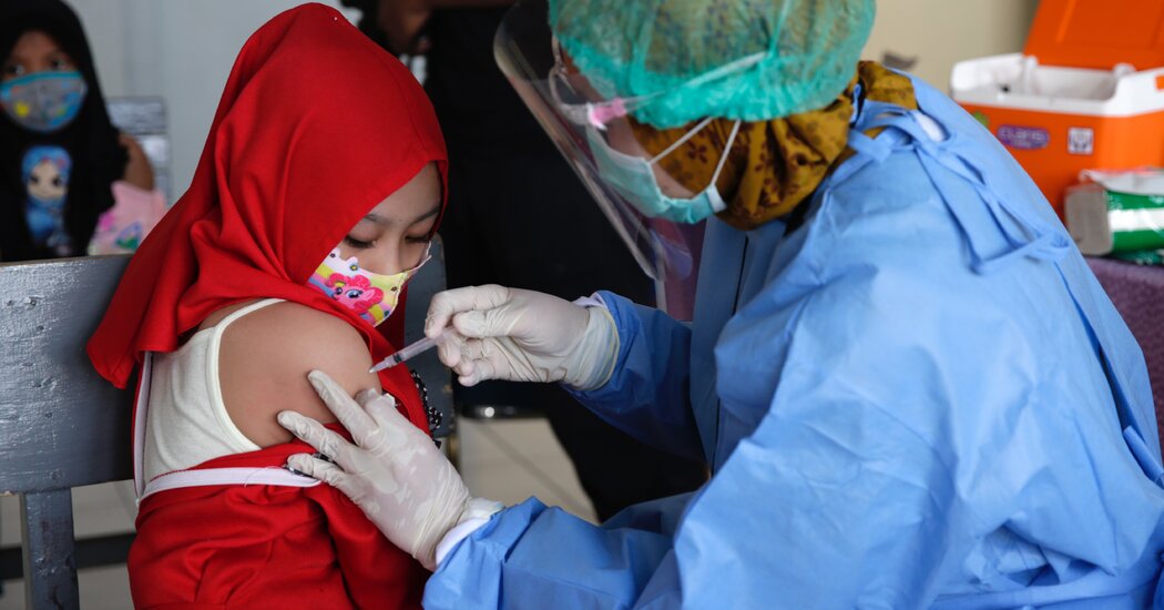
The pandemic dealt a serious setback to global efforts to immunize children against diseases like measles and polio, the Centers for Disease Control and Prevention reported on Thursday, reducing worldwide coverage for some vaccines to levels not seen since more than a decade ago.The proportion of eligible children who received a polio vaccine fell to 83 percent in 2020 from 86 percent the year before, as did coverage with the third dose of the diphtheria-tetanus-pertussis vaccine, known as DTP3. Coverage with the measles vaccine also dipped slightly, to 84 percent last year from 86 percent in 2019.Those setbacks, while seemingly small, meant that millions more children missed out on routine immunizations during the pandemic, putting them and their communities at risk.Globally, nearly 23 million children targeted for the DTP3 shot were not vaccinated in 2020, compared with 19 million in 2019, the C.D.C. said. The vast majority of those had not received a single dose of diphtheria-tetanus-pertussis vaccine. Not since 2009 had coverage with that vaccine been so low.The C.D.C. scientists involved in the report called for action to be taken to address the immunity gaps of preventable diseases in countries already saddled with Covid outbreaks. Scientists from the World Health Organization and UNICEF also were co-authors on the study.The decline in vaccinations follows a decade of stagnant immunization levels. In 2019, measles deaths swelled to their highest levels in 23 years, a consequence of what public health experts described as insufficient vaccination coverage. Scientists said that the pandemic had hampered the tracking of measles outbreaks.The pandemic also disrupted immunization programs, the C.D.C. report said, interrupting the supply of basic vaccines and making it more difficult to administer them.Immunization levels for diphtheria-tetanus-pertussis and measles were lowest in much of Africa, the report said. The W.H.O. also said on Thursday that only five of Africa’s 54 nations were expected to reach a year-end goal of vaccinating 40 percent of their people against Covid. UNICEF, a United Nations agency working to distribute coronavirus vaccines, warned of a shortfall next year in syringes for both Covid and routine vaccinations.

Read more →

How COVID-19 alters the immune system

COVID-19 reduces the numbers and functional competence of certain types of immune cells in the blood, say LMU researchers. This could affect responses to secondary infections.
The SARS-CoV-2 coronavirus causes moderate to severe disease in 3-10% of those infected. In such cases, the immune system overreacts to the virus, triggering an aberrant innate immune response that is characterized by systemic inflammation, intravascular blood clotting and damage to the cardiovascular system. A team led by immunology professor Anne Krug at LMU’s Biomedical Center (BMC), which included many researchers based at the BMC and the LMU Medical Center, has carried out a comprehensive study of this phenomenon, and uncovered hitherto unknown effects of the virus on the immune system. In the journal PLOS Pathogens, they report that, following infection with SARS-CoV-2, the numbers of immune cells called dendritic cells in the circulation decline, while the functionality of the remaining fraction is impaired. The authors believe that this could make patients more susceptible to secondary infections during, and immediately after recovery from a bout of COVID-19.
Dendritic cells (DCs) are responsible for initiating immune responses against invasive pathogens. They do so by activating helper T cells, which in turn stimulate B cells to secrete antibodies directed against the invader. Krug and her colleagues set out to determine the effects of moderate to severe coronavirus infection on this process. They analyzed blood samples obtained from 65 COVID-19 patients who had been treated at the LMU Medical Center. They found that there were fewer DCs in these samples than in the blood of healthy controls. Furthermore, DCs isolated from the blood of patients showed a reduced ability to activate T cells. “We had actually expected that DCs isolated from patients infected with SARS-CoV-2 would activate T cells more potently than DCs obtained from healthy donors,” says Krug. “However, we discovered that, in the course of the disease, the proteins present on the surface of the DCs in patients’ blood were altered in a way that made them more likely to inhibit T cell responses.” In spite of this, by 15 days after diagnosis 90% of these patients had generated antibodies directed against the SARS-CoV-2 spike protein, and many of them had also activated a T cell response. — these responses are the hallmarks of a robust immune reaction against the virus. “So, the drop in the numbers and reduced functionality of DCs does not seem to have a negative impact on the immune response to the coronavirus itself,” Krug says.
However, she is convinced that the reduced number and altered function of DCs is significant. It is conceivable that this might cause the immune system to react less strongly than expected to bacterial or other viral infections following recovery from COVID-19, but this possibility will require further clinical investigation.
What might account for the depletion of DCs in the blood and the decrease in their capacity to stimulate T cells? Krug has several hypotheses to offer. It could in fact represent an appropriate regulatory process, she suggests. COVID-19 is often associated with vigorous inflammation reactions — so the phenomenon might be part of an attempt to downregulate inflammatory processes. Dendritic cells might migrate from the blood into inflamed tissues, such as the lung, which could explain the fall in the numbers of DCs in the circulation. “However, we also found that the regeneration of dendritic cells is delayed,” Krug points out. The authors of the study believe that this phenomenon could weaken the ability of patients to mount effective immune responses to other pathogens during, and in the immediate aftermath of a symptomatic COVID-19 infection. The team will now explore this issue further in an effort to determine whether the effects of SARS-CoV-2 on DCs play a role in long-term COVID.
Story Source:
Materials provided by Ludwig-Maximilians-Universität München. Note: Content may be edited for style and length.

Read more →

Covid: Double vaccinated can easily spread virus at home

SharecloseShare pageCopy linkAbout sharingImage source, Getty ImagesDouble jabbed people are catching Covid and passing it on to those they live with, warn experts who have studied UK household cases. Individuals who have had two vaccine doses can be just as infectious as those who have not been jabbed. Even if they have no or few symptoms, the chance of them transmitting the virus to other unvaccinated housemates is about two in five, or 38%. This drops to one in four, or 25%, if housemates are also fully vaccinated. The Lancet Infectious Diseases work shows why getting even more people vaccinated and protected is important, they say.Unvaccinated people cannot rely on those around them being jabbed to remove their risk of getting infected, they warn.Vaccines do an excellent job of preventing serious Covid illness and deaths, but are less good at stopping infections, particularly since the emergence of the more infectious Delta variant which is dominant in the UK. And over time, the protection offered by vaccines wanes and needs boosting with further doses. Since households are where most Covid transmission occurs, making sure every member who is eligible for a vaccine has had one and is up to date with their doses makes sense, say experts. According to the study, which ran from September 2020 to September 2021 and included 440 households in London and Bolton doing PCR Covid tests: People who are double jabbed have a lower, but still appreciable, risk of becoming infected with the Delta variant compared with unvaccinated peopleThey also appear to be just as infectiousVaccinated people clear the infection more quickly, but their peak viral load – when people are most infectious – is similar to that seen in unvaccinated peopleThis may explain why they can still readily pass on the virus in household settingsProf Ajit Lalvani, of Imperial College London, UK, who co-led the study, said: “The ongoing transmission we are seeing between vaccinated people makes it essential for unvaccinated people to get vaccinated to protect themselves from acquiring infection and severe Covid-19, especially as more people will be spending time inside in close proximity during the winter months.”We found that susceptibility to infection increased already within a few months after the second vaccine dose – so those eligible for booster shots should get them promptly.”Co-lead Dr Anika Singanayagam, also from Imperial, said: “Our findings provide important insights into the effect of vaccination in the face of new variants, and specifically, why the Delta variant is continuing to cause high Covid case numbers around the world, even in countries with high vaccination rates. “Continued public health and social measures to curb transmission – such as mask wearing, social distancing, and testing – thus remain important, even in vaccinated individuals.” The Lancet Infectious DiseasesThe BBC is not responsible for the content of external sites.

Read more →

Horse riding confirmed as a viable mobility treatment for cerebral palsy

Cerebral Palsy (CP) is a common disability among children, characterized by abnormal gait patterns and the inability to maintain posture and balance. While the condition is incurable, physical therapy treatments can go a long way in improving movement and balance. One such treatment approach is hippotherapy (HPOT), which uses horse riding to improve functional mobility in children with CP. Although supported by scientific studies as an effective treatment approach for (CP), there is, unfortunately, little data concerning how HPOT results in improvement.
Recently, a team of researchers from Korea and the United States addressed this question, investigating physical interaction metrics between horses and children with CP during HPOT. “My original research interests lie in the rehabilitation of people with neurological impairment, specifically gait and balance. However, I did not know about hippotherapy until rather recently in 2016. After realizing how effective it is in treating children with CP, I was motivated to explore it further,” explains Dr. Pilwon Hur who headed the study from the Gwangju Institute of Science and Technology (GIST) in Korea. This paper was made available online on September 6, 2021, and was published in Volume 18 Issue 132 of the Journal of NeuroEngineering and Rehabilitation.
The research team studied four children with CP over the course of eight physical therapy sessions. They placed sensors on the horses and children to record their movements and track their acceleration and angular velocity. They found that the data from the horses and children began to resemble each other as time progressed, indicating a synchronization between the horse and the rider. They also gave the children mobility tests after each session and observed improvement in their motor skills at the end of the experiment.
“We found that physical interaction between the children with CP and the horses, characterized by the children adapting to the horse’s movement and vice versa, is extremely important for the rehabilitation to be effective,” says Dr. Hur.
Excited by these findings, the team hopes their work will provide a baseline for further research on HPOT. “To the best of my knowledge, ours is the first study to quantify these interactions and relate them to effectiveness,” says Dr. Hur. “Such an understanding would help us optimize physical therapy programs, improving the quality of life for children with CP.”
Story Source:
Materials provided by GIST (Gwangju Institute of Science and Technology). Note: Content may be edited for style and length.

Read more →

Cleveland Clinic study links gut microbiome and aggressive prostate cancer

Cleveland Clinic researchers have shown for the first time that diet-associated molecules in the gut are associated with aggressive prostate cancer, suggesting dietary interventions may help reduce risk. Findings from the study were published in Cancer Epidemiology, Biomarkers & Prevention.
While more research will be necessary, the study’s lead author Nima Sharifi, M.D., says findings from the team’s analysis of nearly 700 patients may have clinical implications for diagnosing and preventing lethal prostate cancer.
“We found that men with higher levels of certain diet-related molecules are more likely to develop aggressive prostate cancer,” said Dr. Sharifi, director of Cleveland Clinic’s Genitourinary Malignancies Research Center. “As we continue our research in this area, our hope is that one day these molecules can be used as early biomarkers of prostate cancer and help identify patients who can modify their disease risk by making dietary and lifestyle changes.”
In this study, Dr. Sharifi and his collaborators — including Stanley Hazen, M.D., Ph.D., and Eric Klein, M.D. — analyzed data from patients previously enrolled in the National Cancer Institute’s Prostate, Lung, Colorectal and Ovarian (PLCO) Cancer Screening Trial.
They studied baseline levels of certain dietary nutrients and metabolites (byproducts produced when a substance is broken down in the gut) found in patients’ blood serum prior to prostate cancer diagnosis. They compared serum levels between healthy patients and those who later received a prostate cancer diagnosis and died from the disease.
The researchers found that men with elevated levels of a metabolite called phenylacetylglutamine (PAGln) were approximately two or three times more likely to be diagnosed with lethal prostate cancer. This metabolite is produced when microbes in the gut break down phenylalanine, an amino acid found in many plant- and animal-based protein sources like meat, beans and soy.
In addition to PAGln, researchers also discovered that elevated levels of two nutrients abundant in animal products, including red meat, egg yolks and high-fat dairy products, called choline and betaine, also were linked with increased risk for aggressive prostate cancer.
While these nutrients and gut metabolites have been studied previously in heart disease and stroke, this is the first time that gut microbiome metabolites have been studied clinically in relation to prostate cancer outcomes.
Dr. Hazen was the first to identify PAGln’s association with increased cardiovascular disease risk. The findings were published in 2020 in Cell. “Interestingly, we found that PAGln binds to the same receptors as beta blockers, which are drugs commonly prescribed to help lower blood pressure and subsequent risk of cardiac events,” said Dr. Hazen, director of Cleveland Clinic’s Center for Microbiome & Human Health and chair of Lerner Research Institute’s Department of Cardiovascular & Metabolic Sciences. “This suggests that part of beta blockers’ potent efficacy may be due to blocking the metabolite’s activity.”
“New insights are emerging from large-scale clinical datasets that show use of beta blockers is also associated with lower mortality due to prostate cancer,” said Dr. Sharifi, who is a staff physician in Lerner Research Institute’s Department of Cancer Biology. “We will continue to work together to investigate the possible mechanisms linking PAGln activity and prostate cancer disease processes in hopes of identifying new therapeutic targets for our patients.”
The research team also will continue to explore the reliability of using choline, betaine and PAGln as biomarkers of aggressive prostate cancer and how dietary interventions can be used to modulate their levels and reduce patients’ subsequent disease risk.
Chad Reichard, M.D., a urologic oncologist at Urology of Indiana and a previous urology resident at Cleveland Clinic, and Bryan Naelitz, previously a medical student in Dr. Sharifi’s lab and now a urology resident, are co-first authors on the study. Dr. Klein is a urologist and emeritus chair of Glickman Urological & Kidney Institute at Cleveland Clinic. The research was supported by the National Cancer Institute and the National Heart, Lung, and Blood Institute (both parts of the National Institutes of Health), as well as the Prostate Cancer Foundation.
Story Source:
Materials provided by Cleveland Clinic. Note: Content may be edited for style and length.

Read more →

Best water treading technique to prevent drowning

Unless you’re a water-polo player, the “egg-beater” technique may mean little to you.
But new research from the University of Otago has found it may be the most effective skill you can learn to prevent drowning.
Dr Tina van Duijn, from the University’s School of Physical Education, Sport and Exercise Sciences, says it is important during an emergency to be able to perform a movement in the water that is economic — both energetically and cognitively.
“You should still be able to perform it while in panic, or while assessing the environment, making decisions, or even alerting people who might be able to help you.”
Her new research “A Multidisciplinary Comparison of Different Techniques Among Skilled Water Treaders”, funded by the Swiss National Science Foundation, is the first step in identifying what pattern of water treading is best to prevent drowning.
The study involved monitoring the physical and cognitive demands of four common treading techniques on 21 people experienced in water treading, who were chosen to ensure that all the techniques were performed correctly.

Read more →

These hips don’t lie: 3D imaging of a pelvis suggests social care for saber-tooths

You can’t spell ‘Smilodon fatalis’ without ‘fatal’, but researchers at La Brea Tar Pits may have found a softer side to saber-toothed cats along with a connection to our own feline and canine companions.
Published in Scientific Reports, a new study led by Dr. Mairin Balisi, Postdoctoral Fellow at La Brea Tar Pits, in collaboration with orthopaedic surgeons at Cedars-Sinai hospital, used 3D image reconstruction of external and internal bone morphology of a deformed Smilodon hip bone to reveal this saber-toothed cat suffered from hip dysplasia, suggesting a social structure that helped members survive to adulthood even when they couldn’t hunt for themselves due to this birth defect.
Part of the richness of La Brea Tar Pits’ collection are the fossils exhibiting signs of injury and disease-the more than 8,000 specimens that make up the pathology collection. These damaged bones are incredibly valuable for better understanding extinct animals’ behavior. For instance, the lower-back trauma found (and reported in an earlier article by Dr. Balisi) in many Smilodon vertebrae points to a hunting style that includes grappling with large Ice Age prey, like bison. For more than a century, paleontologists thought the massive destruction of the pelvis examined in this study was caused by trauma or infection that eventually led to the animal’s death, but a look inside the bone using modern medical technology told a different story.
Using computed tomography, the same technology common in hospitals (and veterinarian offices), the pelvis and matching thigh bone were scanned, and the resulting images were used to create 3D models of the inside of the bones. “To quote Roy Moodie in his 1930 study: this pelvis is ‘the most strikingly pathological object in the collection of Rancho La Brea fossils’. And so if we were to CT-scan a specimen-with CT being a resource-intensive method- then we had to start with this one,” says Dr. Balisi.
“Understanding the 3D shape of a skeleton is fascinating to me,” says co-author Dr. Robert Klapper, orthopaedic surgeon and sculptor. Seeing the sheer amount of skulls on the Tar Pits’ dire wolf wall over a decade ago led to talking his way into the collection. “When I saw the incredible display of bones at La Brea Tar Pits, I immediately asked to meet (then Collections Manager and study co-author) Chris Shaw. I asked him where were the abnormal joints that I knew must have existed. Chris took me to the stacks and showed me the three pelvises and one femur that he was studying. That’s when we decided to collaborate and began the analysis of the etiology of the end-stage degeneration of the saber-tooth hips.”
This scanned cat’s CAT scan upended the previous interpretation of its hip bones. After careful analysis of the bones’ internal structure, the team concluded that the damage wasn’t the result of an injury suffered on the hunt, but congenital hip dysplasia.

Read more →

Gene signature identifies drivers of treatment resistance in metastatic breast cancer

A multi-institution team of researchers led by scientists at Baylor College of Medicine has discovered a genetic signature that can identify drivers of poor outcomes in advanced estrogen-receptor positive (ER+) breast cancer, which could one day lead to personalized treatment for patients.
The 24-gene signature detects the presence of mutations and translocations in the ER gene that confer the tumor the ability to grow independently from estrogen and therefore unsusceptible to current therapies directed at disrupting estrogen-fueled cancer growth. The findings, published in Cancer Research, a journal of the American Association for Cancer Research, provide a new strategy to refine breast cancer diagnosis that could help guide a more precise selection of tumor-specific treatments.
“About 80% of all breast cancers depend on the hormone estrogen to grow. The hormone promotes tumor growth by binding to the ER,” said corresponding author Dr. Matthew J. Ellis, professor and director of the Lester and Sue Smith Breast Center and a McNair scholar at Baylor.
Disrupting the estrogen-ER interaction is a key therapeutic approach. Drugs such as tamoxifen and fulvestrant target the ER this way, but tumor cells learn to evade this attack and become resistant to these drugs.
“One of the predominant ways ER+ breast cancer cells evade treatment is by creating mutant ERs that no longer can be recognized and targeted by ER-targeting cancer drugs,” said first author Xuxu Gou, a graduate student in the Ellis lab.
The team has been studying ESR1 gene translocations, which refer to the ER gene swapping a part of its sequence with genetic information from another gene. ER gene translocations create chimeric ER proteins, meaning the protein contains only half of the ER protein and the other half comes from different protein. Some of the ER chimeras are extreme versions of mutant ERs because the drug-binding region, which is the same region estrogen binds to, is completely replaced with a region derived from another protein, to which neither the drug nor estrogen can bind. These ER chimeras trigger cancer activity in the absence of hormone.
“Not all ER translocations were active — some drive metastasis and resistance to treatment, but others do not,” Gou said. “To be able to determine whether any particular ESR1 translocation can promote disease progression, we developed a diagnostic genetic signature that detects the presence of an active ESR1 chimeric protein.”
With support from the National Cancer Institute’s PDXnet program, the team used genomics and transcriptomics to annotate 20 mouse models of ER+ patient-derived tumors that demonstrated different degrees of dependence on estrogen for growth. In this data set, a 24-gene signature detected the presence of an active ESR1 fusion, but interestingly also common point mutations in ESR1. These findings were replicated in data from a human metastatic breast cancer cohort. The team therefore called their 24 gene signature the MOTERA score for “Mutant or Translocated Estrogen Receptor Alpha.”
The findings are significant in the area of precision medicine, as they can provide specific details about the tumor that could guide physicians in the selection of more effective treatments.
“In the future, a patient’s cancer cells could be analyzed and, once the MOTERA score indicates the presence of an ER mutation or translocation, then the tumor cells would be further studied to more precisely determine what kind of ER mutant or translocation is present. This would help guide the selection of a personalized, optimal treatment,” said co-author, Dr. Charles E. Foulds, assistant professor at Baylor’s Lester and Sue Smith Breast Center.
Ellis and Foulds are members of the Dan L Duncan Comprehensive Cancer Center at Baylor. Other contributors to this work include Meenakshi Anurag, Jonathan T. Lei, Beom-Jun Kim, Purba Singh, Sinem Seker, Diana Fandino, Airi Han, Saif Rehman, Jianhong Hu, Viktoriya Korchina, Harsha Doddapaneni, Lacey E. Dobrolecki, Nicholas Mitsiades, Michael T. Lewis, Alana L. Welm, Shunqiang Li and Adrian V. Lee. The authors are affiliated with one or more of the following institutions: Baylor College of Medicine, Yonsei University Wonju College of Medicine, University of Cambridge, University of Utah, Washington University School of Medicine in St. Louis, University of Pittsburgh and University of Michigan.

Read more →

Nurses as parents exemplify link between poor sleep and daily stress

A new paper in the peer-reviewed Journal of Sleep Research details how nurses who also are parents might be more susceptible than other groups to daily stress aggravated by poor sleep. The consequences of the sleep-stressor link in nurses with children may affect their caregiving at home and on the job, where nurses represent the largest population of front-line health care workers.
“We were really interested in looking at how the sleep-stressor relationship is different for nurses who are parents and nurses who are not parents,” said lead author Taylor Harris, doctoral student in counseling psychology at the University of Kansas. “We also wanted to look at how many children parents have further influences the relationship between sleep and stress in those working parents, because caregiving at work and at home can be particularly difficult — sometimes we don’t always look at that intersection specifically in the most prominent health care profession, which is nursing.”
Harris and her co-authors, Taylor F. D. Vigoureux and Soomi Lee of the University of South Florida, recruited 60 nurses — both parents and nonparents — and asked them to track their sleep and stress levels on smartphones for two weeks between May and September 2019.
“We had a background survey that was distributed during the informed-consent meetings including just very basic demographic and work characteristics — nurses were full-time inpatient workers, either on the day shift or night shift,” Harris said. “Then, for 14 days, participants responded to these brief surveys on their smartphones. We had them answer questions upon waking and then three times daily in order for us to collect the data we needed. Sleep questions were included in the upon-waking survey, and the daily stressor questions were included in the three following surveys.”
The research confirmed a long-established truth in such studies: Parents are more likely to suffer from worse sleep compared with people without kids.
“We all kind of know that people who are parents have poorer overall sleep,” Harris said. “That could be from the demands of having to take children to appointments, or in general to attend to needs outside of their own — that can be changing diapers, feeding, eating or worrying about children that are older and getting into college to worrying about how children progress from childhood to adulthood. People who are parents do tend to have that additional burden of having a harder time sleeping.”
The study showed this is especially the case with nurses: Poorer-than-usual sleep quality was associated with more frequent and more severe stressors the following day for parent nurses with one child and two or more children, but not for nonparents. Further, the link between sleep quality and daily stressors became greater as the number of children at home increased.

Read more →

How two people controlled HIV after stopping treatment

Research led by scientists at the National Institutes of Health has identified two distinct ways that people with HIV can control the virus for an extended period after stopping antiretroviral therapy (ART) under medical supervision. This information could inform efforts to develop new tools to help people with HIV put the virus into remission without taking lifelong medication, which can have long-term side-effects.
The study, published today in the journal Nature Medicine, was led by Tae-Wook Chun, Ph.D., chief of the HIV Immunovirology Section in the Laboratory of Immunoregulation at the National Institute of Allergy and Infectious Diseases (NIAID), part of NIH; and by Anthony S. Fauci, M.D., NIAID director and chief of the Laboratory of Immunoregulation.
The study involved two adults with HIV who began ART soon after acquiring the virus and continued with treatment for more than six years, successfully suppressing HIV. The individuals then joined an HIV clinical trial and stopped taking ART under medical supervision. The study team followed one of these people for four years and the other for more than five years, with study visits roughly every two to three weeks.
The investigators monitored the timing and size of viral rebounds in each participant, that is, times when the amount of HIV in their blood became detectable. One participant suppressed the virus with intermittent rebounds for nearly 3.5 years, at which point he began taking suboptimal ART without telling the study team. The other participant almost completely suppressed HIV for nearly 4 years, at which point the virus rebounded dramatically because he became infected with a different HIV strain, a phenomenon known as “superinfection.”
In the first participant but not the second, the scientists found high levels of HIV-specific immune cells called CD8+ T cells that can kill virus-infected cells, indicating that different mechanisms of control were at work in each person. The researchers also found that the second participant, who had a weaker CD8+ T cell response against HIV, had a very strong neutralizing antibody response throughout the follow-up period until the sudden viral rebound. According to the scientists, this suggests that neutralizing antibodies may have played a significant role in facilitating near-complete HIV suppression in this individual until he newly acquired a different strain of the virus.
The researchers emphasized that to avoid the emergence of viral resistance and prevent potential misinterpretation of scientific data in studies like this one, it is important to conduct routine antiretroviral drug testing of people with HIV who halt treatment for extended periods. In addition, the researchers identified HIV superinfection as a potential cause of sudden virologic breakthrough in people with HIV who halt treatment, especially when the breakthrough occurs after a prolonged period of virus suppression.
Story Source:
Materials provided by NIH/National Institute of Allergy and Infectious Diseases. Note: Content may be edited for style and length.

Read more →