The effects of pediatric critical illness on absenteeism

Children who survive critical illness and their parents commonly experience physical, emotional, and cognitive conditions as a result of the critical illness. These effects can also include prolonged absences from school and/or work. What has not been fully understood is the rate and duration of school absences among these children and work absences among their caregivers.
A secondary analysis of a randomized trial of pediatric patients hospitalized for acute respiratory failure has shed important light on the subject. The study found that nearly 70% of pediatric patients missed an average of two five-day school weeks post hospital discharge and half of their primary caregivers missed an average of eight workdays post hospital discharge. The findings suggest a risk for negative downstream educational, financial, and health outcomes for patients and added stress and financial risk for their parents.
“This study suggests that post-PICU school absenteeism is an important target for future interventions including understanding the barriers to school participation, development of interventions to mitigate absenteeism, and to help children catch up on missed school,” says Martha A.Q. Curley, PhD, RN, FAAN, Professor of Nursing at the University of Pennsylvania School of Nursing (Penn Nursing) and the senior researcher of the study. “In addition, given the magnitude of missed work found in our study and the hardships described by parents in prior studies, there is a great need for programs and policies to support families during and after pediatric hospitalization.”
The results of the study have been published in the JAMA Network.
Coauthors of the article include Erin F. Carlton, MD, MSc, and Ryan P. Barbaro, MD, MSc, both of the University of Michigan; John P. Donnelly, PhD, of the University of Michigan Medical School; Hallie C. Prescott, MD, MSc, of the Veterans Affairs Center for Clinical Management Research; Lisa A. Asaro, MS, of Boston Children’s Hospital. And R. Scott Watson, MD, MPH, of the University of Washington, Seattle.
Story Source:
Materials provided by University of Pennsylvania School of Nursing. Note: Content may be edited for style and length.

Read more →

Zika vaccine shows promising results in preclinical studies

A Zika virus vaccine candidate is effective at preventing the Zika virus passing from mother to fetus in preclinical animal studies, according to a new study in the journal npj Vaccines.
The research is a collaboration between Trudeau Institute, Texas Biomedical Research Institute’s Southwest National Primate Research Center (SNPRC), and Walter Reed Army Institute of Research (WRAIR), where the vaccine was developed.
“The vaccine has been shown to be safe for non-pregnant humans, but of course we need to know if it is safe and effective for the people at greatest risk: pregnant women and their fetuses,” says In-Jeong Kim, PhD, a viral immunologist at Trudeau Institute and the first paper author. “Our proof-of-concept studies conducted at Trudeau and Texas Biomed show very promising results that the vaccine given before pregnancy will provide high levels of protection for mothers and babies.”
The 2015-2016 Zika outbreak in Brazil and other countries in the Americas caused a surge in miscarriages and a constellation of birth defects, called Congenital Zika Syndrome, including abnormally small heads and neuro-developmental disorders. This prompted the World Health Organization to declare the Zika outbreak a public health emergency of international concern.
“It’s important to test vaccines before the next large outbreak, because there will be another,” says Jean Patterson, PhD, a virologist at Texas Biomed and a senior paper author. “Zika is part of a family of viruses known to go through cycles. These viruses tend to spread rapidly through naïve populations that have never been exposed to the virus before, then infections drop down for years because most people have been exposed. As more and more people are born, there is a new group of naïve individuals in which the virus can once again wreak havoc. We want to help break that cycle.”
The purified, inactivated Zika vaccine (ZPIV) candidate was developed by a team at WRAIR using the same technology they used to develop a Japanese encephalitis vaccine. The vaccine has been tested in non-pregnant animals, showing it effectively clears the virus from blood. In Phase 1 human trials, it has been shown to be safe and elicit a protective immune response.

Read more →

Altered gene expression and cell interactions involved in COPD

Researchers at Baylor College of Medicine, Yale University and other institutions have identified previously unrecognized changes in gene expression and cellular interactions in distinct cell populations in chronic obstructive pulmonary disease (COPD). The findings highlight the complexity and diversity of cellular injury and inflammation in COPD providing a better understanding of the factors and processes involved in the disease.
Published in Nature Communications, the study used single-cell RNA sequencing to analyze gene expression profiles of lung tissue obtained from patients with COPD and from mice exposed to cigarette smoke. The researchers separated all the cells within the lung and measured the gene expression profile of each individual cell. Then, they organized this information in a cell atlas that is available to researchers interested in exploring gene expression in individual cells in lungs with the disease.
“Our analysis identified novel changes in gene expression and cellular interactions in three distinct cell populations commonly implicated in COPD: epithelial (in the lungs), endothelial (in blood vessels) and macrophage cells (part of the immune system),” said Dr. Ivan Rosas, senior author of the study and section chief of pulmonary, critical care and sleep medicine in the Department of Medicine at Baylor.
The researchers identified a subpopulation of epithelial cells in lungs with COPD that has abnormal expression of genes involved in metabolic, antioxidant and cellular stress responses, when compared to controls.
“This highly innovative translational study is a continuation of a successful long-standing collaboration with Dr. Naftali Kaminski and colleagues at Yale University, which has led to the description of multiple lung cell atlases in health and disease that are now publicly available. In the current study, Drs. Maor Sauler and John McDonough of Yale University, the corresponding authors, demonstrate that endothelial cells in capillary blood vessels in COPD lungs are inflamed, and that a subpopulation of macrophages expressing high levels of metallothioneins, proteins that regulate the balance of certain metals in the body, seems to contribute to the disease,” Rosas said.
Future studies evaluating outcomes of specific alterations identified in this study will provide further insights into mechanisms that contribute to COPD and having a large and accessible data set of COPD cells, such as the cell atlas here, is an essential tool to begin developing therapies to target the condition.
The research team continues to focus on developing innovative genomic datasets to better understand the pathologic underpinnings of chronic lung disease. They are currently in the process of completing the largest COVID-19 lung fibrosis cell atlas.
Story Source:
Materials provided by Baylor College of Medicine. Note: Content may be edited for style and length.

Read more →

Dr. Johan Hultin Dies at 97; His Work Helped Isolate 1918 Flu Virus

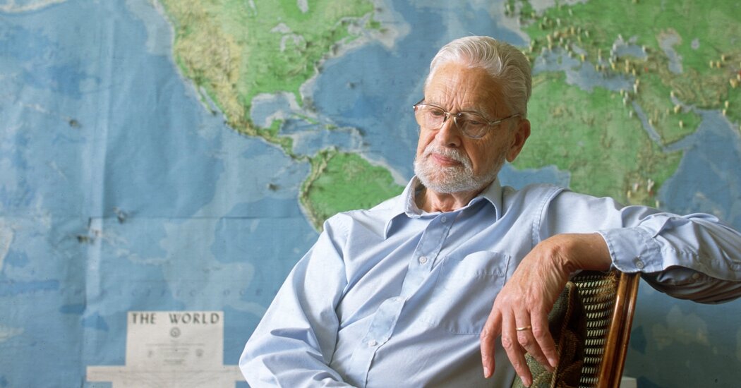
Dr. Hultin’s discovery of a frozen victim of the 1918 pandemic gave scientists the opportunity to map the virus’s genetic material.Dr. Johan V. Hultin, a pathologist whose discovery of victims of the 1918 flu pandemic buried in Alaskan permafrost led to a critical understanding about the virus that caused the outbreak, died on Saturday at his home in Walnut Creek, Calif. He was 97.The death was confirmed by his wife, Eileen Barbara Hultin.Dr. Hultin’s discovery was crucial to finding the genetic sequence of the virus, allowing researchers to examine what made it so lethal and how to recognize it if it came again. The virus, which was 25 times more deadly than ordinary flu viruses, killed tens of millions of people and infected 28 percent of Americans, dropping the average life span in the United States by 12 years.Dr. Hultin’s quest to find victims of the 1918 flu was sparked in 1950 by an offhand remark over lunch with a University of Iowa microbiologist, William Hale. Dr. Hale mentioned that there was just one way to figure out what caused the 1918 pandemic: finding victims buried in permafrost and isolating the virus from lungs that might be still frozen and preserved.Dr. Hultin, a medical student in Sweden who was spending six months at the university, immediately realized that he was uniquely positioned to do just that. The previous summer, he and his first wife, Gunvor, spent weeks assisting a German paleontologist, Otto Geist, on a dig in Alaska. Dr. Geist could help him find villages in areas of permafrost that also had good records of deaths from the 1918 flu.After persuading the university to provide him with a $10,000 stipend, Dr. Hultin set off for Alaska. It was early June 1951.Three villages seemed like they might have what he wanted, but when he arrived at the first two, the victims’ graves were no longer in permafrost.The third village on his list, Brevig Mission, was different. The flu had devastated the village, killing 72 out of 80 Inuit residents. Their bodies were buried in a mass grave with a large wooden cross at either end.When Dr. Hultin arrived and politely explained his mission, the village council agreed to let him dig. Four days later, he saw his first victim.“She was a little girl, about 6 to 10 years old. She was wearing a dove gray dress, the one she had died in,” he recalled in an interview in the late 1990s. The child’s hair was braided and tied with bright red ribbons. Dr. Hultin called for help from the University of Alaska Fairbanks, and the group eventually found four more bodies.They stopped digging. “We had enough,” Dr. Hultin said.Dr. Hultin was a resident in surgical pathology at the Mayo Clinic in Rochester, Minn., in 1957.via Hultin familyHe removed still-frozen lung tissue from the victims, closed the grave and took the tissue back to Iowa, keeping it frozen on dry ice in the passenger compartment of a small plane.Back in the lab, Dr. Hultin tried to grow the virus by injecting the lung tissue into fertilized chicken eggs — the standard way to grow flu viruses. He was caught up in the excitement of his experiment, he said, and had not thought about the possible danger of introducing a deadly virus into the world.“I remember the sleepless nights,” he said. “I couldn’t wait for morning to come to charge into my lab and look at the eggs.”But the virus was not growing.He tried squirting lung tissue into the nostrils of guinea pigs, white mice and ferrets, but again he failed to revive the virus.“The virus was dead,” he said.Dr. Hultin never published his results but bided his time, working as a pathologist in private practice in San Francisco and hoping for another opportunity to resurrect that virus.His chance came in 1997, when, sitting by a pool on vacation with his wife in Costa Rica, he noticed a paper published in Science by Dr. Jeffery K. Taubenberger, now chief of the viral pathogenesis and evolution section at the National Institute of Allergy and Infectious Diseases.It reported a remarkable discovery. Dr. Taubenberger had searched a federal repository of pathology samples dating to the 1860s and found fragments of the 1918 virus in snippets of lung tissue from two soldiers who had died in that pandemic. The tissue had been removed at autopsy, wrapped in paraffin and stored in the warehouse.Dr. Hultin immediately wrote to Dr. Taubenberger, telling him about his trip to Alaska. He offered to return to Brevig to see if he could find more flu victims.“I remember getting that letter and thinking: ‘Gosh. This is really incredible. This is amazing,’” Dr. Taubenberger said in an interview this week. He thought the next step would be to apply for a grant for Dr. Hultin to return to Brevig. If all went well, Dr. Hultin might go back in a year or two.Dr. Hultin had a different idea.“I can’t go this week, but maybe I can go next week,” he told Dr. Taubenberger.He added that he would go alone and pay for the trip himself so that there would be no objections from funding agencies, no delays, no ethics committees and no publicity.Mrs. Hultin told her husband that the village council would never allow him to disturb the grave again. “I told him it was a fool’s errand,” she recalled on Tuesday.Dr. Hultin, though, found an ally in a council member, Rita Olanna, whose relatives had died during the flu pandemic and were buried in that grave. Her grandmother had met Dr. Hultin when he arrived in 1951. Ms. Olanna told Dr. Hultin, “My grandmother said you treated the grave with respect.”Dr. Hultin during his second trip to the Brevig Mission burial ground, in 1997.via Hultin familyHe was allowed to open the grave again. This time, four young men from the village helped him dig.At first, every body they found had decomposed. Then, toward the end of the afternoon, when the hole was seven feet deep, they saw the body of a woman that was mostly intact, with lungs that were still preserved. He extracted lung tissue and placed it in a preservative solution.After closing the grave, he made two wooden crosses to replace the original ones, which had rotted. Later, he had two brass plaques made with the names of the Brevig flu victims, which had been recorded, and returned to the village to attach them to the new crosses flanking the grave.When he returned to San Francisco, Dr. Hultin sent the lung tissue to Dr. Taubenberger in four packages — two with Federal Express, one with UPS and one more with the U.S. Postal Services’s Express Mail. He didn’t want to take any chances of losing the tissue.Dr. Taubenberger got all of the packages. The lung tissue from the Brevig woman was invaluable, he said, because the snippets of lung from the soldiers had so little virus that, with the technology at the time, the effort to get the complete viral sequence would have been delayed by at least a decade.Using the tissue Dr. Hultin provided, Dr. Taubenberger’s group published a paper that provided the genetic sequence of a crucial gene, hemagglutinin, which the virus had used to enter cells. The group subsequently used that tissue to determine the complete sequence of all eight of the virus’s genes.Dr. Hultin in 1951 at the site of a mass grave of victims of the 1918 flu pandemic.via Hultin familyJohan Viking Hultin was born on Oct. 7, 1924, into a wealthy Stockholm family. His father, Viking Hultin, had inherited an export business. When Johan was 10, his parents divorced and his mother, Eivor Jeansson Hultin, married Carl Naslund, a pathologist and virologist at the Karolinska Institute in Stockholm.He had two sisters; one died of sepsis at 6, and the other died in auto accident at 32. After high school, Johan went to Uppsala University to study medicine.He married his childhood sweetheart, Gunvor Sande, when he was completing medical school. The couple divorced in 1973, and he married Eileen in 1985.Along with his wife, Dr. Hultin is survived by his children, Peder Hultin, Anita Hultin and Ellen Swensen; three stepdaughters, Christine Peck, Karen Hill and Deborah Kenealy; 12 grandchildren; and seven great-grandchildren.Before results from the study of the Brevig woman’s virus were published, Dr. Hultin asked the villagers if they wanted the village to be identified in a news release and a journal article. They might be besieged by media. “Maybe you won’t like that,” he warned them.The Brevig residents came to a consensus: Publish the paper and identify the village. Dr. Hultin was listed as a co-author.

Read more →

'Heartburn' helps bacteria to survive antibiotic treatment

Even at high concentrations, antibiotics won’t kill all bacteria. There are always a few survivors, even in a bacterial population that is genetically identical. Scientists at the KU Leuven (Belgium) and the University of Groningen (the Netherlands) discovered that these survivors share a common feature: they accumulate acid in their cells, which shuts down their protein synthesis. These results could lead to improved antimicrobial treatments, and were published in Nature Communications on 27 January.
Bacteria and other micro-organisms can become resistant to antimicrobials, which is a growing problem in medicine. Although new antimicrobial drugs are one solution, we can also help prevent resistance developing by adopting better treatment regimens using existing antimicrobials. But the very first step in the development of resistance is not very well understood, explains Bram Van den Bergh, postdoc at the Center for Microbiology at VIB — KU Leuven (Belgium): ‘Cells have to survive an initial antibiotic treatment before they become resistant to future treatments. I wanted to understand why these cells survive.’
Mutations
In previous work, Van den Bergh noted that in clonal cultures of bacteria (where all cells are genetically identical), there are always a few cells that survive a treatment with antibiotics. ‘Maybe one in a thousand cells, or even only one in a hundred thousand, will survive. We cultured these survivors and treated them again, to evolve bacteria in the lab that are more antibiotic-tolerant.’ After only a few days of this directed evolution via treatment and recovery, already up to 50% of all cells would survive the antibiotics.
In the current work, genetic analysis showed that these survivors share mutations in one crucial cellular system: Respiratory Complex I. This is a vital part in the machinery that drives energy production in bacteria and beyond. Specifically, the mutations blocked the ability of this complex to pump protons out of the cells. This can lead to serious heartburn, as the cells can’t get rid of accumulating acid.
Acidity
‘We wondered how this could lead to increased survival’, explains Van den Bergh. The solution started partially by chance during a meeting at a conference in Switzerland between his supervisor Jan Michiels and Matthias Heinemann, a specialist in cell metabolism and professor of molecular systems biology at the University of Groningen. Michiels gave a talk on cellular acidification, Heinemann presented data that showed that a problem in metabolism can lead to tolerance against antibiotics. The two scientists concluded they were studying the same phenomenon but from different perspectives.

Read more →

Psychiatric disease associated with increased risk of death from cardiovascular disease and diabetes

Among patients with chronic, non-communicable diseases, the risk of death is more than doubled if they also have a psychiatric comorbidity, according to a new study publishing January 27th in PLOS Medicine by Seena Fazel of the University of Oxford, UK, and colleagues.
Non-communicable diseases such as diabetes and heart disease are a global public health challenge accounting for an estimated 40 million excess deaths annually. In the new study, researchers used national registers in Sweden to investigate more than 1 million patients born between 1932 and 1995 who had diagnoses of chronic lung disease, cardiovascular disease, and diabetes. More than a quarter (25-32%) of people in the analysis had a co-occurring lifetime diagnosis of any psychiatric disorder.
Within 5 years of diagnosis, 7% (range 7.4%-10.8%; P

Read more →

Genetic clues link lipoprotein A to prostate cancer risk

A new analysis has uncovered a potential link between higher prostate cancer risk and genetic variants associated with higher bloodstream levels of the cholesterol-transporting molecule lipoprotein A. Anna Ioannidou of Imperial College London, U.K., and colleagues present these findings in the open-access journal PLOS Medicine.
Some factors associated with higher risk of prostate cancer cannot be modified, such as older age and being of African descent. Meanwhile, other risk factors for the aggressive form of the disease, such as smoking and obesity, can potentially be modified. Previous research suggests that higher blood levels of lipids might also be associated with increased risk. If so, lipid-lowering drugs could theoretically reduce prostate cancer risk. However, the existing evidence for associations between blood lipids and prostate cancer has been inconclusive.
To better understand these possible associations, Ioannidou and colleagues analyzed links between prostate cancer risk and several blood lipids: namely, lipoprotein A, low-density lipoprotein cholesterol, high-density lipoprotein cholesterol, triglycerides, and apolipoproteins A and B. They drew on two large research initiatives, U.K. Biobank and the PRACTICAL consortium, in order to analyze genomic and prostate cancer-risk data for hundreds of thousands of individuals.
The study employed a method known as Mendelian randomization, which harnesses the inherent randomness of the genetic process of meiosis to boost the validity of an analysis. So, instead of considering direct measurements of lipids in the bloodstream, the researchers evaluated variations in individuals’ DNA sequences that are associated with different blood levels of the lipids. Then, they analyzed if these genetic variants were statistically linked to prostate cancer risk.
The analysis showed that genetic variants that predict higher blood levels of lipoprotein A were associated with a higher overall risk of prostate cancer, and also a higher risk of advanced or early-age-onset prostate cancer. The researchers did not find any significant associations for any of the other blood lipids.
These findings suggest the possibility that lipoprotein A-lowering drugs could be developed or repurposed to lower risk of prostate cancer for some individuals. More research will be needed to confirm the associations observed in this study and to clarify the underlying biological mechanisms.
The authors add, “Our study suggests that individuals with higher lipoprotein A blood levels, which is a protein that transports cholesterol in the blood, may have a greater risk of developing prostate cancer.”
Story Source:
Materials provided by PLOS. Note: Content may be edited for style and length.

Read more →

Why the human brain is more vulnerable to disease

With the help of cerebral organoids, IMBA scientists were able to ascertain that Tuberous Sclerosis, a rare neurodevelopmental genetic disorder, arises developmentally rather than only genetically. With these patient-derived laboratory models of the human brain, they pinpointed the origin of the disease to progenitor cells specific to humans. The findings, now published in Science, further show that the pathology of diseases affecting the human brain could only be well understood using human-derived brain organoid models.
The complexity of the human brain is largely due to its development involving processes unique to humans, many of which are still lurking in the darkest corners of our current scientific knowledge. Tuberous Sclerosis Complex (TSC) is no exception in this respect, as it has long been described as a chiefly genetic disorder based on data obtained from animal models. Now, breakthrough research from the Knoblich lab at IMBA — Institute of Molecular Biotechnology of the Austrian Academy of Sciences — uses patient-derived cerebral organoid models to pierce the mysteries of this rare neurodevelopmental disease. “Our findings on the root cause of TSC led us to a progenitor cell type specific to the human brain. This explains why the pathology of this disease could not be well established with other laboratory models,” explains IMBA Scientific Director Jürgen Knoblich, co-corresponding author on the publication.
In many affected patients, TSC manifests in the form of severe epilepsy and psychiatric symptoms like autism and learning difficulties. Morphologically, TSC is characterized by well-described signs often found in the brains of patients. Among those are benign tumors present in a defined area of the brain, as well as lesions in the brain cortex, or “cerebral mantle,” called “tubers.” For a long time, both morphological aberrations have been attributed to a genetic cause. However, results from the analysis of patient samples diverged from the prevalent theory, mainly with regards to tubers. “To study Tuberous Sclerosis, we developed cerebral organoid models of the disease: three-dimensional cell cultures that we use to model the brain and that we can derive from any patient,” explains co-corresponding author Nina Corsini, Research Associate in the Knoblich Group at IMBA.
For the study led by Corsini and Knoblich, the team grew brain organoids from several affected patients, a method that allows to investigate molecular and cellular mechanisms that existed in the patients’ brains at some point during development. “With this approach, we found that, like in the patients’ brains, the organoids grew tumors and had disorganized areas that resembled patient tubers,” explains Oliver Eichmüller, the first author on the study. However, recapitulating the pathophysiology of a disease is only the first step towards designating the culprit: “By digging further into the causes, we found that both of these abnormalities were triggered by the excessive proliferation of a cell type specific to the human brain,” states Eichmüller. These cells were termed Caudal Late Interneuron Progenitors, or CLIP cells. They are cells found during the developmental stage of human brains but not in animals like mice. “Our study shows that our brain is very complex — much more complex than the brains of most animals.,” says Corsini.
The scientists draw parallels to other neurodevelopmental and neuropsychiatric diseases, but also to malignant diseases affecting human brains, speculating that these could also be caused by human-specific developmental processes. “Our findings on human-specific principles in brain development and pathology could also apply to other known diseases for which no therapies exist to this date,” states Knoblich.
Having made headlines worldwide in 2013 for establishing human brain organoids at IMBA, the Knoblich lab already adapted this revolutionary technology to studying hidden processes of human brain development, as well as several diseases affecting the human brain. With their current findings, the team is now able to shed light on one of the shady slopes of neuroscience and medicine. “We will clearly not stop here!,” exclaims Knoblich. “As a next step, we aim to investigate further neuropsychiatric diseases by adapting our technology further. We are confident that this human-derived laboratory model will finally help us identify human-specific mechanisms that have been overlooked for far too long!”

Read more →

Researchers identify proteins that could predict liver transplant rejection

Northwestern University scientist have discovered families of proteins in the body that could potentially predict which patients may reject a new organ transplant, helping inform decisions about care.
The advancement marks the beginning of a new era for more precise study of proteins in specific cells.
Scientists tend to look at shifting patterns of proteins as if through goggles underwater, taking in just a fraction of available information about their unique structures. But in a new study to be published January 27 in the journal Science, scientists took a magnifying glass to these same structures and created a clarified map of protein families. They then held the map up in front of liver transplant recipients and found new indicators in immune cell proteins that changed with rejection.
The result, the Blood Proteoform Atlas (BPA), outlines more than 56,000 exact protein molecules (called proteoforms) as they appear in 21 different cell types — almost 10 times more of these structures than appeared in similar previous studies.
Scratching the surface of potential
“We’re working to create the protein equivalent of the Human Genome Project,” said Neil Kelleher, a leading expert in proteomics and co-corresponding author of the paper. “The BPA is a microcosm of that, including a specific-use case.”
Kelleher is the Walter and Mary Glass Professor of Molecular Biosciences and professor of chemistry in Northwestern’s Weinberg College of Arts and Sciences and a professor of medicine in Northwestern University Feinberg School of Medicine. He is also the director of the Chemistry of Life Processes Institute (CLP) and faculty director of Northwestern Proteomics, a center of excellence within CLP that develops novel platforms for drug discovery and diagnostics.

Read more →

'Cell atlas' of brain vasculature connects stroke with novel immune cells

In work that will enhance the study of such disparate diseases as stroke and dementia, researchers at UC San Francisco have catalogued all the cells that form the blood vessels of the human brain, along with their locations and the genes transcribed in each.
The atlas characterizes more than 40 previously unknown cell types, including a population of immune cells whose communication with the brain’s vascular cells contributes to the bleeding of a hemorrhagic stroke. This devastating form of stroke accounts for 10-15 percent of all strokes in the U.S., mostly among younger people. About half of hemorrhagic strokes are fatal.
The findings will serve as a foundation for new research on brain vasculature globally, the scientists said.
“This research gives us the map and the list of targets to start developing new therapies that could change the way we treat a lot of cerebrovascular diseases,” said Ethan Winkler, MD, PhD, a neurosurgeon and research associate at the UCSF Weill Institute for Neurosciences and one of the lead authors of the study, which appears in the Jan. 27 issue of Science.
Tangles in the brain’s vasculature
The team, headed by Adib Abla, MD, associate professor of neurological surgery and Daniel Lim, MD, PhD, professor of neurological surgery, both members of the UCSF Weill Institute for Neuroscience, along with Tomasz Nowakowski, PhD, analyzed cells in arteriovenous malformations, or AVMs, tangles of poorly formed arteries in the brain that are often the source of a hemorrhagic stroke. They compared the AVMs with samples of normal brain vasculature from five volunteers who were already undergoing brain surgery for epilepsy.

Read more →