Compounds made from 'digested' molecules feeds appetite for greener pharmaceuticals and agrochemicals

A method of producing vital chemical building blocks for use in the pharmaceutical and agrochemical industries that mimics how plants manufacture them has been developed by University of Warwick scientists.
Using enzymes in the same way that plants do, the scientists have created bacteria that ‘digest’ molecules to synthesise new compounds in a process that is reusable and produces minimal waste products. Their results are published in a new study in the journal ACS Catalysis and could help the pharmaceutical and agrochemical industries in making their manufacturing process more environmentally friendly.
The scientists were particularly interested in reproducing a process in plants called the indole-3-acetamide (IAM) pathway, that allows the plant to produce compounds such as indolic amides, carboxylic acids and auxins. These compounds have a number of agrochemical and pharmaceutical applications but are difficult for industry to manufacture except by using chemical catalysts, which produce a lot of toxic chemical waste.
While scientists have been aware of how nature produces these molecules for decades, until now the technology did not exist that could take advantage of it. Now for the first time, a team based at the Warwick Integrative Synthetic Biology Centre has developed a process that uses enzymes in a series of cascade reactions to break down molecules and synthesise them into the required compounds, in the same way a plant would.
The study, funded by UK Research and Innovation and the Royal Society, was led by Dr Binuraj Menon at the University of Warwick School of Life Sciences, now based at the University of Portsmouth. He said: “We knew that multiple pathways to Auxin molecules exist in plants. Also, some plant pathogenic bacteria utilize these routes to infect and grow in plant roots and galls. However, reconstructing it in an industrial and friendly microbial host always we have encountered several functional issues.
“By engineering these enzymes, we can adapt the process for the purpose of large-scale production, making it easily accessible, purifiable and compatible. The advantage here is the applicability of these enzymes, as the existing enzymatic solutions to make amide and carboxylic acid are challenging, time consuming and require many expensive components
Enzymes serve a variety of purposes in living organisms and are often best known for forming part of the human digestive system by breaking down food. They are involved in many other functions as biocatalysts, in accelerating chemical reactions. Enzymes are being investigated as alternatives to current chemical methods, cutting industrial emissions and resources, an ideal solution for moving towards greener and environmentally friendly industrial production.
To produce the enzymes for this study, the scientists used non-pathogenic bacteria that were engineered to overexpress them. These enzymes can be separated for reactions in mild and aqueous conditions or the bacteria can be directly used for reactions.
The bacterial cells reproduce quickly and chemicals are produced from cheaper components like glucose, making it easily scaled up and reused with little waste or environmental impact as is often encountered in chemical catalysts. By redesigning microbes and enzymes, they can be engineered to have new abilities and applications.
Dr Menon added: “We are basically harnessing the power of nature to solve many problems in the chemical, pharmaceutical, agriculture and manufacturing industries via engineering microbes and enzymes. Synthetic Biology is essentially using biology for synthetic purposes, and here we have displayed how blending and mixing it with different enzymes can be used with many similar molecules.
“In the near future, additional engineering and lab-based evolution of these enzymes will allow us to prepare bespoke molecules and targeted chemicals. The engineered bacteria could also be used to coat seeds for healthy germination and root development, or as a weed killer by tuning the auxins, with many direct applications and possibilities.”
Story Source:
Materials provided by University of Warwick. Note: Content may be edited for style and length.

Read more →

Instability of brain activity during sleep and anesthesia underlies the pathobiology of Alzheimer's disease, study finds

A new study at Tel Aviv University revealed a pathological brain activity that precedes the onset of Alzheimer’s first symptoms by many years: increased activity in the hippocampus during anesthesia and sleep, resulting from failure in the mechanism that stabilizes the neural network. The researchers believe that the discovery of this abnormal activity during specific brain states may enable early diagnosis of Alzheimer’s, eventually leading to a more effective treatment of a disease that still lacks effective therapies.
The study was led by Prof. Inna Slutsky and doctoral students Daniel Zarhin and Refaela Atsmon from the Sackler Faculty of Medicine and the Sagol School of Neuroscience at Tel Aviv University. Additional participants in the study include: Dr. Antonella Ruggiero, Halit Baeloha, Shiri Shoob, Oded Scharf, Leore Heim, Nadav Buchbinder, Ortal Shinikamin, Dr. Ilana Shapira, Dr. Boaz Styr, and Dr. Gabriella Braun, all from Prof. Slutsky’s laboratory. Collaborations with the laboratory teams of Prof. Yaniv Ziv of the Weizmann Institute, and Prof. Yuval Nir of TAU were essential for the project. Prof. Tamar Geiger, Dr. Michal Harel, and Dr. Anton Sheinin of Tel Aviv University, as well as researchers from Japan, also contributed to the study. The article was published in the scientific journal Cell Reports.
Prof. Slutsky: “According to the recent study published this month in the Lancet Public Health journal, the number of people with dementia worldwide will increase from 50m in 2019 to more than 150m in 2050, growing by ~370% in North Africa and the Middle East. In Israel, a 145% increase is predicted, compared to ~74% in Western Europe. This huge increase in the prevalence of Alzheimer’s due to the expected rise in population growth and in life expectancy will continue unless we develop effective treatments. This is clearly an alert for investing in dementia research and its most frequent form — Alzheimer’s disease.”
“Innovative imaging technologies developed in recent years have revealed that amyloid deposits, a hallmark of Alzheimer’s disease pathology, are formed in patients’ brains as early as 10-20 years before the onset of typical symptoms such as memory impairment and cognitive decline. Unfortunately, most efforts to treat Alzheimer’s disease by reducing the amount of amyloid-beta proteins and their aggregation have failed. If we could detect the disease at the pre-symptomatic stage, and keep it in a dormant phase for many years, this would be a tremendous achievement in the field. We believe that identifying a signature of aberrant brain activity in the pre-symptomatic stage of Alzheimer’s and understanding the mechanisms underlying its development is a key to effective treatment.”
The researchers used animal models for Alzheimer’s, focusing on the hippocampal region of the brain, which plays a key role in memory processes, and is known to be impaired in Alzheimer’s patients. At first, they measured cell activity in the hippocampus when the model animal was awake, active, and exploring its surroundings. For this they used advanced methods that measure brain activity at a resolution of single neurons.
Daniel Zarhin: “Previous studies have examined cell activity in the brains of anesthetized animals in a model for Alzheimer’s and found overactivity in the hippocampus and cortex. To my surprise, when I examined the model animals, I found no difference between the activity of neurons and synapses in their hippocampus and corresponding activity in the control group of healthy animals.”
In light of these findings, the researchers decided to examine activity in the hippocampus in other states of consciousness — under anesthesia and during natural sleep. Refaela Atsmon: “It is known that neuronal activity of the hippocampus decreases during sleep in healthy animals. But when I examined model animals in early stages of Alzheimer’s, I found that their hippocampal activity remained high even during sleep. This is due to a failure in the physiological regulation, never before observed in the context of Alzheimer’s disease.” Daniel Zarhin found similar dysregulation in model animals under anesthesia: neuronal activity does not decline, the neurons operate in a manner that is too synchronized, and a pathological electrical pattern is formed, similar to ‘quiet’ seizures in epileptic patients. Halit Baeloha, who is researching sleep problems related to Alzheimer’s disease, emphasizes that the discovered disruption begins before the onset of the typical sleep disturbances observed in Alzheimer’s patients.

Read more →

Response to exercise is key to novel device therapy for the most common type of heart failure

Heart failure with preserved ejection fraction (HFpEF), also called diastolic heart failure, affects 3 million Americans. Despite being the most common type of heart failure in the United States, effective treatments remain elusive, leading to high morbidity and mortality.
“HFpEF makes up half all heart failure cases, yet we have very limited treatment options,” said Sanjiv Shah, MD, director of research at the Bluhm Cardiovascular Institute and director of the Northwestern Medicine HFpEF Program. “Most standard therapies for heart failure are ineffective in this condition, leaving a major unmet need for a large patient population.”
This type of heart failure occurs when the left ventricle is unable to relax, limiting the amount of blood filling into the heart, which causes fluid to build up in the lungs and the body, causing symptoms including shortness of breath, fluid retention, irregular heartbeat, and exercise intolerance.
A Northwestern Medicine-led study published in The Lancet suggests that some patients with HFpEF may benefit from a novel, minimally invasive cardiac implant device called an atrial shunt. The study also offers new insight into the role exercise plays in understanding, diagnosing and treating this type of heart failure.
“While the overall trial was neutral, in our subgroup analyses, we found that what happens in the heart and lungs during exercise is of prime importance in this type of heart failure,” said Dr. Shah, international principal investigator of the trial. “The normal response to exercise is relaxation of the blood vessels in the lungs. Patients with HFpEF who are able to relax the blood vessels in their lungs appear to do well with the device, whereas those whose blood vessels can’t relax appear to do worse when an atrial shunt is implanted.”
An atrial shunt is placed through a catheter, creating a small hole between the left and right atria allowing blood to flow from the stiff left atrium to the normal right atrium, potentially lowering pressure in the left atrium and reducing the symptoms of HFpEF. The procedure is minimally invasive, low-risk and patients go home the next day.
“What we saw in this study is encouraging and suggests that future clinical trials should specifically investigate the subgroup of patients with HFpEF whose pulmonary blood vessels respond normally to exercise,” said Dr. Shah. “If future trials validate what we found, the potential is enormous. This subgroup comprises two thirds of people with this type of heart failure — that is 2 million people could benefit from this innovative therapy. This simple, one-time procedure could reduce hospitalizations and significantly improve quality of life.”
While cardiovascular conditions such as coronary artery disease are routinely diagnosed with exercise testing, clinical assessment for HFpEF is done at rest — something that Dr. Shah hopes will change following this trial.
“This has potential to change the way we evaluate patients with this condition and should guide how future clinical trials are conducted and the criteria for enrollment,” said Dr. Shah.
The Corvia REDUCE LAP-HF II pivotal trial is the largest device study ever conducted in HFpEF and the first pivotal trial of interatrial shunts completed. The results were also presented today at the Cardiology Research Foundation (CRF)’s Technology and Technology and Heart Failure Therapeutics meeting.
Story Source:
Materials provided by Northwestern Memorial HealthCare. Note: Content may be edited for style and length.

Read more →

COVID-19 less severe in fully vaccinated, study finds

The clinical and imaging characteristics of COVID-19 breakthrough infections in fully vaccinated patients tend to be milder than those of partially vaccinated or unvaccinated patients, according to a new multicenter study published in the journal Radiology.
The number of confirmed COVID-19 cases worldwide now exceeds 270 million with an overall mortality rate of approximately 2%.
COVID-19 vaccines are effective and critical tools for bringing the pandemic under control. However, vaccines are not 100% effective at preventing illness. Breakthrough infections are defined as the detection of severe acute respiratory syndrome coronavirus 2 (SARS-CoV-2) ribonucleic acid (RNA) or antigen in a respiratory specimen collected from a person 14 days or more after receiving all recommended doses of COVID-19 vaccines.
Breakthrough cases are on the rise with the highly transmissible Omicron variant. Therefore, it is important to know how vaccination impacts not only COVID-19 disease severity but also clinical data and medical imaging results.
“Although the risk of infection is much lower among vaccinated individuals, and vaccination reduces the severity of illness, clinical and imaging data of COVID-19 breakthrough infections have not been reported in detail,” said the study’s senior author, Yeon Joo Jeong, M.D., Ph.D., from the Department of Radiology and Biomedical Research Institute at Pusan National University Hospital in Busan, South Korea. “The purpose of this study was to document the clinical and imaging features of COVID-19 breakthrough infections and compare them with those of infections in unvaccinated patients.”
In this retrospective multicenter cohort study, Dr. Jeong and colleagues analyzed data from adult patients registered in an open data repository for COVID-19 — Korean Imaging Cohort for COVID-19 (KICC-19) — between June and August 2021. Hospitalized patients with baseline chest X-rays were divided into three groups, according to their vaccination status. The researchers evaluated differences between clinical and imaging features and analyzed associations between clinical factors — including vaccination status — and clinical outcomes.
Of the 761 hospitalized patients with COVID-19, the mean age was 47 years, and 385 (51%) were women. Forty-seven patients (6.2%) were fully vaccinated (breakthrough infection), 127 were partially vaccinated (17%), and 587 (77%) were unvaccinated. Chest CT scans were performed on 412 (54%) of the patients during hospitalization. Of patients undergoing CT, the proportion of CT scans without pneumonia was 22% (71/326) of unvaccinated patients, 30% (19/64) of partially vaccinated patients, and 59% (13/22) of fully vaccinated patients. Fully vaccinated status was associated with a lower risk of requiring supplemental oxygen than unvaccinated status, as well as lower risk of intensive care unit (ICU) admission.
The results also showed associations between the risk of severe disease and clinical characteristics such as higher age, history of diabetes, lymphocytopenia, thrombocytopenia, elevated LDH (lactate dehydrogenase), and elevated CRP (C-reactive protein). Notably, age was also found to be an important predictor of more severe disease in COVID-19 patients, even in those with a breakthrough infection.
The researchers noted that observed differences in clinical characteristics may reflect differences in vaccination priorities based on underlying comorbidities. During the study period, high-risk groups, such as individuals over 65 years old, health care workers and people with disabilities were priority targets for COVID-19 vaccination. Therefore, elderly patients and patients with at least one comorbidity were more common in the vaccinated group than in unvaccinated group in the study.
“Despite these differences, mechanical ventilation and in-hospital death occurred only in the unvaccinated group,” Dr. Jeong said. “Furthermore, after adjusting for baseline clinical characteristics, analysis showed that fully vaccinated patients were at significantly lower risk of requiring supplemental oxygen and of ICU admission than unvaccinated patients.”
Although additional research will be needed as different variants emerge, this study sheds light on the clinical effectiveness of COVID-19 vaccination in the context of breakthrough infections.

Read more →

New computational tool predicts cell fates and genetic perturbations

Imagine a ball thrown in the air: it curves up, then down, tracing an arc to a point on the ground some distance away. The path of the ball can be described with a simple mathematical equation, and if you know the equation, you can figure out where the ball is going to land. Biological systems tend to be harder to forecast, but Whitehead Institute Member Jonathan Weissman, postdoc in his lab Xiaojie Qiu, and collaborators at the University of Pittsburgh School of Medicine are working on making the path taken by cells as predictable as the arc of a ball. Rather than looking at how cells move through space, they are considering how cells change with time.
Weissman, Qiu, and collaborators Jianhua Xing, professor of computational and systems biology at the University of Pittsburgh School of Medicine, and Xing lab graduate student Yan Zhang have built a machine learning framework that can define the mathematical equations describing a cell’s trajectory from one state to another, such as its development from a stem cell into one of several different types of mature cell. The framework, called dynamo,can also be used to figure out the underlying mechanisms — the specific cocktail of gene activity — driving changes in the cell. Researchers could potentially use these insights to manipulate cells into taking one path instead of another, a common goal in biomedical research and regenerative medicine.
The researchers describe dynamo in a paper published in the journal Cell on February 1. They explain the framework’s many analytical capabilities and use it to help understand mechanisms of human blood cell production, such as why one type of blood cell forms first (appears more rapidly than others).
“Our goal is to move towards a more quantitative version of single cell biology,” Qiu says. “We want to be able to map how a cell changes in relation to the interplay of regulatory genes as accurately as an astronomer can chart a planet’s movement in relation to gravity, and then we want to understand and be able to control those changes.”
How to map a cell’s future journey
Dynamo uses data from many individual cells to come up with its equations. The main information that it requires is how the expression of different genes in a cell changes from moment to moment. The researchers estimate this by looking at changes in the amount of RNA over time, because RNA is a measurable product of gene expression. In the same way that knowing the starting position and velocity of a ball is necessary to understand the arc it will follow, researchers use the starting levels of RNAs and how those RNA levels are changing to predict the path of the cell. However, calculating changes in the amount of RNA from single cell sequencing data is challenging, because sequencing only measures RNA once. Researchers must then use clues like RNA-being-made at the time of sequencing and equations for RNA turnover to estimate how RNA levels were changing. Qiu and colleagues had to improve on previous methods in several ways in order to get clean enough measurements for dynamo to work. In particular, they used a recently developed experimental method that tags new RNA to distinguish it from old RNA, and combined this with sophisticated mathematical modeling, to overcome limitations of older estimation approaches.

Read more →

Study links lead in childhood well water to teen delinquency

Exposure to lead in drinking water from private wells during early childhood is associated with an increased risk of being reported for delinquency during teenage years, according to a new study by Indiana University, Duke University and other researchers that uses data from Wake County.
The study, appearing this week in Proceedings of the National Academy of Sciences, found that children who get their water from private wells before age 6 have higher blood lead levels and, as a result, have a 21% higher risk of being reported for any delinquency after age 14, and a 38% increased risk of having a record for a serious complaint, such as felony property or weapons offenses and misdemeanor assault.
“We know that lead exposure early in life has been linked to lower IQ, reduced lifetime earnings and an increased risk for behavioral problems and criminal activity,” said Jackie MacDonald Gibson, lead author of the study and chair of the Department of Environmental and Occupational Health at the IU School of Public Health-Bloomington.
“This research highlights the need to recognize the risks to children relying on private well water and for new programs to ensure they have access to clean drinking water. Failing to do so imposes burdens not just on the affected children and their families but also on society at large,” Gibson said.
Researchers analyzed a 20-year dataset linking blood lead measurements for 13,580 children under the age of 6 to their drinking water source before age 6, and to reported juvenile delinquency records after the children reached age 14.
The dataset was drawn from records of all children who were tested for blood lead levels in Wake County, NC, between 1998 and 2011 and juvenile delinquency reports of the children in the sample from the NC Department of Public Safety database.

Read more →

How HIV elimination is within Australia's reach

SharecloseShare pageCopy linkAbout sharingImage source, Getty ImagesAustralian HIV diagnoses have hit an all-time low, with the country in sight of eliminating transmissions. It’s a legacy of Australia’s early, effective response, reports Gary Nunn from Sydney. During the outbreak in the 1980s, a political friend of then Health Minister Neal Blewett had a word in his ear.”Look mate, there are no votes in collaborating with these [people],” he said.He was referring to gay men, sex workers and people who inject drugs – those heavily impacted by the virus.Australia’s exceptional HIV/Aids response owes much, experts say, to politicians and other powerful decision-makers offering these communities seats at the table. It was an extraordinary “leap of faith”, says author Nick Cook.”Homosexuality was still illegal in some Australian states, so all three groups were stigmatised criminals; society’s most hated,” he says.”It was also smart strategy: the government couldn’t be seen spending money on telling gay men how to have sex and drug users how to inject safely, during an epidemic. But they could funnel money into trusted community groups who could.”‘A model country’As the 40th anniversary of Australia’s first HIV diagnosis approaches, two recent books detail what set apart the country’s lauded public health response.Cook’s Fighting for Our Lives charts the collaboration mentioned above, while In The Eye Of The Storm, by three Australian academics, tells the under-reported stories of individuals who volunteered in vast numbers to ease suffering.By the end of the 1980s, Australia was hailed by the World Health Organization as a prevention model for other countries to emulate.It was one of the few nations that avoided an epidemic among injecting drug users, with rates five to 10 times less than some European countries and parts of the US. Image source, Getty ImagesInfections among Australian female sex workers were negligible. Fifty per cent of people with HIV worldwide are women; in Australia, it’s about 10%.”[Then] Prime Minister Bob Hawke’s swift introduction of a needle exchange was way ahead of most countries,” says Eamonn Murphy, deputy executive director for programmes, UNAIDS.”Engaging the most affected population – especially gay men – at all stages, from design and implementation to evaluation, research and funding – made the Australian response one of the most effective.”Galvanised communityCook says Australia’s isolated geography provided a “head start” – the virus arrived later.There was also a recently co-ordinated and emboldened community, ready to step up: in 1978, the first Sydney Mardi Gras protest march united several LGBTQ+ community groups.This created the conditions for people to volunteer in such high numbers, says Dr Shirleene Robinson.”The infrastructure existed: publications, connections and organisations that could be directed towards the epidemic,” the co-author of In the Eye Of The Storm says.How violent first Mardi Gras spurred changeVolunteers – many gravely ill or experiencing deep grief – provided in-home care for the sick and dying, staffed needle exchanges and telephone helplines, produced educational resources, served on boards of management, and provided friendship and practical support.They helped those with HIV/Aids navigate a hostile medical system that had, in preceding decades, treated gay men as mentally ill and requiring curing. The Victorian Aids Council ran training sessions on how to care for dying people for those who’d never done so.You might also be interested in: The man who saved thousands of people from HIVThe misinformation in HIV still circulating in 2021This video can not be playedTo play this video you need to enable JavaScript in your browser.”They still didn’t know how the virus was transmitted, yet there was an overwhelming sense of needing to do more,” Dr Robinson says. She gives special mention to the lesbians who helped despite being a low-impacted population: “As part of the marginalised LGBTQ+ community, they empathised,” she says.Dr Robinson calls for a memorial to these volunteers: “They’ve been undervalued when compared with heroic images of iconic Australian volunteers: surf lifesavers and volunteer firefighters. They saved lives, too.”There were some drawbacks, however. Aids councils were hamstrung on how far they could push governments who funded them. In 1991, direct action group ACT UP Australia – its terminally ill members impatient for expedited and equal access to early treatment drug AZT – staged a “die-in” at the Department of Health. Some leapt from the public gallery onto parliament’s floor, chanting the name of the new health minister: “Aids drugs now, Brian Howe.”Elimination in sightAt the time, Britain and America had governments whose position on HIV and gay equality has widely been characterised as hostile. Australia had a Labor government committed to a bipartisan approach on HIV/Aids and, significantly, a conservative opposition which supported their swift measures.Eamonn Murphy says this bipartisan support continues today, meaning Australia continues leading the world in this area.”Its PrEP programme [the daily HIV prevention pill] is a model we use at UNAIDS for other countries,” he says.”Australia rolled out one of the first large-scale implementations. They put it on their pharmaceutical benefits scheme relatively early, making it free. They combined researchers’ and community expertise, rather than a top-down approach. You don’t see that in other public health responses.” Image source, Getty ImagesThe results, Mr Murphy says, speak for themselves. “Australia is one of a small number of countries with the three 90s: 90% are diagnosed; 90% of those are on treatment and 90% of those have an undetectable viral load, meaning they can’t transmit HIV sexually.”In December the Kirby Institute reported Australia’s lowest recorded number of new HIV diagnoses in a year since 1984. At 633 cases, a six-year downward trend was continued, although experts believe the substantial drop from 901 diagnoses in 2019 is due to Covid-19 restrictions.ACON’s sexual health director, Matthew Vaughan says there’s a remarkable upshot to this ongoing collaboration.”We’re on track to end HIV in NSW, Australia’s most populous state, by 2030.”

Read more →

Pfizer to Ask FDA to Authorize Covid-19 Vaccine for Children Under 5

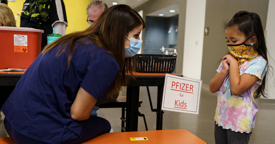
Pfizer and its partner BioNTech are expected as soon as Tuesday to ask the Food and Drug Administration to authorize a coronavirus vaccine for children under 5 years old as a two-dose regimen while they continue to research how well three doses work.Federal regulators are eager to review the data in hopes of authorizing shots for young children on an emergency basis as early as the end of February, according to multiple people familiar with the discussions, who were not authorized to speak publicly. If Pfizer waited for data on a three-dose regimen, the data would not be submitted until late March and the vaccine might not be authorized for that age group until late spring.Federal officials and Pfizer executives had been suggesting for days that an application for emergency authorization of a vaccine for children as young as 6 months was in the works. Every age group above that is eligible for shots, and the highly contagious Omicron variant has led to a sharp rise in infections among all ages, including children. There are more than 19 million Americans under 5 years old.Scott Gottlieb, a Pfizer board member and former commissioner of the Food and Drug Administration, told CBS News on Sunday that the vaccine might be authorized as soon as March for the youngest age group. The development was first reported by The Washington Post. Parents of younger children have been eager to see Pfizer and federal regulators move to make the shots available. Many families have said they are in limbo waiting out the Omicron wave and grappling with day care closures and child care crises.Federal officials are eager to begin a vaccination program for young children because the Pfizer-BioNTech studies showed two doses provided a notable level of protection against Covid-19 with no safety concerns, according to people who have been briefed on the data. Officials expect the regimen will eventually include three doses.Pfizer and BioNTech in mid-December announced that children 6 months to two years old who were given two doses of the vaccine at a tenth of the strength of adult doses produced an immune response comparable to that of young people ages 16 to 25. But among children ages 2 to 4, the response was less robust.The setback prompted the companies to test a third low dose of the shot in young children. Rather than wait for those results, federal regulators took the unusual approach of encouraging Pfizer to apply for authorization for a two-dose regimen to begin protecting children from the Omicron variant and other possible subsequent mutations, according to four people familiar with the strategy.Even though young children largely do well in combating the virus, the American Academy of Pediatrics says that the Omicron variant has intensified the threat of Covid-19 in children under 5.“Sadly, we are seeing the rates of hospitalizations increasing for children 0 to 4, children who are not yet currently eligible for Covid-19 vaccination,” Dr. Rochelle Walensky, the director of the Centers for Disease Control and Prevention, told reporters this month. She said the high rate of transmission of the Omicron variant was likely to blame.The Coronavirus Pandemic: Key Things to KnowCard 1 of 4Omicron in retreat.

Read more →

‘Decoy’ Protein Works Against Multiple Coronavirus Variants in Early Study

The NIH continues to support the development of some very innovative therapies to control SARS-CoV-2, the coronavirus that causes COVID-19. One innovative idea involves a molecular decoy to thwart the coronavirus.

How’s that? The decoy is a specially engineered protein particle that mimics the 3D structure of the ACE2 receptor, a protein on the surface of our cells that the virus’s spike proteins bind to as the first step in causing an infection.

The idea is when these ACE2 decoys are administered therapeutically, they will stick to the spike proteins that crown the coronavirus (see image above). With its spikes covered tightly in decoy, SARS-CoV-2 has a more-limited ability to attach to the real ACE2 and infect our cells.

Recently, the researchers published their initial results in the journal Nature Chemical Biology, and the early data look promising [1]. They found in mouse models of severe COVID-19 that intravenous infusion of an engineered ACE2 decoy prevented lung damage and death. Though more study is needed, the researchers say the decoy therapy could potentially be delivered directly to the lungs through an inhaler and used alone or in combination with other COVID-19 treatments.

The findings come from a research team at the University of Illinois Chicago team, led by Asrar Malik and Jalees Rehman, working in close collaboration with their colleagues at the University of Illinois Urbana-Champaign. The researchers had been intrigued by an earlier clinical trial testing the ACE2 decoy strategy [2]. However, in this earlier attempt, the clinical trial found no reduction in mortality. The ACE2 drug candidate, which is soluble and degrades in the body, also proved ineffective in neutralizing the virus.

Rather than give up on the idea, the UIC team decided to give it a try. They engineered a new soluble version of ACE2 that structurally might work better as a decoy than the original one. Their version of ACE2, which includes three changes in the protein’s amino acid building blocks, binds the SARS-CoV-2 spike protein much more tightly. In the lab, it also appeared to neutralize the virus as well as monoclonal antibodies used to treat COVID-19.

To put it to the test, they conducted studies in mice. Normal mice don’t get sick from SARS-CoV-2 because the viral spike can’t bind well to the mouse version of the ACE2 receptor. So, the researchers did their studies in a mouse that carries the human ACE2 and develops a severe acute respiratory syndrome somewhat similar to that seen in humans with severe COVID-19.

In their studies, using both the original viral isolate from Washington State and the Gamma variant (P.1) first detected in Brazil, they found that infected mice infused with their therapeutic ACE2 protein had much lower mortality and showed few signs of severe acute respiratory syndrome. While the protein worked against both versions of the virus, infection with the more aggressive Gamma variant required earlier treatment. The treated mice also regained their appetite and weight, suggesting that they were making a recovery.

Further studies showed that the decoy bound to spike proteins from every variant tested, including Alpha, Beta, Delta and Epsilon. (Omicron wasn’t yet available at the time of the study.) In fact, the decoy bound just as well, if not better, to new variants compared to the original virus.

The researchers will continue their preclinical work. If all goes well, they hope to move their ACE2 decoy into a clinical trial. What’s especially promising about this approach is it could be used in combination with treatments that work in other ways, such as by preventing virus that’s already infected cells from growing or limiting an excessive and damaging immune response to the infection.

Last week, more than 17,500 people in the United States were hospitalized with severe COVID-19. We’ve got to continue to do all we can to save lives, and it will take lots of innovative ideas, like this ACE2 decoy, to put us in a better position to beat this virus once and for all.

References:

[1] Engineered ACE2 decoy mitigates lung injury and death induced by SARS-CoV-2 variants.Zhang L, Dutta S, Xiong S, Chan M, Chan KK, Fan TM, Bailey KL, Lindeblad M, Cooper LM, Rong L, Gugliuzza AF, Shukla D, Procko E, Rehman J, Malik AB. Nat Chem Biol. 2022 Jan 19.

[2] Recombinant human angiotensin-converting enzyme 2 (rhACE2) as a treatment for patients with COVID-19 (APN01-COVID-19). ClinicalTrials.gov.

Links:

COVID-19 Research (NIH)

Accelerating COVID-19 Therapeutic Interventions and Vaccines (NIH)

Asrar Malik (University of Illinois Chicago)

Jalees Rehman (University of Illinois Chicago)

NIH Support: National Heart, Lung, and Blood Institute; National Institute of Allergy and Infectious Diseases

Read more →

Lifestyle more likely to affect a child’s BMI than the weight of their mother

Researchers from the University of Bristol and Imperial College London have found that a high Body Mass Index (BMI) of a mother before and during pregnancy is not a major cause of high BMI in their offspring — indicating that childhood and teen obesity is more likely to be a result of lifestyle factors.
The study was published today (1st February 2022) in BMC Medicine used data from two longitudinal studies — Children of the 90s (also known as the Avon Longitudinal Study of Parents and Children) based at the University of Bristol, and Born in Bradford based in Bradford Teaching Hospitals NHS Foundation Trust.
It is known that greater maternal BMI before or during pregnancy is associated with a higher BMI in children, however the extent to which the mother’s weight causes obesity in childhood, or whether this is caused by environmental and lifestyle factors post conception and birth is unclear.
The research team from the University of Bristol used a method called Mendelian randomisation, which measures variation in genes to determine the effect of an exposure on an outcome. They looked at birthweight and BMI at age 1 and 4 years in both Children of the 90s and Born in Bradford participants, and then also BMI at age 10 and 15 years in just the Children of the 90s participants. They found that there was a moderate causal effect between maternal BMI and the birth weight of children, however in most older age groups they did not find a strong causal effect.
Lead author Dr Tom Bond, Senior Research Associate at the University of Bristol explained: “We found that if women are heavier at the start of pregnancy this isn’t a strong cause of their children being heavier as teenagers. This is important to know. Supporting women and men at all ages to keep a healthy weight will be needed to prevent obesity. It isn’t enough to just focus on women entering pregnancy. Despite this, there is good evidence that maternal obesity causes other health problems for mothers and babies (aside from offspring obesity). So prospective mothers should still be encouraged and supported to maintain a healthy weight. It will be important to broaden this work to investigate other characteristics of mothers and fathers during pregnancy and a child’s early life that might affect children’s weight, and also to look at the offspring when they are in adulthood and are old enough to begin showing early signs of heart disease risk.”
Story Source:
Materials provided by University of Bristol. Note: Content may be edited for style and length.

Read more →