Avian Influenza Is Affecting Wild Mammals
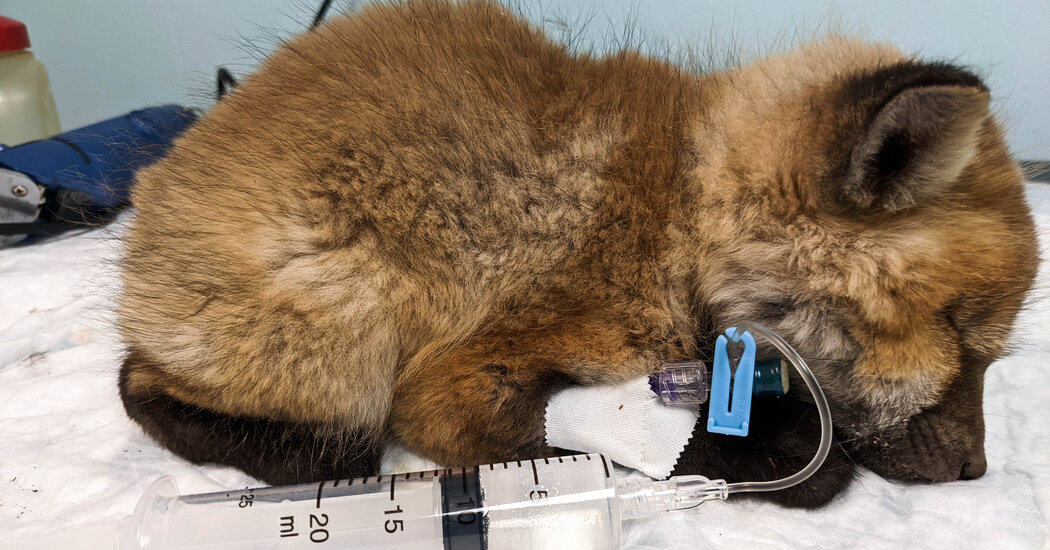
As a new version of bird flu spread through North America this spring, scientists began finding the virus in red foxes, bobcats and other mammals.Something was wrong with the foxes. That was what callers to the Dane County Humane Society in Wisconsin kept saying in April, as they reported fox kits, or young foxes, behaving in strange ways: shaking, seizing or struggling to stand. The kits, which were often lethargic and wandering by themselves, also seemed unusually easy to approach, showing little fear of humans.“We just kept getting calls,” said Erin Lemley, a wildlife veterinary technician at the humane society’s wildlife center. “And the foxes started coming in.”Some of the kits that were admitted for treatment were quiet and withdrawn, she said. Others stumbled around or had seizures, their heads ticcing, their eyes flicking rhythmically. After the staff ruled out rabies, low blood sugar and other potential causes, laboratory testing revealed a surprising culprit: a highly virulent strain of avian influenza.“It was not a fun surprise,” said Dr. Shawna Hawkins, a zoo and wildlife veterinarian at the University of Wisconsin-Madison.The virus, a type of bird flu known as Eurasian H5N1, has been spreading rapidly in the United States this spring, infecting flocks of farmed poultry in 36 states and prompting mass culls of domestic birds.But this version of the virus appears to be taking a much greater toll on wild birds than previous lineages have, finding its way into ducks, geese, gulls and terns, among many others. That, in turn, means that the virus poses an elevated danger to mammals that prey on those birds, including wild red foxes.At least seven U.S. states have detected the virus in red fox kits, to which the pathogen appeared to be particularly lethal. Two bobcats in Wisconsin, a coyote pup in Michigan and skunks in Canada have also tested positive for the virus, as have foxes, otters, a lynx, a polecat and a badger in Europe. (Two human cases, one in the United States and one in Britain, have been reported as well, both of which were in people who had close contact with birds.)The Fascinating World of BirdsAncient Swans: Paleontologists were able to reconstruct what a flightless bird that prowled the seas of Japan millions of years ago looked like.Confounding Creature: Nature seems to have a strong preference for bilateral bodies. But a parrot that walks on three limbs defies the expectations.Avian Vagrants: Birds traveling outside their native range might not have lost their way. They could be adapting to environmental changes.Transfixing Beauty: Each spring and fall, the skies in Denmark come to life with the swirling displays of European starlings.There is no evidence that mammals play a significant role in spreading the virus, and the risk to humans remains low, experts said. “This is very much still an avian virus,” said Richard Webby, an influenza virologist at St. Jude Children’s Research Hospital in Memphis.But evolution is a numbers game, he said, and the more mammals the virus infects, the more opportunities it has to pick up new mutations that could help it spread among foxes, bobcats or even humans.“What it’s going to take for this virus to transition from being a duck or a chicken virus to being a mammalian virus is more chances to replicate in those mammalian hosts,” Dr. Webby said. “So that’s why when we see these mammals being infected by this virus, we do take notice.”Twitching foxesA rescued fox kit at the Dane County Humane Society. The H5N1 virus has appeared to be especially dangerous to young foxes.Dane County Humane SocietyThe new lineage of the virus spread through Europe, Africa, the Middle East and Asia last year, sparking outbreaks in wild and domestic birds. It also showed up in a handful of wild mammals, including fox kits in the Netherlands in the spring of 2021.By the end of the year, the virus had made its way to North America. As it raced through the migrating American bird population this spring, reports began to emerge of infected fox kits — first in Ontario and subsequently in Wisconsin, Minnesota, Michigan, Iowa, Alaska, Utah and New York.  In some bird species, the virus caused obvious neurological symptoms, and many infected foxes displayed abnormal behaviors, too. They twitched, walked in circles and salivated excessively. In the most severe cases, the foxes developed seizures; death often followed shortly after, experts said.Post-mortem examinations revealed that many of the kits had pneumonia, said Dr. Betsy Elsmo, a diagnostic pathologist at the Wisconsin Veterinary Diagnostic Laboratory who performed the necropsies. When she examined the animals’ brain tissue under a microscope, Dr. Elsmo saw clear signs of damage.“There was a lot of inflammation in the brain microscopically,” she said. “The pattern of injury that I saw was consistent with a viral lesion.”So far, the virus appears to be taking a greater toll on fox kits than adult foxes, potentially because the young animals do not yet have fully developed immune systems, experts said.But the overall infection and mortality rate is unknown. “We’re just getting kind of anecdotal reports in nature right now,” said Michelle Carstensen, the wildlife health program supervisor for the Minnesota Department of Natural Resources.Wisconsin officials also detected the virus in two adult bobcats this spring. “Both bobcats showed reduced to no fear of humans,” Dr. Lindsey Long, wildlife veterinarian for the Wisconsin Department of Natural Resources, said in an email. “They were noted sitting on porches and in close proximity to human activity without the usual fear response.”One bobcat seemed to be shivering, while the other appeared to be having trouble breathing, she added. The bobcats, which were euthanized, had microscopic brain lesions that were “pretty much identical” to those in the affected foxes, Dr. Elsmo said.The virus was also recently detected in a coyote pup in Michigan, said Dr. Megan Moriarty, the wildlife veterinary specialist at the state’s Department of Natural Resources.Scientists suspect that the animals are acquiring the virus by eating infected birds. In a laboratory study, researchers had previously demonstrated that red foxes that were fed infected bird carcasses could contract, and then shed, the virus.Although it is possible that the virus has evolved in ways that make it better at infecting mammals, scientists say that the most probable explanation for the sudden rise in infected mammals is that this lineage is infecting enormous numbers of wild birds, increasing the odds that hunters and scavengers might stumble across infected food sources.So far, the virus does not appear to be causing enough illness or death in wild mammals to put these species at risk, experts said. And there is no evidence of sustained mammal-to-mammal transmission. “Mammals are generally considered to be dead-ends for highly pathogenic avian influenza,” Dr. Moriarty said.An early analysis of viral genomes from the Wisconsin fox kits suggests that the infections are essentially a series of one-offs — the result of individual foxes coming into contact with infected birds rather than foxes transmitting the virus to each other. “The preliminary data that we have suggests that these are all independent spillover events,” Dr. Elsmo said.But much remains unknown, including whether the virus will establish itself in wild birds for the long haul, which could pose a sustained risk to mammals.And even isolated mammalian infections provide the virus with new opportunities to evolve. “There’s a risk of it adapting to and then transmitting between mammals, and then you have a new problem,” said Dr. Jolianne Rijks, a veterinarian at the Dutch Wildlife Health Center.Some state officials said that they had started more routinely testing sick mammals for the virus, especially ones with neurological symptoms. Animals that test positive should also have samples of their virus sequenced so scientists can monitor for any potentially worrisome changes, Dr. Webby said.Experts also encourage members of the public to report any wild animals that appear to be acting strangely. “That’s how all this started,” Dr. Elsmo said, “as citizens seeing abnormally behaving kits and reporting them.”

Read more →

Uterine Cancer Is on the Rise, Especially Among Black Women

The cancer eventually will become the third most common type among women, experts say. The mortality rate is highest among Black Americans.Linda Collins had been in menopause for almost a decade when she started bleeding again. The bleeding was light — occasional spotting, really — and she barely gave it a passing thought.When she finally went in for a checkup, her doctor refused to let her leave until she underwent a biopsy. Within days, Ms. Collins learned she had cancer of the uterus — and an especially aggressive form of it.“I had no pain, no other symptoms, and I didn’t think seriously about it,” said Ms. Collins, 64, a retiree in the Bronx. “That was a mistake.”Cancer of the uterus, also called endometrial cancer, is increasing so rapidly that it is expected to displace colorectal cancer by 2040 as the third most common cancer among women, and the fourth-leading cause of women’s cancer deaths.The mortality rate has been increasing by almost 2 percent a year overall, with even sharper spikes among Asian, Hispanic and Black women, according to a recent study in JAMA Oncology. Despite the increase, there has been little public attention to the disease.Overall survival rates are high when uterine cancer is detected early, but few women are aware that a change in menstrual bleeding, before or after menopause, is one of the main warning signs, along with pelvic pain and painful urination and intercourse.Uterine cancer was long believed to be less common among Black women. But newer studies have confirmed that it is not only more likely to strike Black women, but also more likely to be deadly.Black women die of uterine cancer at twice the rate of white women, according to a report in March from an expert panel convened by the American College of Obstetricians and Gynecologists.The gap is one of the largest racial disparities observed for any cancer, the report said. Black women are also more likely to develop a form called non-endometrioid uterine cancer, which is more aggressive.Among all populations, uterine cancer is being detected more often in younger women who are still in their childbearing years as well as in women who don’t have any of the known risk factors, such as obesity, infertility and never having been pregnant, said Dr. Shannon Westin, a gynecologic oncologist at the University of Texas MD Anderson Cancer Center in Houston.When she first started caring for women with uterine cancer, she said, there were about 39,000 new cases a year. Now, there are more than 65,000 — and she has been in practice only 15 years.“That’s a clear indicator we should be ringing the alarm bells,” Dr. Westin said.Dr. Carol Brown, a gynecologic oncologist at Memorial Sloan Kettering Cancer Center in New York, called the surge in cases an epidemic.“The striking statistic is that right now, in the year 2022, the number of women who will lose their lives to endometrial cancer in the U.S. is almost the same as those who will die of ovarian cancer, which is unbelievable to those of us in practice for the last 30 years,” said Dr. Brown, who is the center’s senior vice president and chief health equity officer.Adrienne Moore, a respiratory therapist in the Atlanta area, was diagnosed with uterine cancer at 45. Doctors initially dismissed her symptoms as perimenopause.Lynsey Weatherspoon for The New York TimesThe JAMA Oncology study analyzed racial trends in uterine cancer while correcting for hysterectomy rates. That’s important, because Black women have a higher rate of hysterectomies, said Megan A. Clarke, the lead author. (Women who have had a hysterectomy do not have a uterus, so they cannot develop endometrial cancer; including them in the calculus artificially lowers the incidence rate.)“By making the correction for hysterectomy rates, we are more confident — the increase is real,” said Dr. Clarke, an epidemiologist at the National Cancer Institute.Black women represented just under 10 percent of the 208,587 uterine cancer cases diagnosed in the United States between 2000 and 2017, but they made up almost 18 percent of the nearly 16,797 uterine cancer deaths during that period, Dr. Clarke’s study found.The uterine cancer death rate for Black women is 31.4 per 100,000 women ages 40 and up, compared with 15.2 per 100,000 for white women in the same age group, Dr. Clarke reported. (Comparable death rates for Asian American women were nine per 100,000, and for Hispanic Americans, 12.3 per 100,000.)That makes uterine cancer an outlier, since progress has been made toward narrowing the racial gap in death rates from most cancers over the past two decades. Another National Cancer Institute report, published in JAMA Oncology in May, found that overall, death rates from cancer have declined steadily among Black Americans between 1999 and 2019, though they continue to be higher than those of other racial and ethnic groups.The reasons for the increase in uterine cancer cases are not well understood. The most common form, endometrioid cancer, is associated with estrogen exposure, which is higher when obesity is present, and obesity rates have been rising in the United States.But non-endometrioid cancer has increased in prevalence, too, and it is not linked to excess weight. Dr. Clarke’s study found that Black women are more likely to have this aggressive form of uterine cancer. They are less likely to be diagnosed early in the course of the illness, and their survival rates are worse no matter when they are diagnosed and what subtype of the cancer they have.“At every stage of diagnosis, there are different outcomes,” said Dr. Karen Knudsen, chief executive of the American Cancer Society. “Are they getting access to the same quality of cancer care?” She has called for more research into the factors driving the trends.The expert panel convened by the American College of Obstetricians and Gynecologists found racial and ethnic disparities in the care given to Black and Hispanic women with uterine cancer. They were less likely than white women to undergo hysterectomy, less likely to have their lymph nodes properly biopsied to see if the cancer had spread, and less likely to receive chemotherapy, even for a more threatening cancer.Dr. Carol Brown, a gynecologic oncologist at Memorial Sloan Kettering Cancer Center in New York, has tracked the rise in uterine cancer and called it an epidemic.Michelle V. Agins/The New York TimesAdrienne Moore, a respiratory therapist who is Black and lives in the Atlanta area, was 45 when she found out she had uterine cancer, but she said it took her almost a year to obtain the diagnosis. She went from one doctor to another complaining of heavy, very painful monthly bleeding that had started after several months without periods at all.Ms. Moore, who is now 51, was uninsured at the time, and doctors she consulted dismissed her complaints as symptoms of perimenopause or fibroids.After Ms. Moore purchased health insurance under the Affordable Care Act, she saw a gynecologist who performed another scan. It showed a thickening of the uterine wall, which can be suggestive of cancer, and the doctor immediately ordered a biopsy.Three days later, the doctor called her to the office. “I knew what it was then. I knew it was cancer,” Ms. Moore said.Ms. Moore said she is alive today because she was persistent. “If your body is telling you something, you’ve got to listen to it, and then you’ve got to get other people to listen to you,” she said.Dr. Kemi Doll, a gynecologic oncologist at the University of Washington School of Medicine in Seattle, has been doing research for years to figure out why so many Black women die of endometrial cancer.She has found that ultrasound scans that measure the thickness of the uterine wall are less accurate when patients have the more-lethal non-endometrioid type of uterine cancer, which is more common among Black women.The scans are also less effective when women have uterine fibroids, which obscure the scanner’s view, she found. That may explain why Black women, many of whom suffer from uterine fibroids, are more often diagnosed later in the disease process, Dr. Doll said.If thickening is not discovered, physicians will generally not go on to perform a biopsy to test for the cancer.New Developments in Cancer ResearchCard 1 of 7Progress in the field.

Read more →

New pediatric obesity program makes treatment more accessible

Pediatric obesity remains a serious health problem in the United States, where one in five children are affected. The U.S. Preventive Services Task Force now recommends that pediatricians screen for obesity during primary care visits and refer families to behavioral intervention programs. While there is growing interest in providing these services, accessibility continues to challenge their success.
To address this, researchers at University of California San Diego School of Medicine conducted a randomized clinical trial comparing the leading treatment program, Family-Based Treatment (FBT), with a new Guided Self-Help (GSH) program designed to provide similar resources in a less intensive and more accessible way.
The study, publishing June 17, 2022 in Pediatrics, found that GSH and FBT were similarly effective in supporting pediatric weight loss, but families were more likely to maintain attendance in GSH.
In the clinical trial, 164 children and their parents were randomly assigned to one of the two programs. Participants were recruited from two clinic sites in San Diego County, which primarily serve Latino families. The prevalence of childhood obesity in these neighborhoods (Escondido and Chula Vista) is 38 percent.
The traditional FBT program consists of 20 one-hour group sessions over six months. FBT is held at academic research centers, which adds geographical constraints. Attrition rates in these programs are high, with many parents noting scheduling issues, transportation difficulties and competing work and family responsibilities as contributing factors.
In response to these challenges, the GSH model was developed to provide shorter treatment sessions and greater scheduling flexibility. The new program consists of 14 visits, each 20 minutes in length and held at the child’s primary care clinic. Families are given material to practice between sessions in a self-directed manner, and then meet individually with a health coach to review and troubleshoot strategies.

Read more →

Neuroscientists create maps of the brain after traumatic brain injury

Scientists from the University of California, Irvine have discovered that an injury to one part of the brain changes the connections between nerve cells across the entire brain. The new research was published this week in Nature Communications.
Every year in the United States, nearly two million Americans sustain a traumatic brain injury (TBI). Survivors can live with lifelong physical, cognitive and emotional disabilities. Currently, there are no treatments.
One of the biggest challenges for neuroscientists has been to fully understand how a TBI alters the cross-talk between different cells and brain regions.
In the new study, researchers improved upon a process called iDISCO, which uses solvents to make biological samples transparent. The process leaves behind a fully intact brain that can be illuminated with lasers and imaged in 3D with specialized microscopes.
With the enhanced brain clearing processes, the UCI team mapped neural connections throughout the entire brain. The researchers focused on connections to inhibitory neurons, because these neurons are extremely vulnerable to dying after a brain injury. The team first looked at the hippocampus, a brain region responsible for learning and memory. Then, they investigated the prefrontal cortex, a brain region that works together with hippocampus. In both cases, the imaging showed that inhibitory neurons gain many more connections from neighboring nerve cells after TBI, but they become disconnected from the rest of the brain.
“We’ve known for a long time that the communication between different brain cells can change very dramatically after an injury,” said Robert Hunt, PhD, associate professor of anatomy and neurobiology and director of the Epilepsy Research Center at UCI School of Medicine whose lab conducted the study, “But, we haven’t been able to see what happens in the whole brain until now.”
To get a closer look at the damaged brain connections, Hunt and his team devised a technique for reversing the clearing procedure and probing the brain with traditional anatomical approaches.

Read more →

Rethinking the rabies vaccine

Rabies virus kills a shocking 59,000 people each year, many of them children. Some victims, especially kids, don’t realize they’ve been exposed until it is too late. For others, the intense rabies treatment regimen is out of the question: treatment is not widely available and the average $3,800 expense poses unthinkable economic burden for most people around the world.
Rabies vaccines, rather than treatments, are much more affordable and easier to administer. But those vaccines also come with a massive downside:
“Rabies vaccines don’t provide lifelong protection. You have to get your pets boosted every year to three years,” says LJI Professor Erica Ollmann Saphire, Ph.D. “Right now, rabies vaccines for humans and domestic animals are made from killed virus. But this inactivation process can cause the molecules to become misshapen — so these vaccines aren’t showing the right form to the immune system. If we made a better shaped, better structured vaccine, would immunity last longer?”
Saphire and her team, in collaboration with a team led by Hervé Bourhy, Ph.D., at the Institut Pasteur, may have discovered the path to better vaccine design. In a new study, published in Science Advances, the researchers share one of the first high-resolution looks at the rabies virus glycoprotein in its vulnerable “trimeric” form.
“The rabies glycoprotein is the only protein that rabies expresses on its surface, which means it is going to be the major target of neutralizing antibodies during an infection,” says LJI Postdoctoral Fellow Heather Callaway, Ph.D., who serves as the study’s first author.
“Rabies is the most lethal virus we know. It is so much a part of our history — we’ve lived with its specter for hundreds of years,” adds Saphire, who also serves as LJI’s President and CEO. “Yet scientists have never observed the organization of its surface molecule. It is important to understand that structure to make more effective vaccines and treatments — and to understand how rabies and other viruses like it enter cells.”
Rabies the shapeshifter

Read more →

Math model predicts efficacy of drug treatments for heart attacks

Researchers used mice to develop a mathematical model of a myocardial infarction, popularly known as a heart attack.
The new model predicts several useful new drug combinations that may one day help treat heart attacks, according to researchers at The Ohio State University.
Typically caused by blockages in the coronary arteries — or the vessels that supply blood to the heart — these cardiovascular events are experienced by more than 800,000 Americans every year, and about 30% end up dying. But even for those who survive, the damage these attacks inflict on the muscles of the heart is permanent and can lead to dangerous inflammation in the affected areas of the heart.
Treatment to restore blood flow to these blocked passages of the heart often includes surgery and drugs, or what’s known as reperfusion therapy. Nicolae Moise, lead author of the study and a postdoctoral researcher in biomedical engineering at Ohio State, said the study uses mathematical algorithms to assess the efficacy of the drugs used to combat the potentially lethal inflammation many patients experience in the aftermath of an attack.
“Biology and medicine are starting to become more mathematical,” Moise said. “There’s so much data that you need to start integrating it into some kind of framework.” While Moise has worked on other mathematical models of animal hearts, he said that the framework detailed in the current paper is the most detailed schematic of myocardial infarctions in mice ever made.
The research is published in the Journal of Theoretical Biology.
Represented by a series of differential equations, the model Moise’s team created was made using data from previous animal studies. In medicine, differential equations are often used to monitor the growth of diseases in graph form.

Read more →

A smart jumpsuit tracks infants' motor development

New wearable technology creates new possibilities for assessing the neurological development of young children. Early motor assessment is essential for supporting the early detection of neurodevelopmental problems and their therapeutic interventions.
A Finnish research group at the BABA Center, Helsinki Children’s Hospital, has developed a novel wearable for infants for the reliable assessment of motor abilities during early development. The smart jumpsuit MAIJU (Motor Assessment of Infants with a Jumpsuit) is a wearable medical device equipped with multiple movement sensors, which assist in assessing and predicting children’s neurological development.
In their recent study, the scientists measured infants at 5 to 19 months of age using the MAIJU jumpsuit during spontaneous playtime, mostly in the children’s own home environment. In the beginning of the study, the postures and movements of the infants were identified visually from a video recording using a motility description scheme custom developed for this study. This information was then used to train an algorithm based on machine learning to recognise the same postures and movements for every second of each child’s playtime at an accuracy that equals visual assessment by a trained expert.
“The development of the MAIJU wearable required a technical breakthrough in the development of machine learning algorithms for this purpose. This was achieved by combining a new kind of motility description with state-of-the-art deep learning solutions,” states the leader of technical development, Dr Manu Airaksinen.
New methods for studying neurological development
The MAIJU jumpsuit and the associated analytical solutions allow both the assessment and tracking of child’s motor maturation at an unprecedented accuracy. This advance can be exploited in many ways, for instance in early clinical diagnostics and other developmental assessments, and it also makes it possible to measure efficacy in various kinds of medical treatments and therapies.

Read more →

More than one way to skin a tumor

If cancerous cells started wreaking havoc in your body, like terrorists in a building, your immune system would act like a SWAT team to combat the enemy.
But what if the building entrance were blocked by a mysterious conspirator? The terrorists would surely be able to continue their evil deeds inside the building.
Now, researchers at Kyoto University have identified the mechanism by which active inflammation occurs in parallel with immunosuppression — the conspirator that blocks the entrance — in the tumor microenvironment or TME.
Single-cell RNA sequencing was used to analyze the action mechanism of the inhibitors of prostaglandin E2 receptor in mouse models. Results showed reduced gene expression of regulatory T cell or Treg, a strong immunosuppressive cell, involved in immunosuppression.
“We discovered that EP2/EP4 inhibitors suppressed tumor growth in mouse models by allowing Treg to infiltrate the tumor and activate there,” explains lead author Dean Thumkeo of the Department of Drug Discovery Medicine.
To put the results into human perspective, publicly accessible data indicate that some patients with either lung, ovarian, breast, or liver cancers have high EP2/EP4 expression levels.
“What is important is that EP2/EP4 expression was positively correlated with the expression of genes related to Treg recruitment and activation in these cancer patients,” adds Thumkeo.
This correlation suggests to the researchers that patients with high EP2/EP4 expression suffer from a TME that highly suppresses the immune system, suggesting that cancer treatment employing EP2/EP4 inhibitors might benefit some patients with these cancers.
Currently, immune checkpoint inhibitors, or ICIs, which target the immune system rather than the cancer cells, appear to be the new go-to cancer treatment. Typically, about a quarter of cancer patients show a significant response to ICIs. For the remaining portion of patients, EP2/EP4 inhibitors may be a potential alternative.
“For now, the discovery of the anti-immunosuppressants gives us a new way forward in exploring cancer therapeutics beyond ICIs,” concludes Thumkeo.
Story Source:
Materials provided by Kyoto University. Note: Content may be edited for style and length.

Read more →

Transparent face masks protect while facilitating communication, study finds

Commercially available transparent face masks allow for the perception of facial expressions while suppressing the dispersion of respiratory droplets that spread the SARS-CoV-2, and thus have a clear advantage over surgical face masks.
In the two years since the start of the COVID-19 pandemic, the use of facemasks in public as protection has increased across the globe. The use of masks is effective in controlling the spread of the virus, but it is not without negatives. In particular, as masks cover the lower face, they conceal facial expressions; hence, it is sometimes difficult to read the expressions of individuals wearing masks, affecting communication and interaction. The use of transparent face masks may mitigate the effects of surgical face masks.
A team of researchers from Japan, led by Professor Jun I. Kawahara at Hokkaido University and Associate Professor Yuki Miyazaki at Fukuyama University have investigated the effects of wearing a transparent mask on the perception of facial expressions. Their findings were published in the journal i-Perception.
Compared to a surgical face mask, it is most likely that the transparent masks would allow for the better perception of facial expressions. In addition, the vast majority of studies on the effects of face masks on perceptions on facial expressions have been conducted on populations in the Western countries. The cues used for comprehending facial expressions differ from East to West, and the authors wished to see if studies from the West could be applied to the East.
The team conducted two separate experiments, one using images of female faces and the other using images of male faces, expressing a range of expressions. The images were taken from a published database, and were manipulated so that each image was available in three versions: without a mask, with a surgical mask and with a transparent mask. Volunteers viewed a random series of images, one at a time, and were asked to determine the facial expression in each image.
In both experiments using male and female faces, they were able to confirm that the use of surgical masks tended to disrupt the perception of facial expressions, making them harder to recognise. The ability to recognise fear and happiness were the most affected, with happy faces being primarily misclassified as neutral, and fearful faces being misclassified as angry or surprised. However, in both experiments, the use of transparent masks did not cause any negative effect on perception of facial expressions. In both experiments, the perception of facial emotion was not influenced by the sex of the participants or the face models.
The study shows that transparent face masks, which suppress respiratory droplets that spread the SARS-CoV-2 virus, do not occlude facial expressions. There were also some limitations to the study, due to the digital manipulation of the images used: first, the effect of transparent masks under various real-world lighting conditions may differ; and second, people may communicate differently while wearing face masks. Further studies are needed to determine if cultural factors play a role in perception of facial expression obscured by masks.
Story Source:
Materials provided by Hokkaido University. Note: Content may be edited for style and length.

Read more →

A new hope for a therapy against retinitis pigmentosa

Retinitis pigmentosa, a degenerative genetic disease of the eye, is characterized by progressive vision loss, usually leading to blindness. In some patients, structural defects in the photoreceptor cells have been observed, but the molecular mechanisms involved are not understood. A team from the University of Geneva (UNIGE), in collaboration with the University of Lausanne (UNIL), has identified the essential role played by a molecular zipper formed by four proteins. The absence of this zipper leads to cell death in retinal cells. This discovery could lead to the development of therapeutic approaches for retinitis pigmentosa. This work can be read in the journal PLOS Biology.
Retinitis pigmentosa is the most common hereditary retinal disease in humans, with a prevalence of one in every 4,000 people worldwide. The first symptoms usually appear between the ages of 10 and 20 with a loss of night vision. Thereafter, the visual field narrows into a “tunnel vision” to finally lead to blindness around the age of 40. This disease is characterized by a degeneration of the light sensitive cells, the photoreceptors.
These specialized neuronal cells of the retina are responsible for the conversion of light into a nerve signal. The outer segment of the cell is made up of stacks of discs on which the light-sensitive pigments are located. The inner segment contains all the metabolic machinery essential to the functioning of the cell and is linked to the outer segment by the connecting cilium.
A molecular zipper
Mutations in the genes of four proteins located in this connecting cilium are all associated with retinal pathologies presenting degeneration of photoreceptors. These four proteins had been identified by the laboratory of Paul Guichard and Virginie Hamel of the Department of Molecular and Cellular Biology of the Faculty of Science. They are located in centrioles, cylindrical structures made of microtubules and present in all animal cells.
“In the centriole, these proteins ensure the cohesion of the different microtubules by acting like a zipper. We wondered if they did not play the same role in the tubular structures of the connecting cilium,” explains Virginie Hamel, last author of the study.
Observations with unprecedented precision
Thanks to an expansion microscopy technique optimized by the group of Virginie Hamel and Paul Guichard, which allow cells to be inflated without deforming them, the scientists were able to observe retinal tissue with a resolution never achieved. The biologists focused on the structure of connecting cilia from mice that had — or did not have — a mutation in the gene for one of the four mentioned proteins. These observations were conducted at different life stages. “In the absence of the mutation, we found that these proteins ensure, just as we had previously seen in centrioles, the cohesion between microtubules by forming a zipper that closes as development proceeds,” explains Olivier Mercey, researcher in the Department of Molecular and Cellular Biology and first author of the study.
On the other hand, when the gene for this protein is mutated, although the structure of the microtubules appears normal in the first days, the microtubules gradually become less and less attached to each other. In adulthood, the affected mice have microtubules that are no longer “zipped” together at all and eventually collapse, leading to cell death of the photoreceptors.
Restoring the “molecular zipper” to prevent cell death
This work, supported by the European Research Council (ERC) and the Pro Visu Foundation, has led to a better understanding at the molecular and structural level of retinitis pigmentosa, which allows to consider therapeutic treatments that act upstream of cell degeneration.
“By injecting the protein into patients suffering from certain types of retinitis pigmentosa, we can imagine that the molecular zipper could be restored to ensure the structural integrity of the microtubules of the connecting cilia, thus preventing the death of photoreceptor cells. We are evaluating this approach in collaboration with our colleagues from UNIL and the Jules-Gonin Ophthalmic Hospital, Yvan Arsenijevic and Corinne Kostic,” concludes Paul Guichard, coauthor of the study.
Story Source:
Materials provided by Université de Genève. Note: Content may be edited for style and length.

Read more →