Simulating cuts and burns reveals wound healing and clearing power of fibroblasts

Burn wounds are notoriously prone to bacterial infection and typically lead to a larger amount of scar tissue than laceration wounds.
In APL Bioengineering, by AIP publishing, researchers from Boston University and Harvard University created a biomimetic model to study wound healing in burn and laceration wounds. They discovered that fibroblasts — normally considered building cells that give shape and strength to tissues and organs — clear away damaged tissue before depositing new material. This part of the healing process is slower in burn wounds, where more tissue damage is present.
Cell biologists identify four phases of wound healing: bleeding stoppage, inflammation, new tissue formation, and tissue strengthening. During the inflammation and formation stages, immune cells are thought to clear bacteria and dead cells from the wound. They also activate fibroblasts and blood vessels to begin repairs.
“Depending on the injury, the extent and duration of these four phases can wildly vary across different wound types,” said author Jeroen Eyckmans. “Given that laceration wounds are well perfused with blood, they tend to heal well. However, in burns, the blood vessels are cauterized, preventing blood from entering the wound bed and slowing down the healing process. Severe burn wounds also have large amounts of dead tissue that physically block new tissue formation.”
To study how the mode of injury impacts the healing rate of wounds, the team designed an in vitro model system made of fibroblasts embedded in a collagen hydrogel. Wounds were created in this microtissue using a microdissection knife to mimic laceration or a high-energy laser to simulate a burn.
Although both wound types were equal in size, laser ablation caused more cell death and tissue damage next to the wound margins compared to knife wounds.
“During healing, we found that the fibroblasts first cleared the damaged material from the wound before depositing new material,” said Eyckmans. “This was a surprising finding because removal of dead tissue has been attributed to specialized immune cells such as macrophages, and fibroblasts have been considered to be tissue-building cells, not tissue-removal cells.”
Given that there was more tissue damage in the laser ablation wounds, it took fibroblasts more time to remove the damage, ultimately delaying tissue healing.
Based on these findings, therapies that promote wound clearance could accelerate healing. Genetically engineered white blood cells, designed to remove dead tissue, could be particularly useful for reaching injured organs and tissues deep in the body.

Read more →

Gene essential to making DNA appears to be a good target in minimizing pulmonary hypertension

The vascular smooth muscle cells that normally give blood vessel walls strength and flexibility proliferate and become destructive in pulmonary hypertension, a typically rapidly progressing condition that makes it hard to get blood inside our lungs and oxygen to our bodies.
Now scientists have found that inhibiting a gene essential to making DNA so the cells can take on this uncharacteristic growth, can significantly reduce the destructive cell proliferation and disease progression, they report in the European Heart Journal.
The findings point toward a treatment target for a condition that can inexplicably affect females ages 30 to 60 and that currently does not have great treatment options, says Yuqing Huo, MD, PhD, director of the Vascular Inflammation Program in the Vascular Biology Center at the Medical College of Georgia.
Pulmonary hypertension is basically high blood pressure in the lungs that can make breathing difficult and damage or destroy the right side of a heart which has to pump against the abnormally high pressures. It’s characterized by remodeling of the pulmonary arteries that feed oxygen-depleted blood to the lungs where it can take up oxygen and lose carbon dioxide, a byproduct of oxygen use. But there is much to be learned about why and how the cells manage the unusual growth and where therapy might best intervene.
The Vascular Biology Center team led by Huo reported late in 2022 in the journal Circulation that the vascular smooth muscle cells that encase blood vessels of the heart have the same adverse reaction when fat and cholesterol start getting deposited on their lining. The resulting abnormal proliferation and growth of vascular smooth muscle cells in this scenario worsens heart disease by prompting a thickening of the muscular wall of the blood vessels further narrowing the passageway for blood out to the body, where fat and cholesterol have already taken a stand.
Essential to cell growth, including this unhealthy proliferation, is more DNA, RNA and the proteins they produce. Key to that is purine, one of two chemical compounds in the body used to make the building blocks DNA and RNA. Key to purine production, is the gene ATIC, and in this case, more of it.

The new studies found that, as with the unhealthy proliferation in coronary arteries, when the scientists deleted ATIC from either the vascular smooth muscle cells or body wide, it reduced the development and progression of pulmonary hypertension in their mouse model of the breath-taking condition. It even mitigated a very severe form of the condition.
“If we block this process, the blood vessel wall will stay relatively normal and the blood will still pass through,” Hou says.
When the scientists looked in mouse models as well as human pulmonary arteries and lung tissue, they found a similar scenario: Genes essential to production of purine are increased and so is actual purine production, and expression of the ATIC gene.
When they first looked at human genetic data, they had clues that is what they might find: They saw proliferation as well as increased expression of genes that indicated the cells were making purine from scratch, so called de novo purine synthesis, rather than from recycling, which is the body’s other option. That’s the same high energy purine producing process they found vascular smooth muscle cells on coronary arteries were using.
Huo and his colleagues note that when vascular smooth muscle cells begin to proliferate and become resistant to death, they start resembling cancer cells. What the scientists have now found underlines the similarities between metabolic shifts that occur in both the early development of pulmonary hypertension and cancer, Huo and his colleagues write. In fact, making purine from scratch also is increased in many cancer cells, which may make ATIC a logical treatment target there as well, Huo and his colleagues write. For example, one way the drug methotrexate, used to slow the growth of cancer cells, is thought to work is by inhibiting ATIC. Also, newer ATIC inhibitors for cancer are under study.
Huo also notes that more specific inhibitors are needed to treat pulmonary hypertension and that the same inhibitors likely would also work in the coronary arteries.
The lungs are extremely vascular and pulmonary arteries branch like a tree in the lungs until they eventually become smaller vessels called arterioles and eventually even smaller, single-layer blood vessels called capillaries, which surround the millions of air sacs in the lungs. The capillaries are where carbon dioxide escapes from the blood and where oxygen from the air we breathe moves in. Huo notes capillaries don’t have the typical layer of smooth muscle cells only the endothelial cells that normally just line blood vessels.
Right heart failure is a leading cause of death in people with pulmonary hypertension and is even tougher to treat than left heart failure, which can result from more common problems like coronary artery disease, heart attack and high blood pressure, Huo says.
Postdoctoral Fellow Qian Ma, PhD, is first author on the new paper. The research was supported by the American Heart Association and the National Institutes of Health.

Read more →

AI model helps atopic dermatitis patients diagnose complications and malignant diseases

A team of dermatologists has developed an artificial intelligence (AI) model that empowers atopic dermatitis (AD) patients to detect complications from bacterial or viral infections and distinguish between eczema and skin lesions caused by a type of blood cancer.
The AI model is detailed in a paper published in the Journal of Dermatological Science on January 11, 2023.
AD is a chronic disease that affects around 12 percent of people and often begins in childhood. Patients with AD typically have a suppressed skin immune barrier, which reduces their protection against microbial pathogens, leading to complications of the eczema from bacterial or viral infections. This may include herpes simplex, impetigo, and Kaposi varicelliform eruption (eczema herpeticum).
Recognizing whether AD has led to any of these complications can be challenging for patients as the symptoms’ appearance on the skin is very similar to AD itself. Moreover, mycosis fungoides, a type of blood cancer that causes skin lesions, can also exhibit similar symptoms to AD and may co-exist with AD. Some medications for AD can even worsen infections or mycosis fungoides.
Proper and early diagnosis of complications and malignant diseases is critical for appropriate treatment and better outcomes. However, patients cannot always recognize any abnormal symptoms and visit a doctor as soon as possible due to the similarity of symptoms.
To address this issue, the team trained their convolutional neural network (CNN) model on non-standard images of AD, impetigo, mycosis fungoides, herpes simplex, and Kaposi varicelliform eruption. They then compared the AI’s diagnosis accuracy to a set of non-standard images manually cropped and diagnostically annotated by dermatologists. They found that their system achieved a diagnostic accuracy almost equal to the manually assessed image set.
The team is currently developing an AI-powered smartphone app to translate their system, enabling patients to manage their skin diseases remotely with just their phone’s camera. They are also experimenting with AD patients to improve the app’s usability.
Yuta Yanagisawa, a researcher with the Tohoku University School of Medicine and co-author of the paper, said, “A dermatologist would of course be able to spot the difference, but it is incredibly impractical for an AD patient to visit a dermatologist every day. If only there were some handy, low-cost mechanism that replicated that dermatologist’s knowledge and could be used during a patient’s daily regimen of checking their skin.”
The team believes that this technology will help patients with skin diseases to manage their conditions effectively and efficiently, resulting in better health outcomes.

Read more →

Fats help tag medical implants as friend or foe

Medical implants can be lifesaving or significantly improve quality of life, but to our immune system they can appear as intruders.
Rice University bioengineer Omid Veiseh and collaborators have found that lipid deposition on the surfaces of implants can play a mediating role between the body and implants, with some lipids acting as peacekeepers while others stir up conflict.
“We learned that as immune cells crawl on an implanted biomaterial, they leave lipid vesicles that signal to the host immune system whether the biomaterials should be ignored or walled off from the body,” said Veiseh, a Rice assistant professor of bioengineering and Cancer Prevention and Research Institute of Texas scholar.
This knowledge could help scientists develop biomaterials or coatings for implants that deflect host immune system aggression, reducing malfunction rates for biomedical devices such as pacemakers, cerebrospinal fluid shunts, coronary stents, surgical mesh, drug delivery pumps, biosensors and more.
The study is published in Advanced Materials.
“A major problem in all biomedical implants is that the immune system attacks them,” said Christian Schreib, a Rice graduate student and lead author on the study. “Essentially, it encapsulates them in a fibrotic capsule that destroys their functionality and makes them not work anymore.”
“Our team was able to develop a chemical surface modification that preferentially recruits macrophages which leave behind a ‘do-not-attack’ lipid-vesicle signature allowing implants to exist in the body without being recognized as foreign,” Veiseh said.

Fibrosis, or scarring, is the accumulation of excess tissue at the site of an injury. The fibrotic response to implants has traditionally been associated with the deposition of proteins on the implanted surface.
“In our research, we realized that, while proteins are important, fat molecules also play a significant role in the fibrotic process,” Schreib said. “We identified two lipid profiles, fatty acids and phospholipids. Fatty acids are more likely to provoke an immune response, while phospholipids are more likely to fly under the radar and not irk the immune system.
“Now that we understand this, we can use this knowledge to engineer materials for use in implants that are less likely to trigger an immune response. We could, say, engineer a material that pulls in phospholipids to it, so that when you implant the material, the phospholipids naturally deposit onto it and help it evade the immune system. We might also want to look at taking those fat molecules like the phospholipids and chemically functionalize them to the device surface before implantation.”
When an immune response is triggered in the body, immune cells are mobilized at the site of the injury or intrusion. The increased traffic of immune cells near the implant leads to a greater accumulation of fibrotic tissue.
“A thick layer of cells deposited on the implant is likely to stop it from working,” Schreib said. “But if you have a layer of lipids on the atomic scale, that’s not going to affect its functionality to the same extent.”
Optimizing implant performance is most critical for patient groups who rely on them for the management of chronic and potentially life-threatening conditions such as hydrocephalus, a disorder that involves an excess buildup of cerebrospinal fluid (CSF) in the brain. For many patients, the only effective management strategy is the placement of a CSF shunt that diverts excess fluid to a different body cavity. Pediatric hydrocephalus patients face particularly high rates of implant failure, which can result in headaches, vomiting, loss of vision, brain injury and death if not addressed quickly.

“As a pediatric neurosurgeon, it’s safe to say that shunt malfunctions are the bane of my existence,” said Dr. Brian Hanak, assistant professor of neurosurgery at Loma Linda University Children’s Hospital in California who is a co-author on the study. While CSF shunt malfunction can occur in any age group, malfunction rates are much higher in young children. “Most of us working in this field feel that is likely related to the fact that the brain’s innate immune system is particularly revved up in young children,” he said.
“In young children and babies, shunt malfunction rates are in the ballpark of 40%-50% at two years post-implantation. Frankly, I’m embarrassed to routinely implant the most failure-prone life-sustaining device in modern medicine. If you developed a pacemaker with a 40% to 50% failure rate at two years, it would never get approval from the U.S. Food and Drug Administration, because that’s appalling. But that’s unfortunately the industry standard for CSF shunts.”
Hanak said many brain implants could benefit from a reduced innate immune response.
“One in particular that always comes to mind for me is brain-computer interface technology,” he said. “It’s been about 20 years now that we’ve had proof-of-concept that you can implant a microelectrode array in someone’s brain and have them use that array to control a robotic arm.
“You might ask, if that’s the case, then why is this technology not something that every paralyzed person can use to improve their independence and quality of life? The reason is that the immune response mounted to those implanted electrode arrays makes them unable to record neural activity beyond two to three years in vivo. At the moment, with our current state of technology, it’s not really a viable solution, certainly not a long-term solution for paralyzed patients.”
The National Institutes of Health (R01 DK120459), the Defense Advanced Research Projects Agency (D20AC00002), the Rice University Academy Fellowship, the Shared Equipment Authority at Rice and the National Science Foundation (CBET1626418) supported the research.

Read more →

New, non-invasive imaging tool maps uterine contractions during labor

Researchers funded by the National Institutes of Health have developed a new imaging tool, called electromyometrial imaging (EMMI), to create real-time, three-dimensional images and maps of contractions during labor. The non-invasive imaging technique generates new types of images and metrics that can help quantify contraction patterns, providing foundational knowledge to improve labor management, particularly for preterm birth. The small study is supported in part by NIH’s Eunice Kennedy Shriver National Institute of Child Health and Human Development (NICHD) through its Human Placenta Project and other programs. The findings are published in Nature Communications.
“EMMI has the potential to answer critical questions about uterine contractions and will help us better understand what occurs during pregnancy and labor,” said Diana W. Bianchi, M.D., NICHD Director. “With additional research, the tool may potentially predict who is at risk to deliver prematurely or whose labor pattern will eventually result in the need for a cesarean section delivery. This will also help care providers evaluate whether a treatment or intervention is working.”
The study team, led by Yong Wang, Ph.D., and Alan Schwartz, M.D., Ph.D., at Washington University in St. Louis, and Alison Cahill, M.D., at the University of Texas at Austin, initially developed EMMI using a sheep model and reported their findings in Science Translational Medicine. In the new study, the team tailored EMMI for human clinical use and tested it among a group of 10 women with healthy pregnancies. Current clinical methods to measure contractions (i.e., tocodynamometry and an intrauterine pressure catheter) can only provide limited details, such as contraction duration and intensity, while also being invasive.
EMMI integrated two types of non-invasive scans — a fast anatomical MRI to obtain an image of the uterus (which can be taken during early term pregnancy, or 37 weeks gestation), and a multi-channel surface scanning electromyogram that uses sensors placed along the belly to measure contractions during labor. These data are then combined and processed into three-dimensional uterine maps, with warm colors denoting areas of the uterus that are activated earlier in a contraction, cool colors indicating areas that are activated later and gray areas showing inactive regions. A sequence of maps is generated over time, creating a visual timelapse that shows where contractions start, how they spread and/or synchronize, and potential patterns that are associated with a typical pregnancy versus one with complications.
EMMI maps were also used to develop metrics to describe uterine contractions. The maximal activation ratio, for example, measures the total surface area of the uterus that becomes electrically active during an individual contraction.The activation curve slope measures the rate of uterine electrical activation. The fundal early activation ratio helps quantify the region that generates contractions to dilate the cervix.
Results from the pilot study also bring clarity on a longstanding question on how contractions begin — EMMI data suggest there is no fixed, pacemaker-like region in the uterus that initiates labor. The study team observed varied patterns of contractions and metrics among the 10 study participants, with some similarities between women who had never given birth and those who had. However, more research is needed to confirm and expand upon these observations.
EMMI offers new possibilities for better understanding human labor and facilitating the development of optimized, patient-specific interventions. The authors note that an EMMI contraction atlas generated from healthy pregnancies can serve as a resource to understand and diagnose preterm labor and possibly identify patients who would benefit from an induction versus those who may need a cesarean section.

Read more →

Understanding sound direction estimation in monaural hearing

The ability to locate sounds in the surrounding environment is a remarkable feature of the human ear. Typically, people with good hearing use both ears to detect and interpret auditory cues. Differences in the loudness or arrival time of sounds at each ear provide us with vital information about the location and direction of the sound source. Interestingly, however, studies have suggested that while binaural cues are sufficient for sound localization, they are not necessary. People with monaural hearing (hearing loss in one ear) can perceive sound location as well.
Fortunately for engineers, this can help remove limitations on the design and positioning of audio recording devices and microphone arrays. Used for source localization and noise reduction, microphone arrays need to be placed at specific intervals and positions to effectively capture and analyze sound from different directions. To avoid poor sound quality resulting from inadequate microphone array design or positioning, the capability to estimate the sound direction using monaural cues is highly desirable as it can help simplify microphone array designs.
In a study made available online on 13 January 2023 and published in the journal Applied Acoustics on 28 February 2023, Prof. Masashi Unoki and his colleagues from the Japan Advanced Institute of Science and Technology (JAIST) and Toyama Prefectural University, Japan, have proposed a method that uses monaural cues to estimate the direction-of-arrival (DOA) of sound signals in three dimensions. “In our work, we propose an estimation method based on monaural modulation spectrum (MMS), which relies on modulation in the frequency spectrum of the received signal to detect the signal DOA. This can help us develop monaural cues for single-channel signal processing,” explains Prof. Unoki.
To determine the monaural DOA, the team simulated sound signals from different directions using artificial amplitude modulation noise and human speech signals while accounting for the effect of the ears, torso, and head in filtering sound. Next, they obtained the MMS of the signals describing their frequency modulations to identify key features that could be tied to the DOA of the signals. To avoid monaural front-back confusion, which occurs when sound sources at the same angle in front of or behind the listener can produce the same estimates for the DOA, the researchers considered the effect of head movement on the MMS to realize a more accurate DOA estimation.
Using the known DOA and the features of the MMS as training data, they then constructed a polynomial regression model that estimated the DOA from the MMS features of the sound signal in terms of the horizontal and the vertical direction of the listener. The model could accurately estimate the DOA of 829,440 speech signals, outperforming even human monaural hearing.
While the team qualifies their findings by suggesting that there is more work to be done to account for background noise and individual differences in ear shape when creating the model, the study demonstrates an impressive advancement in monaural sound localization. Speculating about its implications, the researchers envision their technology’s applications in sound surveillance techniques and hearing aid enhancements. “Our study will help reveal our ability to localize sounds based on monaural hearing, which, in turn, could stimulate various innovations in hearing aid techniques in the long-term,” concludes Prof. Unoki.

Read more →

Researchers develop enhanced genetic animal model of Down syndrome

National Institutes of Health researchers compared a new genetic animal model of Down syndrome to the standard model and found the updated version to be enhanced. The new mouse model shows milder cognitive traits compared to a previously studied Down syndrome mouse model. The results of this study, published in Biological Psychiatry, may help researchers develop more precise treatments to improve cognition in people with Down syndrome.
Scientists found that the new mouse model, known as Ts66Yah, had memory difficulties and behavior traits, but the symptoms were not as severe as seen with the previous mouse model. Scientists often use different strains of mice as animal models to study human diseases because most genes in humans have similar counterparts in mice.
“A mouse model that more precisely captures the genetics of Down syndrome has important implications for human clinical trials that aim to improve cognition,” said Diana W. Bianchi, M.D., director of the Eunice Kennedy Shriver National Institute of Child Health and Human Development, senior investigator in the National Human Genome Research Institute (NHGRI) Center for Precision Health Research and senior author of the study.
About 6,000 newborns are diagnosed with Down syndrome each year in the United States. In most cases, these babies have a third copy of chromosome 21. An additional chromosome 21 adds an extra copy of over 200 protein-coding genes to that person’s genome, which causes difficulties with learning, speech and motor skills.
A previous mouse model, known as Ts65Dn, has been considered the standard for Down syndrome research, used in preclinical studies for nearly 30 years. Along with some successful cognitive treatments, such as a recent hormone-based cognitive treatment, some other treatments that were effective in the mouse model were not as effective in humans.
Importantly, the previous mouse model’s genome contains 45 extra genes that are irrelevant to human Down syndrome, a byproduct of how the model was developed. Humans and mice have very similar genomes, but the chromosomes that make up those genomes do not precisely align across those two species. For example, many of the genes found on human chromosome 21 are found on mouse chromosomes 16 and 17. The previous mouse model has an additional region of mouse chromosome 17 that contains 45 extra genes not found on human chromosome 21. How these 45 extra genes affect the brain and behavior of the previous Ts65Dn mice has not been investigated until now.
To create an enhanced mouse model of Down syndrome, researchers at the University of Strasbourg, France, removed these extra 45 genes using CRISPR gene-editing technology. Dr. Bianchi’s group then compared the two mouse models and found that the extra 45 genes in the previous mouse model were affecting brain development and contributed to more severe difficulties with motor skills, communication and memory.
“There are considerable effects of these extra genes on mouse brain development and behavior,” said Faycal Guedj, Ph.D., staff scientist in NHGRI’s Center for Precision Health Research and first author of the study. “What was previously thought as the best mouse model of Down syndrome has traits derived from genes that are not relevant to human chromosome 21.”
Researchers at the Center for Precision Health Research aim to use cutting-edge genomics tools to foster next-generation healthcare. With this new and improved mouse model, Dr. Bianchi’s group hopes to develop more precise treatments for improving cognition in people with Down syndrome.
“The possibility of treating intellectual disabilities in the context of Down syndrome goes to the core of changing conceptions about the nature of disability, its medical and clinical aspects, and what we, often pejoratively, consider ‘normal’ and ‘desirable’ in the context of medical care and in society,” underscores Christopher R. Donohue, Ph.D., NHGRI senior historian. “As cognitive treatments based on genetic models become more feasible in the future, researchers, in conversation with disability ethicists, those with Down syndrome and other healthcare professionals, should carefully weigh potential benefits versus drawbacks, including contributing to ableism in medicine, and other forms of stigma.”

Read more →

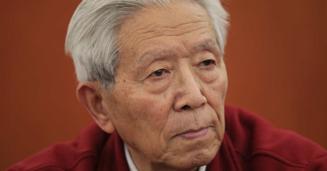
Jiang Yanyong, Who Helped Expose China’s SARS Crisis, Dies at 91

A retired military surgeon, he blew the whistle in 2003 on Beijing’s cover-up of the epidemic. He was later punished for denouncing the Tiananmen Square crackdown.Dr. Jiang Yanyong, a prominent military surgeon who became a national hero for exposing the Chinese government’s cover-up of the SARS epidemic in 2003, but was later punished for denouncing the Tiananmen Square crackdown, died on Saturday. He was 91.His death was widely reported by Chinese-language media in Hong Kong and abroad, as well as by friends in China, who shared a notice on social media saying he had succumbed to pneumonia and other illnesses. Two friends of his family told The New York Times that they had confirmed his death with relatives, but both asked not to be identified, fearing recriminations.Chinese state media have not confirmed the news of Dr. Jiang’s death, which is not uncommon for a politically sensitive figure.In the spring of 2003, alarmed to hear health officials downplaying the threat of severe acute respiratory syndrome, or SARS, Dr. Jiang sent a letter to several news organizations refuting the official story. His revelations prompted China’s top leaders to acknowledge that they had provided false information about the epidemic and to begin a nationwide effort to battle it, saving countless lives.“I was telling the truth,” Dr. Jiang later recalled in a 2013 interview with the state-run Beijing News. “I believed the government would treat me fairly.”It did, at least for a while, hailing Dr. Jiang as a hero. But nearly two decades later, the Chinese authorities would go on to virtually repeat the cover-up with the outbreak of Covid-19.Dr. Jiang was retired in 2003 when he heard that many Beijing hospitals, including the elite military hospital where he had worked, were dealing with a surge in patients infected with the SARS virus. By then, SARS was already a full-blown epidemic, spreading to multiple countries and infecting more than 1,000 people in Hong Kong and southern China alone.So it came as a shock to Dr. Jiang when China’s top health minister, Zhang Wenkang, appeared on television, saying that Beijing had only 12 cases of SARS and three deaths.“You are safe here whether you wear the mask or not,” Mr. Zhang said. “Beijing is perfectly safe to visit for business or pleasure.”Stunned by those blatantly misleading remarks, Dr. Jiang sat down the next day and wrote his letter, saying that there were already over 100 cases in Beijing alone. The lanky, elderly doctor accused Mr. Zhang, who had also been trained as a military doctor, of “abandoning even his most basic standards of integrity as a doctor.”A waitress, left, being admitted to the SARS ward of a hospital in Guangzhou, in southern China, in 2004.China Photo ASW, via ReutersAs a Communist Party member who held a rank corresponding to major general in the United States, Dr. Jiang was taking a huge risk. Nonetheless, he signed his name and sent the letter off to several local media outlets. Foreign journalists soon caught wind of it, and Time magazine broke the news.The impact was immediate.World Health Organization inspectors quickly extended a trip to Beijing to inspect the hospitals where Dr. Jiang said there were hidden cases, putting pressure on the government. Chinese leaders fired Mr. Zhang, the health minister, and the mayor of Beijing.For a brief period, Dr. Jiang received nationwide acclaim. One local magazine called him the “honest doctor.”“His goal wasn’t to make China lose face,” his daughter, Jiang Rui, said later in an interview. “He just saw that he had a chance to save lives.”But the next year, emboldened by his new political capital, he wrote a letter to top Chinese leaders, calling on them to acknowledge that the 1989 crackdown on the Tiananmen Square pro-democracy protests had been wrong, and that the student movement had in fact been a “patriotic movement.”Dr. Jiang wrote about what he’d seen at No. 301 Military Hospital in Beijing on the night that tanks rolled into the square and People’s Liberation Army soldiers began shooting indiscriminately at student protesters. Scores of wounded civilians were rushed to the hospital, he said. Many had been hit by bullets designed to break apart after impact and shred internal organs.It was the bloodiest night of his decades-long career.“My brain buzzed and I almost passed out,” he recalled. “Lying before me this time were our own people, killed by children of the Chinese people, with weapons given to them by the people.”Soon after the letter became public, Dr. Jiang and his wife were detained. He was forced to undergo lengthy interrogation and indoctrination sessions. For years, he was barred from leaving China and periodically subjected to monitoring, harassment and house arrest. He all but disappeared from public view.World Health Organization experts in 2004, checking a building where SARS was suspected to have been spread. China Photos ASW, via ReutersJiang Yanyong was born on Oct. 4, 1931, in the eastern Chinese city of Hangzhou to a wealthy banking family. Growing up in Shanghai, he resolved to become a doctor after watching his aunt succumb to tuberculosis. In 1949, the year Mao Zedong’s Communists took power, he enrolled in Yenching University to study medicine and went on to train at Peking Union Medical College, the country’s most prestigious medical school.Inspired by Norman Bethune, a Canadian doctor who died on the front lines of the Communist resistance to the Japanese occupation in 1939, he enlisted in the People’s Liberation Army and specialized in surgery. In 1957, he was assigned to No. 301 Hospital in Beijing.He became known for his precision, steady hands and willingness to take on difficult cases, earning him the nickname “Brave Jiang.”But his idealism didn’t last long. In 1966, Mao unleashed the Cultural Revolution, the decade-long period of chaos that upended Chinese society. Groups of militant youths known as Red Guards roved the country, determined to root out “class enemies.” Dr. Jiang, whose father had been a banker and whose cousin was an official in the rival Nationalist party in Taiwan, was an easy target.Branded a counterrevolutionary, he was imprisoned, beaten and later sent to a prison farm for five years in the remote deserts of Qinghai Province in western China, away from his wife and children. After he was politically rehabilitated in the early 1970s, he returned to No. 301 hospital, where he eventually worked his way up to chief of surgery.He retired in the early 2000s, but he continued to treat patients and mentor younger doctors. In 2004, he was awarded the prestigious Ramon Magsaysay Award for Public Service in recognition of his “brave stand for truth in China, spurring lifesaving measures to confront and contain the deadly threat of SARS.”In addition to Ms. Jiang, his daughter, he is survived by his wife, Hua Zhongwei, and a son, Jiang Qing.Dr. Jiang never backed down on the subject of Tiananmen Square. In 2019, before the 30th anniversary of the crackdown, he wrote a letter to Xi Jinping, China’s leader, again demanding justice for the “crime” of 1989. Soon after, the 87-year-old doctor was again under house arrest.Like others who challenged Communist Party policy, he was largely erased from the official record, and he was sometimes painted as a wrongdoer for having spoken out.A multiple-choice question posed by a test-prep school in 2017 asked about his decision to come forward about SARS. The “correct” answer was B: Doing so was wrong because it harmed the interests of the nation, the society and the community, and he should be subject to legal punishment.“All I did was say a few honest things,” said Dr. Jiang, pictured in 2004. Hu Jia/Associated PressSuch priorities still seemed to hold sway in late 2019, when a new coronavirus was emerging in China. A doctor in the central city of Wuhan, Li Wenliang, posted a warning to a group of fellow doctors about the still-unidentified disease, which he said resembled the SARS virus.The government reprimanded Dr. Li and forced him to renounce his warning. As the epidemic grew, government officials continued to underplay the coronavirus’s threat, delaying efforts to contain it, with global ramifications.When Dr. Li died of Covid, he was mourned across China as someone who had spoken truth to power, as Dr. Jiang had done before him.“I’m not a hero,” Dr. Jiang said in the 2013 interview. “All I did was say a few honest things.”“If everyone spoke the truth, then there would be nothing to hide,” he added. “If everyone spoke the truth, the country would naturally be better off.”Chris Buckley

Read more →

Heartbeat May Shape Our Perception of Time, Study Shows

Researchers have long assumed that the brain controls our sense of time. A new study suggests the heart plays an important role.It is a truism that time seems to expand or contract depending on our circumstances: In a state of terror, seconds can stretch. A day spent in solitude can drag. When we’re trying to meet a deadline, hours race by.A study published this month in the journal Psychophysiology by psychologists at Cornell University found that, when observed at the level of microseconds, some of these distortions could be driven by heartbeats, whose length is variable from moment to moment.The psychologists fitted undergraduates with electrocardiograms to measure the length of each heartbeat precisely, and then asked them to estimate the length of brief audio tones. The psychologists discovered that after a longer heartbeat interval, subjects tended to perceive the tone as longer; shorter intervals led subjects to assess the tone as shorter. Subsequent to each tone, the subjects’ heartbeat intervals lengthened.A lower heart rate appeared to assist with perception, said Saeedeh Sadeghi, a doctoral candidate at Cornell and the study’s lead author.“When we need to perceive things from the outside world, the beats of the heart are noise to the cortex,” she said. “You can sample the world more — it’s easier to get things in — when the heart is silent.”The study provides more evidence, after an era of research focusing on the brain, that “there is no single part of the brain or body that keeps time — it’s all a network,” she said, adding, “The brain controls the heart, and the heart, in turn, impacts the brain.”Interest in the perception of time has exploded since the Covid pandemic, when activity outside the home came to an abrupt halt for many and people around the world found themselves facing stretches of undifferentiated time.A study of time perception conducted during the first year of the lockdown in Britain found that 80 percent of participants reported distortions in time, in different directions. On average, older, more socially isolated people reported that time slowed, and younger, more active people reported that it sped up.“Our experience of time is affected in ways which mirror, generally, our well-being,” said Ruth S. Ogden, a psychology professor at Liverpool John Moores University and the author of the lockdown study. “People with depression experience a slowing of time, and that slowing of time is experienced as being a worsening factor of the depression.”The new Cornell study addresses something different: how we perceive the passage of microseconds. Understanding those mechanisms may help us to manage trauma, in which instantaneous experiences are remembered as drawn out, Dr. Ogden said.When trying to assess the importance of an experience, she said, “our brain just looks back and says, Well, how many memories did we make?”She added, “When you have this really rich memory, richer than you would normally get in a 15-minute period of your life, that’s going to trick you into thinking that it was long.”Research into perception of time has focused, until recently, on different areas of the brain, said Hugo Critchley, a professor of psychiatry at Brighton and Sussex Medical School who has studied how heartbeats affect memory for words and fear responses.“I think there’s much greater appreciation that cognitive functions are intimately linked, perhaps even grounded in, the control of the body, whereas most of the psychology up to the 1990s dismisses the body as being something controlled at the level of the brain stem,” said Dr. Critchley, who was not involved in the Cornell heartbeat study.Previous research has investigated how physical arousal is connected to stress processing, and emotional states like anxiety and panic, Dr. Critchley said. The new study expands on that by focusing on the role of the heart in a nonemotional function, the perception of time, which can be linked to larger distortions in thinking.“You can’t look at cognitive function in isolation,” he said. “Even understanding how the brain develops and starts representing internal mental states, people are looking at the primacy of the inescapable internal information you need to control to keep alive.”One reason that the body may be closely involved in the perception of time is that time is closely related to metabolic needs, said Adam K. Anderson, a professor of psychology at Cornell and a co-author of the new study.“Time is a resource,” Dr. Anderson said. “If the body was a battery, or a gas tank, it’s trying in the moment to say, How much energy do we have? We’re going to make things seem shorter or longer in terms of time based on how much bodily energy we have.”

Read more →

Aggressive Medical Care Remains Common at Life’s End

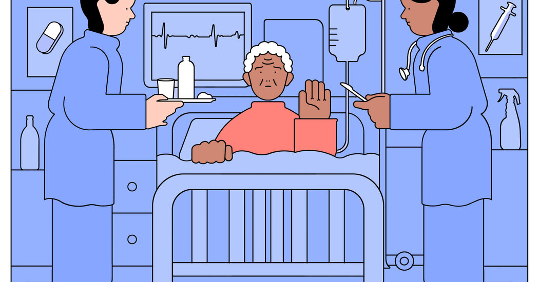
Most older cancer patients received invasive care in the last month of their lives, a new study finds. That may not be what they wanted.In July, Jennifer O’Brien got the phone call that adult children dread. Her 84-year-old father, who insisted on living alone in rural New Mexico, had broken his hip. The neighbor who found him on the floor after a fall had called an ambulance.Ms. O’Brien is a health care administrator and consultant in Little Rock, Ark., and the widow of a palliative care doctor; she knew more than family members typically do about what lay ahead.James O’Brien, a retired entrepreneur, was in poor health, with heart failure and advanced lung disease after decades of smoking. Because of a spinal injury, he needed a walker. He was so short of breath that, except for quick breaks during meals, he relied on a biPAP, a ventilator that required a tightfitting face mask.He had standing do-not-resuscitate and do-not-intubate orders, Ms. O’Brien said. They had discussed his strong belief that “if his heart stopped, he would take that to mean that it was his time.”Listening in on the phone while a hospital palliative care nurse-practitioner talked to her father about his options, Ms. O’Brien provided a blunt translation to an always blunt man: “Dad, your heart and lungs are done.”The next day, he declined surgery to repair his hip. A startled anesthesiologist and an orthopedist called his daughter, apparently expecting her to talk her father into agreeing to the operation. She didn’t try.“He was dying,” she said in an interview. “He’d either die comfortably or, with a big surgical incision, he’d die uncomfortably. Or die of something more complicated — potential infections, bowel obstructions, so many things that can happen.” Mortality rates after hip fractures, though improving, remain high.Her father, who wasn’t cognitively impaired, had decided that surgery was “silly” and unnecessary. She supported his decision and contacted a local hospice.Families often have to run interference in such scenarios, and a new study in JAMA Network Open helps explain why. The authors, most of them at Case Western Reserve University School of Medicine, analyzed five years of data from a cancer registry, nursing home assessments and Medicare claims to look at “aggressive end-of-life care” among 146,000 older patients with metastatic cancer.They compared nursing home residents’ care in the last 30 days of their lives with the care for non-institutionalized patients living in communities, the lead author, Siran Koroukian, a health services researcher at Case Western Reserve, said.The team looked for commonly used markers of aggressive care, including cancer treatment, repeated emergency room visits or hospitalizations, admission to an intensive care unit, lack of hospice enrollment until three days before death, and death in a hospital.“In all probability, hospice should have been considered” for these patients, said Sara Douglas, a co-author and oncology researcher at the Case Western Reserve University School of Nursing.Yet the majority of both groups — 58 percent of community dwellers and 64 percent of nursing home residents — received aggressive treatment in their final 30 days. A quarter underwent cancer treatment: surgery, radiation, chemotherapy.Although studies repeatedly show that most patients want to die at home, 25 percent of the community dwellers and almost 40 percent of the nursing home residents died in hospitals.Hospice leaders, palliative care specialists, health care reformers and advocacy groups have worked for years to try to lower such numbers. “Patients who received this type of aggressive care experience more pain, actually die sooner, have a much poorer quality of life at the end. And their families experience more doubt and trauma,” Dr. Douglas said.Because the researchers used large databases, the study can’t indicate whether some patients actually opted for continued treatment or hospitalization. Some treatments the authors deemed aggressive could instead have been palliative, intended to increase comfort, like radiation to shrink tumors that might impede breathing.Still, “these are really sobering statistics,” said Douglas White, director of the Center for Ethics and Decision Making in Critical Illness at the University of Pittsburgh School of Medicine.A lot of factors contribute to invasive actions in patients’ final days and weeks. Some originate within the health care system itself. Doctors may be reluctant to initiate difficult conversations about what dying patients want, or be poorly trained in conducting them.“The minute you have this conversation, people assume, ‘You’re giving up on me,’” Dr. Douglas said. Even having an advance directive and a Physician Order for Life-Sustaining Treatment, or P.O.L.S.T., doesn’t always ward off aggressive treatment.But studies also show that even when crucial discussions take place, patients and surrogate decision-makers frequently misinterpret them. “Families often leave these conversations with much more optimistic expectations than their doctors meant to convey,” Dr. White said.His research has documented the effects of optimism bias. Surrogates understand positive prognoses more accurately than negative ones. They may grasp that most people in this situation will die, but insist that their particular loved one is different, fiercer, stronger. Misplaced optimism then leads to more aggressive treatment.Sometimes, family demands prevail even over the patient’s own wishes. Jennifer Ballentine, chief executive of the Coalition for Compassionate Care of California, knew that one of her relatives didn’t want high-intensity care if he became terminally ill. But when he developed aggressive prostate cancer at 79, his wife insisted that he pursue treatment.“He refused. He kept saying he just wanted to be in hospice,” Ms. Ballentine recalled. “She kept saying, ‘Absolutely not.’ ” He capitulated until, after three exhausting months of chemotherapy with several hospital stays, he died in hospice care.The health care system could improve end-of-life care. When palliative care is introduced soon after a diagnosis, patients have a better quality of life and less depression, a study of people with metastatic lung cancer found. Though they were less likely to undergo aggressive treatment, they survived longer.Palliative care doctors, skilled in discussions of serious illness, are scarce in some parts of the country, however, and in outpatient practices.Adopting a so-called concurrent care approach to hospice might also ease these transitions. The Medicare hospice benefit requires patients to forgo treatment for their terminal illness; hospice through the Veterans Health Administration system, with more liberal criteria, allows patients to receive both treatment and hospice.A recent study of veterans with end-stage kidney disease, who were likely to die within days if forced to discontinue dialysis, shows the impact of concurrent care. Palliative dialysis — administered less often or for shorter periods than the standard regimen — can help control symptoms like shortness of breath.“Being required to stop a treatment that is helping your quality of life can mean that you won’t sign up for hospice,” said the lead author, Melissa Wachterman, a palliative care doctor at Harvard Medical School.In her study, veterans who discontinued dialysis when they enrolled in hospice received just four days of care before they died, so short a time that even expert hospices would struggle to provide full support. Those receiving concurrent dialysis as hospice patients, almost all through the V.A., averaged 43 days of hospice care.Medicare has authorized pilot studies of concurrent care, but for now, patients and families must often seize the reins to make their end-of-life wishes known and determine how best to fulfill them.Some patients want every possible action taken to extend their lives, even briefly. For those who feel otherwise (former President Jimmy Carter, for instance), asking about palliative care and hospice can open the door to straightforward discussions.James O’Brien was among the latter. His daughter drove 12 hours, from Little Rock to Santa Fe, to spend a quiet day with him. “We had some good time together,” she said. “We talked about what was going to happen.”She was there as the hospice team provided medication to keep him comfortable and withdrew the biPAP. “It was very peaceful,” she said. “I told him I loved him. I knew he could hear me. I stayed with him until he took his last breath.”

Read more →