
Attracting stem cells and facilitating bone regeneration by adhesive protein


Eating a traditional Mediterranean-type diet — rich in foods such as seafood, fruit, and nuts — may help reduce the risk of dementia by almost a quarter, a new study has revealed.
Experts at Newcastle University found that individuals who ate a Mediterranean-like diet had up to 23% lower risk for dementia than those who did not.
This research, published today in BMC Medicine, is one of the biggest studies of its kind as previous studies have typically been limited to small sample sizes and low numbers of dementia cases.
Priority for researchers
Scientists analysed data from 60,298 individuals from the UK Biobank, a large cohort including individuals from across the UK, who had completed a dietary assessment.
The authors scored individuals based on how closely their diet matched the key features of a Mediterranean one. The participants were followed for almost a decade, during which time there were 882 cases of dementia.
The authors considered each individual’s genetic risk for dementia by estimating what is known as their polygenic risk — a measure of all the different genes that are related to the risk of dementia.
Dr Oliver Shannon, Lecturer in Human Nutrition and Ageing, Newcastle University, led the study with Professor Emma Stevenson and joint senior author Professor David Llewellyn.
The research also involved experts from the universities of Edinburgh, UEA and Exeter and was part of the Medical Research Council-funded NuBrain consortium.
Dr Shannon said: “Dementia impacts the lives of millions of individuals throughout the world, and there are currently limited options for treating this condition.
“Finding ways to reduce our risk of developing dementia is, therefore, a major priority for researchers and clinicians.
“Our study suggests that eating a more Mediterranean-like diet could be one strategy to help individuals lower their risk of dementia.”
The authors found there was no significant interaction between the polygenic risk for dementia and the associations between Mediterranean diet adherence. They say this may indicate that even for those with a higher genetic risk, having a better diet could reduce the likelihood of developing the condition.
This finding was not consistent across all the analyses and the authors propose further research is needed to assess the interaction between diet and genetics on dementia risk.
John Mathers, Professor of Human Nutrition, Newcastle University, said: “The good news from this study is that, even for those with higher genetic risk, having a better diet reduced the likelihood of developing dementia.
“Although more research is needed in this area, this strengthens the public health message that we can all help to reduce our risk of dementia by eating a more Mediterranean-like diet.”
Important intervention
The authors caution that their analysis is limited to individuals who self-reported their ethnic background as white, British or Irish, as genetic data was only available based on European ancestry, and that further research is needed in a range of populations to determine the potential benefit.
They conclude that, based on their data, a Mediterranean diet that has a high intake of healthy plant-based foods may be an important intervention to incorporate into future strategies to reduce dementia risk.
Dr Janice Ranson, at the University of Exeter, joint lead author on the paper, said: “The findings from this large population-based study underscore the long-term brain health benefits of consuming a Mediterranean diet, which is rich in fruits, vegetables, whole grains, and healthy fats.
“The protective effect of this diet against dementia was evident regardless of a person’s genetic risk, and so this is likely to be a beneficial lifestyle choice for people looking to make healthy dietary choices and reduce their risk of dementia.
“Future dementia prevention efforts could go beyond generic healthy diet advice and focus on supporting people to increase consumption of specific foods and nutrients that are essential for brain health.”
Every minute of every day, our body adapts to meet the needs of each moment. When we binge on carbs, exercise, or become sick, chemical reactions inside our cells switch on, slow down, or shift strategy so that we have the energy and strength we need.
All this happens without us knowing it, perhaps explaining why so little is understood about how the body senses and responds to these constant demands. Seeking answers to this question, scientists at University of Utah Health led research that opens up a whole new world within our cells. Their study, published in Science, uncovers a vast network of interactions that suggest how cells adjust in real time to withstand stresses on our health.
“We’re discovering how nature has evolved to ‘drug’ its own proteins and pathways,” says Jared Rutter, Ph.D., distinguished professor in the Department of Biochemistry at University of Utah and the study’s corresponding author. “By following nature’s lead, we’re learning how to make better therapeutics.”
These findings — and the technology that made them possible — has become the basis for the biotechnology company Atavistik Bio, co-founded by Rutter. The company is leveraging this new understanding to accelerate drug discovery for metabolic diseases and cancer.
At a more fundamental level, Rutter says, the advance deepens knowledge about how cells and our bodies work.
A New Frontier
The network described in the study represents an underappreciated layer of regulation in cells that comes from an unexpected source. For nearly 20 years, Rutter’s lab has researched metabolism, the chemical reactions that produce energy and build essential components to keep cells running smoothly. Their new research finds that intermediate products of those chemical reactions are more than passive building blocks and sources of fuel for cells, as had long been thought.
Instead, these intermediate products, along with other metabolites, make up an expansive web of sentries that monitor the environment and prompt cells to adapt when needed. They do this by interacting with proteins and modifying how they work. Does a big meal pump too many carbs in the body? Or too much fat? Like a railroad switch that guides a train onto a new track, these protein-metabolite interactions shift metabolic operations to break down those nutrients and steady the course.
The study’s first author Kevin Hicks, Ph.D., developed a new technology, termed MIDAS, that reveals the enormity of the regulatory network that acts as an interface between environmental cues and cell metabolism, called the protein-metabolite interactome. The highly sensitive technique identified interactions that had never been seen. An analysis of 33 human proteins involved in converting carbohydrates into fuel found 830 interactions with metabolites. Given that there are thousands of proteins in the cell, the full scale of the network is predicted to be much larger.
“It’s surprising how little we know about the extent of these interactions,” Hicks says. “We are pushing our understanding of the biological network in new directions.”
Metabolic processes that become derailed can lead to illness and disease. Rutter and Hicks say that shedding light on additional interactions in the network will lead to a better understanding of root causes of diseases — and the development of new therapeutic approaches for getting things back on track.
Yersinia bacteria cause a variety of human and animal diseases, the most notorious being the plague, caused by Yersinia pestis. A relative, Yersinia pseudotuberculosis, causes gastrointestinal illness and is less deadly but naturally infects both mice and humans, making it a useful model for studying its interactions with the immune system.
These two pathogens, as well as a third close cousin, Y. enterocolitica, which affects swine and can cause food-borne illness if people consume infected meat, have many traits in common, particularly their knack for interfering with the immune system’s ability to respond to infection.
The plague pathogen is blood-borne and transmitted by infected fleas. Infection with the other two depends on ingestion. Yet the focus of much of the work in the field had been on interactions of Yersinia with lymphoid tissues, rather than the intestine. A new study of Y. pseudotuberculosis led by a team from Penn’s School of Veterinary Medicine and published in Nature Microbiology demonstrates that, in response to infection, the host immune system forms small, walled-off lesions in the intestines called granulomas. It’s the first time these organized collections of immune cells have been found in the intestines in response to Yersinia infections.
The team went on to show that monocytes, a type of immune cell, sustain these granulomas. Without them, the granulomas deteriorated, allowing the mice to be overtaken by Yersinia.
“Our data reveal a previously unappreciated site where Yersinia can colonize and the immune system is engaged,” says Igor Brodsky, senior author on the work and a professor and chair of pathobiology at Penn Vet. “These granulomas form in order to control the bacterial infection in the intestines. And we show that if they don’t form or fail to be maintained, the bacteria are able to overcome the control of the immune system and cause greater systemic infection.”
The findings have implications for developing new therapies that leverage the host immune system, Brodsky says. A drug that harnessed the power of immune cells to not only keep Yersinia in check but to overcome its defenses, they say, could potentially eliminate the pathogen altogether.
A novel battlefield
Y. pestis, Y. pseudotuberculosis, and Y. enterocolitica share a keen ability to evade immune detection.
“In all three Yersinia infections, a hallmark is that they colonize lymphoid tissues and are able to escape immune control and replicate, cause disease, and spread,” Brodsky says.
Earlier studies had shown that Yersinia prompted the formation of granulomas in the lymph nodes and spleen but had never observed them in the intestines until Daniel Sorobetea, a research fellow in Brodsky’s group, took a closer look at the intestines of mice infected with Y. pseudotuberculosis.
“Because it’s an orally acquired pathogen, we were interested in how the bacteria behaved in the intestines,” Brodsky says. “Daniel made this initial observation that, following Yersinia pseudotuberculosis infection, there were macroscopically visible lesions all along the length of the gut that had never been described before.”
The research team, including Sorobetea and later Rina Matsuda, a doctoral student in the lab, saw that these same lesions were present when mice were infected with Y. enterocolitica, forming within five days after an infection.
A biopsy of the intestinal tissues confirmed that the lesions were a type of granuloma, known as a pyogranuloma, composed of a variety of immune cells, including monocytes and neutrophils, another type of white blood cell that is part of the body’s front line in fighting bacteria and viruses.
Granulomas form in other diseases that involve chronic infection, including tuberculosis, for which Y. pseudotuberculosis is named. Somewhat paradoxically, these granulomas — while key in controlling infection by walling off the infectious agent — also sustain a population of the pathogen within those walls.
The team wanted to understand how these granulomas were both formed and maintained, working with mice lacking monocytes as well as animals treated with an antibody that depletes monocytes. In the animals lacking monocytes “these granulomas, with their distinct architecture, wouldn’t form,” Brodsky says.
Instead, a more disorganized and necrotic abscess developed, neutrophils failed to be activated, and the mice were less able to control the invading bacteria. These animals experienced higher levels of bacteria in their intestines and succumbed to their infections.
Groundwork for the future
The researchers believe the monocytes are responsible for recruiting neutrophils to the site of infection and thus launching the formation of the granuloma, helping to control the bacteria. This leading role for monocytes may exist beyond the intestines, the researchers believe.
“We hypothesize that it’s a general role for the monocytes in other tissues as well,” Brodsky says.
But the discoveries also point to the intestines as a key site of engagement between the immune system and Yersinia.
“Previous to this study we knew of Peyer’s patches to be the primary site where the body interacts with the outside environment through the mucosal tissue of the intestines,” says Brodsky. Peyer’s patches are small areas of lymphoid tissue present in the intestines that serve to regulate the microbiome and fend off infection.
In future work, Brodsky and colleagues hope to continue to piece together the mechanism by which monocytes and neutrophils contain the bacteria, an effort they’re pursing in collaboration with Sunny Shin’s lab in the Perelman School of Medicine’s microbiology department.
A deeper understanding of the molecular pathways that regulate this immune response could one day offer inroads into host-directed immune therapies, by which a drug could tip the scales in favor of the host immune system, unleashing its might to fully eradicate the bacteria rather than simply corralling them in granulomas.
“These therapies have caused an explosion of excitement in the cancer field,” Brodsky says, “the idea of reinvigorating the immune system. Conceptually we can also think about how to coax the immune system to be reinvigorated to attack pathogens in these settings of chronic infection as well.”
Igor E. Brodsky is the Robert R. Marshak Professor and chair of the Department of Pathobiology at the University of Pennsylvania School of Veterinary Medicine.
Rina Matsuda is a doctoral student in the Brodsky Laboratory at Penn’s School of Veterinary Medicine.
Daniel Sorobetea is a research fellow in the Brodsky Laboratory at Penn’s School of Veterinary Medicine.
Brodsky, Matsuda, and Sorobetea coauthored the study with Penn Vet’s Stefan T. Peterson, James P. Grayczyk, Indira Rao, Elise Krespan, Matthew Lanza, Charles-Antoine Assenmacher, Daniel P. Beiting, and Enrico Radaelli and University Hospital Regensburg’s Matthias Mack. Brodsky is senior author, and Matsuda and Sorobetea were co-first authors.
The study was supported by the National Institutes of Health (grants AI128530, AI1139102A1, DK123528, AI160741-01, AI141393-2, and AI164655), Burroughs Wellcome Fund, Foundation Blanceflor Postdoctoral Scholarship, Swedish Society for Medical Research, Sweden-America Foundation J. Sigfrid Edström Award, Mark Foundation, and National Science Foundation GRFP Award.
A team of faculty from Wayne State University has discovered new technology that will quickly and easily detect active Mycobacterium tuberculosis (TB) infection antibodies. Their work, “Discovery of Novel Transketolase Epitopes and the Development of IgG-Based Tuberculosis Serodiagnostics,” was published in a recent edition of Microbiology Spectrum, a journal published by the American Society for Microbiology. The team is led by Lobelia Samavati, M.D., professor in the Center for Molecular Medicine and Genetics in the School of Medicine. Samavati was joined by Jaya Talreja, Ph.D, and Changya Peng, research scientists in Wayne State’s Department of Internal Medicine.
TB remains a global health threat, with 10 million new cases and 1.7 million deaths annually. According to the latest World Health Organization report, TB is the 13th leading cause of death and the second leading infectious killer after COVID-19. Latent tuberculous infection (LTBI) is considered a reservoir for TB bacteria and subjects can progress to active TB. One-third of the world’s population is infected with TB and, on average, 5 to 10% of those infected with LTBI will develop active TB disease over the course of their lives, usually within the first five years after initial infection.
The gold standard tests to determine whether an infection is active TB are the sputum smear and culture tests. However, these methods require collecting sputum, which is time consuming, expensive, requires trained personnel and lacks sensitivity. The current conventional tests differentiating LTBI from uninfected controls — such as tuberculin skin tests (TST) and/or interferongamma release assay (IGRA) — do not differentiate between active TB infection and latent TB. Despite advances in rapid molecular techniques for TB diagnostics, there is an unmet need for a simple inexpensive point-of-care (POC), rapid non-sputum-based test.
Samavati’s research group has worked for more than 15 years to develop technology for detection of antibodies in various respiratory diseases. Her lab has developed a novel non-sputum based technology and has discovered several novel immune-epitopes that differentialy bind to specific immunoglobulin (IgG) in TB-infected subjects. The levels of epitope-specific IgG in seum can differentiate active TB from LTBI subjects, healthy contols and other respiratory diseases. This technology can be used as a simple serum assay non-sputum based serological POC- TB test, which is highly specific and sensitveto diffentiate active TB from LTBI.
“Previously, we developed a T7 phage antigen display platform and after immunoscreening of large sets of serum samples, identified 10 clones that differentially bind to serum antibody (IgG) of active TB patients differentiating TB from other respiratory diseases,” said Samavati. “One of these high-performance clones had homology to the Transketolase (TKT) enzyme of TB bacteria that is an essential enzyme required for the intracellular growth of the bacteria in a host. We hypothesized that abundance of IgG in sera against the identified novel neoantigen that we named as TKTµ may differentiate between active TB, LTBI and other non-TB granulomatous lung diseases such as sarcoidosis. We developed a novel direct Peptide ELISA test to quantify the levels of IgG in serum samples against TKTµ. We designed two additional overlapping M.tb TKT-peptide homologs with potential antigenicity corresponding to M.tb-specifictransketolase (M.tb-TKT1 and M.tb-TKT3) and hence standardized three Peptide ELISA (TKTμ, M.tb TKT1 and M.tb TKT3) for the TB serodiagnosis.”
After development and standardization of a direct peptide ELISA for three peptides, the research team tested 292 subjects, and their TKT-peptide ELISA results show that TB patients have significantly higher levels of TKT-specific antibodies compared to patients who were healthy controls and with LTBI. The increased levels of TKT-specific antibodies is presumably associated with growing M.tb bacteria in active TB patients. TKT plays a key role in the switch from the dormancy to proliferative phase and TKT specific IgG may uncover the differences between active TB and LTBI. Thus, IgG-based serodiagnosis of TB with TKT-peptide ELISA is promising.
Currently, commercially available serological TB tests show poor sensitivity and specificity. The ELISA results obtained with the Wayne State team’s discovered TKT peptides yielded high specificity and sensitivity. Their results show that IgG antibodies against transketolase can discriminate active tuberculosis.
“Our TKT peptide ELISA test requires chemically synthesized TKT peptides to coat the wells in the ELISA plate, less than 100µl blood serum sample from patient, detection reagents and an ELISA plate reader,” said Samavati. “We are extremely enthusiastic about our technology and the fact that with a simple test we can differentiate active TB from LTBI and other respiratory diseases. We believe that our method and TKT peptide ELISA can fit the requirements of the World Health Organization and the Centers for Disease Control and Prevention as a POC screening method.”
The research team has applied a patent application on its technology and is actively seeking companies interested in investing.
This research was supported by the National Heart, Lung and Blood Institute of the National Institutes of Health, grant numbers 113508 and 148089. The Foundation for Innovative New Diagnostics (FIND, Geneva, Switzerland) provided TB and LTBI samples.
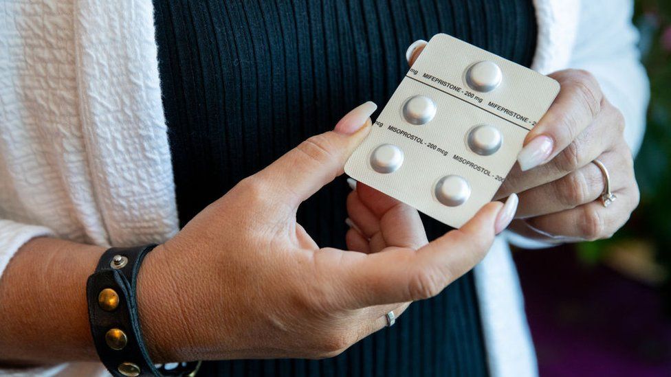
Published14 hours agoShareclose panelShare pageCopy linkAbout sharingImage source, Getty ImagesBy Chloe KimBBC News, New YorkA Texas man is suing three women for helping his now-ex-wife obtain pills to have an abortion last year. The lawsuit says under Texas law, “a person who assists a pregnant woman in obtaining a self-managed abortion has committed the crime of murder”.Marcus Silva’s lawsuit heavily relies on text messages between his ex-wife and the three women. It is the first such lawsuit in the state since the Supreme Court quashed constitutional abortion protections.Mr Silva is seeking $1m (£800,000) in damages. His ex-wife’s friends allegedly texted her information about Aid Access, an international group that provides abortion medication by mail.Text messages reveal Mr Silva’s ex-wife was concerned he would try to make her stay with him if he learned about the pregnancy.One of the women dropped off the pills to Mr Silva’s ex-wife, the lawsuit says.The legal action claims the drug manufacturer will also be named as a defendant once they are identified. There is also a conspiracy charge against the women – the lawsuit alleges they told Mr Silva’s ex-wife to hide the pregnancy and their text messages about the baby from him. Five women sue Texas over abortion accessThe ‘ranch’ for mothers with no place to goMr Silva’s former wife is not named as a defendant – under Texas law, a pregnant person is exempt from prosecution. They have two daughters together, according to the lawsuit. She filed for divorce in May 2022 before the divorce was finalised in February 2023. The lawsuit alleges she learned she was pregnant in July 2022, after the Supreme Court overturned Roe v Wade , but a month before the state’s trigger law banning abortion came into effect, which made providing an abortion a felony punishable by up to life in prison except to save the life of the mother.Even before the trigger ban was in effect, there were a number of other restrictions on abortion in the state.In 2021, Texas passed a law that made most abortions illegal after about six weeks of pregnancy. It gave citizens the right to sue anyone who “knowingly engages in conduct that aids or abets the performance or inducement of an abortion”. A pre-Civil War ban on abortions was also still on the books when Roe v Wade was overturned.Mr Silva is being represented by Republican state representative Briscoe Cain and Jonathan Mitchell, a former solicitor general who helped pen one of Texas’ abortion bans.The defendants did not immediately respond to a request for comment.Last week five women sued the state of Texas over limited abortion access when they had life-threatening health risks. Their lawsuit alleges doctors are refusing to perform the procedure even in extreme cases out of fear of prosecution.A Texas judge is also expected to rule soon in another lawsuit, which takes aim at the Food and Drug Administration’s approval process for the abortion pill, mifepristone. If the judge overturns the FDA’s approval for mifepristone, it could affect the drug’s availability nationwide. You may also be interested in:This video can not be playedTo play this video you need to enable JavaScript in your browser.More on this storyFive women sue Texas over abortion access6 days agoUS fight escalates over abortion pill at pharmacies3 days ago’I waited for my daughter to die so I wouldn’t die’ Video, 00:01:43’I waited for my daughter to die so I wouldn’t die’5 days ago1:4312 US states sue to expand access to abortion pill24 February
Read more →
Scientists from German Cancer Research Center (DKFZ), together with colleagues from Germany, Israel, and the USA, have found that the gut microbiome may modulate the efficacy of CAR-T cellular immunotherpy CAR-T cells in patients with B cell lymphomas. Individualized microbiome information retreaved from patients’ gut microbiomes prior to initiation of CAR T therapy could accurately predict their subsequent responsiveness to therapy, but only in the condition that these patients were not pre-treated with broad spectrum antibiotics.
Increasing evidence from human studies and preclinical experiments suggests that the gut microbiome may modulate the efficacy of T cell-driven cancer immunotherapies, such as immune checkpoint blockade. Immunotherapy with CD19 chimeric antigen receptor (CAR)-T cell has opened up new treatment options for patients with certain forms of refractory and relapsing B-cell leukemias or lymphomas. But the therapy is hampered by considerable heterogeneity in responses. Complete and long-term remission is only achieved in up to 40% of patients.
Researchers from multiple centers in Germany and the United States, led by Eran Elinav, director of the DKFZ-Weizmann Institute of Science Microbiome & Cancer Bridging division, have found that the gut microbiome may modulate the efficacy of CD19 CAR-T cell immunotherapy in patients with B cell leukemias and lymphomas.
This largest prospective study of its kind has followed 172 lymphoma patients previously failing multiple rounds of chemotherapy, from before CAR T immunotherapy initiation until two years later. Interestingly, 20% of patients receiving a subset of broad-spectrum (“high-risk”) antibiotics, such as meropenem, piperacillin-tazobactam or cefepime, featured an altered clinical response to subsequent CAR-T therapy, compared to patients who received other antibiotics and patients who were not treated with antibiotics prior to therapy. However, this antibiotics-associated reduced CAR T therapy response was not driven by the effects of the antibiotics themselves, but rather by the fact that patients treated with “high-risk” antibiotics before initiation of CAR T therapy tended to have higher pre-therapy tumor burden and systemic inflammation as compared to non-antibiotics-treated patients. These adverse pre-treatment conditions rendered subsequent CAR T therapy less effective.
Importantly, exclusion of these confounding “high-risk” antibiotics-treated patients from the analysis enabled the researchers to identify strong and previosuly masked associations between the pre-CAR T therapy gut microbiome and subsequent clinical response to immunotherapy (including patients’ survival). To further strenghen the connections between the starting microbiome and CAR-T efficacy across geography, diet, and and other ‘local’ confounders, the researchers next used machine learning models trained on the German patients and then applied as a validation on the respective American patients. Importantly, the models were able to potently predict therapy outcomes, but only upon exclusion of patients exposed to “high-risk” antibiotics. In other words, the study showed that the pre- therapy gut microbiome in lymphoma patients can help predict their response to subsequent CD19 CAR-T therapy across populations, unless their microbiome is disrupted by wide-spectrum antibiotics.
The researchers identified several key microbiome features enabling the prediction of CAR-T efficacy, including species of Bacteroides, Ruminococcus, Eubacterium, and Akkermansia. Of these, Akkermansia was also associated with higher baseline peripheral T cell levels in these patients.
In all, the study reveals strong associations between microbiomes and CAR-T outcomes, which first author Christoph Stein-Thoeringer (now Professor at the University Medical Center Tübingen, Germany) believes may contribute to the development of microbiome-based predictions of CAR-T cell immunotherapy outcomes. Additionally, findings from this study may enable to better understand the differential CAR-T cell activation, persistence, and clinical efficacy in different patients. The study also highlights the need for further investigation into the causal nature of the relationship between the gut microbiome and cancer immunotherapy outcomes. “These exciting findings,” states Eran Elinav, “exemplify the potential of our unique microbiome signatures to be harnessed as possible markers of disease and treatment responsiveness in multiple human disorders, including cancer. With further research we hope that microbiome-based diagnostics and therapies will be incorporated into the precision oncology field.”
The number of injuries in youth athletics is significantly reduced when coaches and parents have access to digital information on adolescent growth. It also takes twice as long for the first injury to occur. This is shown in a study from Linköping University published in the British Journal of Sports Medicine.
Many promising athletes have had their careers ruined because of injuries. One thing that almost all events in athletics have in common is a high load for a short time, as in jumping, throwing and running. This leads to overuse injuries such as groin pain and sore shoulders but also sudden injuries such as ankle sprain and hamstring tear.
Jenny Jacobsson is a physiotherapist and visiting researcher at the Athletics Research Center at Linköping University. She has worked as a medical coordinator for the Swedish national athletics team for many years and has seen the impact of injuries on athletes.
“Before the 2008 Beijing Olympics, we saw many injuries in our national team and tried to figure out why. At the time, no survey had been done of injury incidence in athletics athletes. But we wanted to find out what was happening among our elite athletes from age 16 and up, including adult elite athletes,” says Jenny Jacobsson.
The survey of injuries in Swedish athletics showed that one of the main causes of injury was prior injury. This means that the earlier an athlete is injured in their career, the higher the likelihood that they will be injured later and more frequently. But causes of injury in youth sports is a complex matter, associated with everything from training amount and load to equipment, and even sleep.
Together with her colleagues at the Athletics Research Center, Jenny Jacobsson has developed a digital health platform containing information for parents and youth coaches on adolescent growth and how this is affected by training, with a focus on athletics athletes aged 12-15.
To investigate whether this type of platform can prevent injuries, the researchers carried out a study where 21 athletics clubs with athletes aged 12-15 were randomised into two groups: an intervention group and a control group. For four months during the early season, the intervention group parents and coaches were given access to the digital information platform, which at the time was not open to outsiders (but is now open to anyone). They were also regularly encouraged to log in and explore its content.
The researchers noted that the clubs given access to the information showed significantly lower injury incidence and that it took twice as long for the first injury to occur. Moreover, the effect was greater in large clubs. The results, published in the British Journal of Sports Medicine, can point the way to more injury-free athletics.
“We haven’t investigated the mechanism leading to change, but we can see that digital information works when it comes to injury prevention. If coaches and parents learn to recognise the problems, it’s possible to reduce the training load in time. Medically we know what is happening in growing bodies, but getting the information out to those who can benefit from it has been a challenge. This platform may bridge that gap,” says Jenny Jacobsson.
The study was financed by the Swedish Research Council for Sport Science (Centrum för idrottsforskning).
We all know how important sleep is for mental health, but a meta-analysis publishing in the journal Current Biology on March 13 found that getting good shut-eye also helps our immune systems respond to vaccination. The authors found that people who slept less than six hours per night produced significantly fewer antibodies than people who slept seven hours or more, and the deficit was equivalent to two months of antibody waning.
“Good sleep not only amplifies but may also extend the duration of protection of the vaccine,” says senior author Eve Van Cauter, professor emeritus at the University of Chicago who, along with lead author Karine Spiegel at the French National Institute of Health and Medicine, published a landmark study on the effects of sleep on vaccination in 2002.
When the COVID-19 pandemic hit, and mass-vaccination became an international priority, Spiegel and Van Cauter set out to summarize our current knowledge about the effect of sleep duration on vaccine response.
To do this, they combed the literature and then combined and re-analyzed the results of seven studies that vaccinated for viral infections (influenza and hepatitis A and B). In their analysis, the team compared the antibody response for individuals who slept a “normal” amount (7-9 hours, as per the National Sleep Foundation’s recommendation for healthy adults) with “short sleepers” who slept less than 6 hours per night. They compared the effect for men versus women and adults over the age of 65 years versus younger adults.
Overall, they found strong evidence that sleeping less than 6 hours per night reduces the immune response to vaccination. When they analyzed men and women separately, though, the result was only significant in men, and the effect of sleep duration on antibody production was much more variable in women. This difference is probably due to fluctuating sex hormone levels in women, the authors say.
“We know from immunology studies that sex hormones influence the immune system,” says Spiegel. “In women, immunity is influenced by the state of the menstrual cycle, the use of contraceptives, and by menopause and post-menopausal status, but unfortunately, none of the studies that we summarized had any data about sex hormone levels.”
The negative effect of insufficient sleep on antibody levels was also greater for adults aged 18-60 compared with people over the age of 65. This was not surprising because older adults tend to sleep less in general; going from seven hours of sleep per night to less than six hours is not as big of a change as going from eight hours to less than six per night.
Some of the studies measured sleep duration directly, either via motion-detecting wristwatches or in a sleep lab, while others relied on self-reported sleep duration. In both cases, short sleep duration was associated with lower levels of antibodies, but the effect was stronger for the studies that used objective measures of sleep, likely because people are notoriously bad at estimating the amount of sleep they have had.
Knowing that sleep duration impacts vaccination might give people some degree of control over their immunity, the authors say. “When you see the variability in protection provided by the COVID-19 vaccines — people who have pre-existing conditions are less protected, men are less protected than women, and obese people are less protected than people who don’t have obesity. Those are all factors that an individual person has no control over, but you can modify your sleep,” says Van Cauter.
However, there’s a lot more to be known about sleep and vaccination, the authors say. “We need to understand the sex differences, which days around the time of vaccination are most important, and exactly how much sleep is needed so that we can give guidance to people,” says Spiegel. “We are going to be vaccinating millions and millions of people in the next few years, and this is an aspect that can help maximize protection.”