Dr. Colin McCord, Who Helped Impose a Smoking Ban, Dies at 94
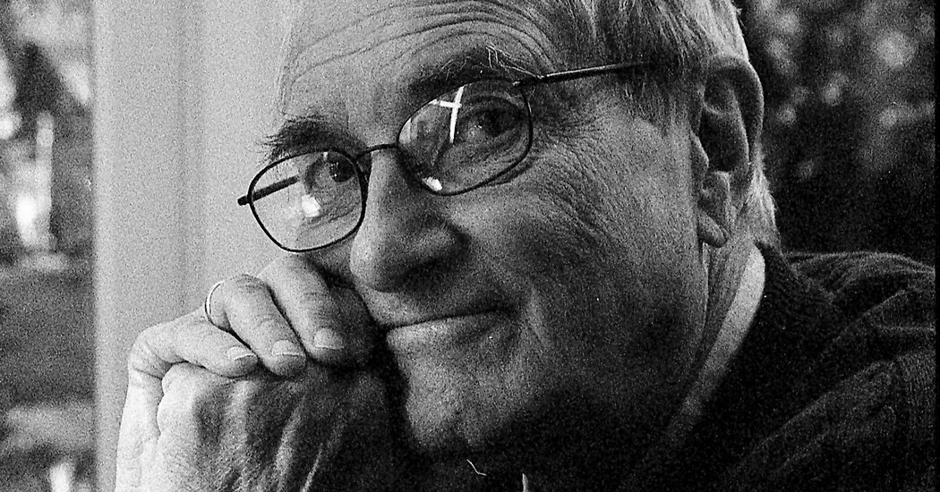
Among his accomplishments in New York City was successfully lobbying to ban smoking indoors, a move that was replicated around the world.Dr. Colin McCord, a surgeon credited with saving countless lives by championing a draconian ban on smoking in New York City and limits on trans fats in processed foods, galvanizing improved health care for Black men in Harlem, and improving maternal and child health globally, died on March 11 at his home in Oxford, England. He was 94.His son, Andy McCord, said the cause was congestive heart failure.Dr. McCord, who was known as Coke, trained lay people as paraprofessional doctors and surgeons in Mozambique and other African nations decimated by the departure of medical personnel; proved the efficacy of oral rehydration to save infants stricken with diarrhea in India and Bangladesh; and helped reduce birthrates in Bangladesh by teaching women to deliver advice on contraception and reproductive health. Those international initiatives probably spared millions of lives.Dr. McCord also exercised a profound impact on public conduct and health policy in New York City.He successfully lobbied for a ban on smoking in workplaces, restaurants and bars while he was an assistant health commissioner in Mayor Michael R. Bloomberg’s administration. The ban, which took effect in 2003, was later expanded and replicated in jurisdictions around the world.New York had banned smoking in most restaurants in 1995, but the city continued to allow smoking in bars and the bar areas of restaurants. As the son of chain smokers who both died of cancer, Dr. McCord described himself as “the heaviest secondhand smoker in New York City.”“It is the most important epidemic of our time,” he said in 2002 when the ban was expanded to include bars. “Each year the Health Department signs death certificates of 10,000 New Yorkers who died because of a tobacco-related cause; 1,000 of these people died because of exposure to secondhand smoke.”Dr. McCord sat next to Mayor Michael R. Bloomberg in 2002 as the mayor signed a law banning smoking in New York City’s bars.Ruth Fremson/The New York TimesYears earlier, Dr. McCord and a fellow researcher created a sensation when they disclosed in a 1990 article in the authoritative New England Journal of Medicine that Black men in Harlem were less likely to live to the age of 65 than men in Bangladesh, which was one of the world’s poorest nation’s when it was created in 1971.The report not only caused a sensation, it also produced results.Dr. McCord was named director of a federally funded prevention program at Harlem Hospital, a division of the city’s Health and Hospitals Corporation. Programs were initiated, as recommended by the report, to address the known causes of early death from chronic disease, including breast cancer screenings and neonatal testing, and investments were made to improve the delivery of health care in an overwhelmed system.Dr. McCord and Dr. Harold P. Freeman of Columbia University and Harlem Hospital, with whom he created the report, concluded that their findings were “not an isolated phenomenon” and that the racial disparities in life expectancy, particularly for Black men and poor people in general, were mirrored elsewhere in the city and around the nation.Dr. Thomas R. Frieden, who had been the city’s health commissioner when Dr. McCord was an assistant commissioner, cited his “clarity of thinking, ethical commitment, and effective action,” which he said saved lives.“Coke’s work on child survival made it more likely that millions of children would survive,” Dr. Frieden said in an email. “His work on surgery saved thousands of mothers and children. And his catalytic thinking on trans fat helped trigger a global movement that will prevent millions of deaths from heart attack.”Dr. McCord in 2013. In addition to his work in New York City, he helped improve health conditions in Mozambique and other African nations, as well as in India and Bangladesh.Dr. Florina SerbanescuDr. McCord was born Colin Wallace Miller on May 15, 1928, in Chicago to Colin Miller, who would become a wire service correspondent and movie producer, and George Lial Mickelberry, who was known as Sis.He was always referred to as Coke, a variation of his father’s nickname, Coco.The couple’s marriage was annulled within a year, and he was raised by his mother, who was a teenager. When Coke was about 4, his mother married A. King McCord, who would become the chairman of the Westinghouse Air Brake Company. He was formally adopted by Mr. McCord when he was 16.He had been enrolled in a military training program at Chicago Harvard School during World War II, but the war ended before the Army could deploy him to the Pacific.After graduating in 1949 from Williams College, where he majored in chemistry, he earned a medical degree from the Columbia University College of Physicians and Surgeons in 1953. He served his residency in surgery at Bellevue Hospital and in thoracic surgery at Bellevue and Presbyterian Hospitals.He married Susan Lewis Hobson in 1954; she died in 2002. He moved to England in around 2004.In addition to his son, he is survived by two daughters, Mary McCord and Anne McCord Wrublewski; his second wife, Susanne Ehrhardt Chowdhury; a stepdaughter, Bristi Chowdhury; a sister, Leslie Danforth; and four grandchildren.After completing his surgical residency, Dr. McCord taught at the University of Oregon, Portland; directed rural health programs in India and Bangladesh for the Department of International Health at Johns Hopkins University; and served as director of surgical services at a hospital in Mozambique from 1981 to 1986.After returning to New York in 1987, he was named associate director of surgery at Harlem Hospital, when he collaborated with Dr. Freeman.The two men found that while a large proportion of the so-called excess deaths among Black men in Harlem resulted from violence and drug abuse, most of the excess was attributable to other causes.“The bottom-line problem is poverty,” Dr. Freeman told The Times in 1990. “People who are intensely poor have other priorities. People think present tense. They don’t think future. They think about making it through the day. People are fighting for their lives.”

Read more →
Study sheds light on how IBD can develop

Inflammatory bowel disease, or IBD, describes Crohn’s disease and ulcerative colitis, two chronic diseases that cause inflammation in the intestines. IBD, which affects about 3 million adults in the United States, is an autoimmune disorder — a condition in which the body’s immune system attacks healthy tissues. Its symptoms include diarrhea, rectal bleeding, fatigue, weight loss, and stomach cramps.
The intestinal epithelium, made up of a layer of cells that lines the intestine, plays an important role in IBD because it can be easily disrupted during gut inflammation. A specialized type of epithelial cells are Paneth cells. The antimicrobial peptides these cells produce help regulate the gut microbiota, or the community of microorganisms that exist in the gut.
A research team led by Declan F. McCole, a biomedical scientist and IBD expert at the University of California, Riverside, reports in their mouse study that reduced activity of the IBD risk gene PTPN2 in intestinal epithelial cells can lead to a decrease in the production of Paneth cell antimicrobial peptides.
The study, published in the journal Cellular and Molecular Gastroenterology and Hepatology, establishes a critical link between PTPN2 and Paneth cells that plays a major role in maintaining normal gut microbe properties.
“This study develops our focus on improving personalized medicine approaches in IBD by understanding how patients with variants in the PTPN2 gene develop IBD,” said McCole, a professor of biomedical sciences in the School of Medicine. “Loss of PTPN2 can lead also to selective loss of Paneth cells in the intestinal epithelium. This loss of PTPN2 causes significant changes in the gut microbiota and increases a particular E. coli.”
Escherichia coli, or E. coli, are bacteria found in the environment, foods, and intestines of people and animals. McCole explained that the E. coli in question, the adherent-invasive E. coli, or AIEC, is increased in IBD and worsens inflammation. First identified in Crohn’s disease patients, AIEC can adhere to and invade epithelial cells as well as immune cells called macrophages.
“AIEC are the strongest candidate for a causal role for bacteria in IBD,” he said.
According to McCole, Paneth cells do not function properly in many patients living with IBD, and this can serve as a marker of disease. The antimicrobial peptides these cells produce are crucially relevant to the intestine’s protective barrier for regulating the relative proportions of bacteria and their interactions with each other. They also help neighboring intestinal stem cells function better.
“We know that in IBD, Paneth cells are often unable to produce sufficient antimicrobial peptides or respond appropriately to gut bacteria,” McCole said. “These functional defects can also be associated with changes in the structure of Paneth cells that reduce their ability to secrete the protective antimicrobial peptides, leading to increases in the populations of bacteria associated with IBD, such as AIEC. These structural changes in the appearance of Paneth cells can also serve as a marker of disease in IBD, especially Crohn’s disease.”
McCole was joined in the study by Vinicius Canale, Marianne R. Spalinger, Rocio Alvarez, Anica Sayoc-Becerra, Golshid Sanati, Salomon Manz, Pritha Chatterjee, Alina N. Santos, Hillmin Lei, Sharon Jahng, Timothy Chu, and Ali Shawki of UCR; Elaine Hanson and Lars Eckmann of UC San Diego; and André J. Ouellette of the University of Southern California.
The study was supported by the Crohn’s and Colitis Foundation; Swiss National Science Foundation; American Gastroenterological Association; Science Without Borders Program; and California Institute of Regenerative Medicine.
“This work sets the foundation for our new research project that will identify pharmacologic agents capable of rescuing Paneth cell function and reducing the contributions of microbes to intestinal inflammation,” McCole said.

Read more →

High blood pressure in your 30s is associated with worse brain health in your 70s

Having high blood pressure in your 30s is associated with worse brain health around age 75, especially for men, according to a new UC Davis study.
The research, published this week in JAMA Network Open, compared magnetic resonance imaging (MRI) brain scans of older adults who had high blood pressure between the ages of 30 to 40 with older adults who had normal blood pressure.
The researchers found that the high blood pressure group had significantly lower regional brain volumes and worse white matter integrity. Both factors are associated with dementia.
The research also showed that the negative brain changes in some regions — such as decreased grey matter volume and frontal cortex volume — were stronger in men. They note the differences may be related to the protective benefits of estrogen before menopause.
“Treatment for dementia is extremely limited, so identifying modifiable risk and protective factors over the life course is key to reducing disease burden,” said first author Kristen M. George, an assistant professor in the Department of Public Health Sciences.
“High blood pressure is an incredibly common and treatable risk factor associated with dementia. This study indicates hypertension status in early adulthood is important for brain health decades later,” George said.

High blood pressure prevalent in U.S.
High blood pressure, also known as hypertension, is blood pressure that is higher than normal. A normal blood pressure level is less than 130/80 mmHg. The Centers for Disease Control and Prevention estimates that 47% of adults in the United States have hypertension.
The rate of high blood pressure varies by sex and race. About 50% of men have high blood pressure compared to 44% of women. The rate of hypertension is about 56% in Black adults, 48% in white adults, 46% in Asian adults and 39% in Hispanic adults. African Americans ages 35 to 64 years are 50% more likely to have high blood pressure than whites.
Data from healthy aging studies
The researchers looked at data from 427 participants from the Kaiser Healthy Aging and Diverse Life Experiences (KHANDLE) study and the Study of Healthy Aging in African Americans (STAR). This provided them with health data from 1964 to 1985 for a diverse cohort of older Asian, Black, Latino and white adults.

They obtained two blood pressure readings from when the participants were between the ages of 30 to 40. This allowed them to determine if they had been hypertensive, transitioning to hypertensive or had normal blood pressure in young adulthood.
MRI scans of the participants conducted between 2017 and 2022 allowed them to look for late-life neuroimaging biomarkers of neurodegeneration and white matter integrity.
A significant reduction in cerebral gray matter volume is seen in both men and women with hypertension but is stronger in men.
Brain scans reveal differences
Compared to participants with normal blood pressure, the brain scans of those transitioning to high blood pressure or with high blood pressure showed lower cerebral gray matter volume, frontal cortex volume and fractional anisotropy (a measure of brain connectivity). The scores for men with high blood pressure were lower than those for women.
The study joins a growing body of evidence that cardiovascular risk factors in young adulthood are detrimental to late-life brain health.
The researchers note that due to the sample size, they could not examine racial and ethnic differences and recommended interpreting results regarding sex differences with caution. They also note that the MRI data was only available from one time-point late in life. This can only determine physical properties like volumetric differences, not specific evidence of neurodegeneration over time.
“This study truly demonstrates the importance of early life risk factors, and that to age well, you need to take care of yourself throughout life — heart health is brain health,” said Rachel Whitmer, senior author of the study. Whitmer is a professor in the departments of Public Health Sciences and Neurology and chief of the Division of Epidemiology. She’s also the associate director of the UC Davis Alzheimer’s Disease Center.
“We are excited to be able to continue following these participants and to uncover more about what one can do in early life to set yourself up for healthy brain aging in late life,” Whitmer said.
Additional authors of the study include Pauline Maillard, Evan Fletcher, Dan M. Mungas and Charles DeCarli, UC Davis; Paola Gilsanz, Kaiser Permanente Division of Research; Rachel L. Peterson, University of Montana, Missoula; Joseph Fong and Elizabeth Rose Mayeda from UCLA; L. Barnes from Rush Medical College; M. Maria Glymour from UCSF.

Read more →

Researchers leverage cell self-destruction to treat brain tumors

Glioblastoma is the most common type of brain tumor in adults. The disease is 100% fatal and there are no cures, making it the most aggressive type of cancer. Such a poor prognosis has motivated researchers and neurosurgeons to understand the biology of tumors with the goal of creating better therapies.
Dominique Higgins, MD, PhD, an assistant professor in the Department of Neurosurgery, has heeded the call. Higgins and a team of researchers at Columbia University have found that glioblastoma tumor cells are particularly sensitive to ferroptosis — a type of cell death that can be triggered by removing certain amino acids from the diet.
“First, we found that when we take away certain amino acids in animal models that the glioblastoma cells are more likely to die by ferroptosis,” said Dr. Higgins. “Secondly, we found that removing these amino acids makes our drugs a lot more effective at inducing ferroptosis in cancer cells.”
Their findings were published in Nature Communications.
Ferroptosis is an iron-dependent type of “programmed cell death” or a biological process that causes cells to “self-destruct” on command. Our bodies don’t need to kill cells unless absolutely necessary, so the process is tightly controlled by certain biological mechanisms. However, researchers are only now beginning to comprehend the process because ferroptosis was recognized only a decade ago.
“The recent discovery of ferroptosis adds to the excitement of it all,” said Higgins, who is a member at the UNC Lineberger Comprehensive Cancer Center. “It is really a rapidly growing body of research, and we are finding that it’s a very important for a lot of biological processes, and not just in cancers.”
Every cell has certain safety features to keep it from going through ferroptosis in an unpredictable way. Two amino acids, cysteine and methionine, are critical for preventing the process from starting in cells. We typically pick up these amino acids through our diet.

Therefore, Higgins’ research team decided to focus their efforts on these components.
By depriving animal models of cysteine and methionine through a customized diet, they found that the glioblastoma cells were significantly more likely to die via ferroptosis. They also found that the diet made their chemotherapy drugs more apt at initiating programmed cell death, meaning that very low doses were able to achieve a more potent effect than before. Ultimately, the animal models had improved survival after going on the diet.
“Now, we need to find a way to eliminate those components through dietary needs, while still maintaining energetic requirements that a patient may have, especially a cancer patient, who has different requirements than the average patient,” said Higgins.
Having proven that the diet is effective in animal models, Higgins is working with colleagues at UNC Lineberger to develop a clinical trial for patients with glioblastoma. He plans to put patients on the diet prior to surgery to understand how it effects the body and the tumor. Once he removes the tumor from the brain, he will analyze it to see how well the tumors responded to the diet.
This type of diet has also shown to be very effective in sarcoma, lung cancers, and pancreatic cancers, so there is hope that this diet can be used to put some extra umph behind chemotherapy and/or surgery to remove tumors throughout the body.

Higgins is also working with Shawn Hingtgen, PhD, professor of pharmacoengineering and molecular pharmaceutics in the UNC Eschelman School of Pharmacy and associate professor in the Department of Neurosurgery, to study the brain’s response to treatment in a more natural setting.
Hingtgen is the overall grant PI for Project Brainslice, a multi-institutional effort to test neurological therapies using tumor samples grown on slices of brain tissue. Higgins maintained that this is a better way to study treatment response than simply observing it in a plastic dish.
Project Brainslice is just one of the many different research tools that researchers have at their fingertips here at UNC’s School of Medicine.
“We have a lot of different research tools unique to UNC, and it is one of the main reasons that I wanted to come to UNC,” said Higgins, who joined UNC Neurosurgery in the fall of 2022. “Just in terms of the ability to study a clinical problem in an accurate animal model, it is one of the few places in the country that has like an established setup to do that.”

Read more →

How Bad Was Covid in NYC? Here’s a 200-Year Timeline of Death Rates

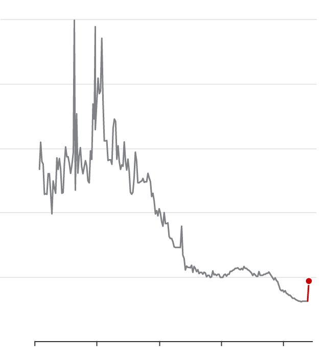
New Yorkers’ life span dropped by 4.6 years in 2020 from the previous year. Health officials say it may take the city years to recover.A wave of illness hit New York City, with little warning. Soon, it was sending the death rate rocketing upward.It was 1834. New York City was just expanding its first railroad line. The penny press was flourishing. Cholera had struck. And smallpox was resurgent.It would be nearly 200 years before another shock that seismic, when the coronavirus pandemic in 2020 caused the death rate in New York City to once again climb about 50 percent over the previous year, according to new data released Friday by the city’s health department.Throughout the 19th century, periodic outbreaks of cholera, smallpox, and other infectious diseases caused the city’s death rate to surge. But by the early 20th century, vaccines, improved sanitation and a variety of public health advances — from the disinfection of drinking water to the pasteurization of milk — had largely subdued this cycle of epidemics. The city’s death rate began to see drops and plateaus, a pattern that largely held for more than a century — until 2020.The story of the city’s declining death rate, and how Covid upended that trend, is instantly communicated in a well-known chart published regularly by New York City’s health department, and now updated to include the first year of the pandemic.Called “The Conquest of Pestilence in New York City,” it showed how strides in public health eventually quelled the epidemics of the 19th century. For the last century or so, the death rate — measured as the number of deaths per 1,000 residents — was relatively flat or declining, until the pandemic’s disastrous first wave in early 2020.

Read more →

The Virtual Vet Will See You Meow

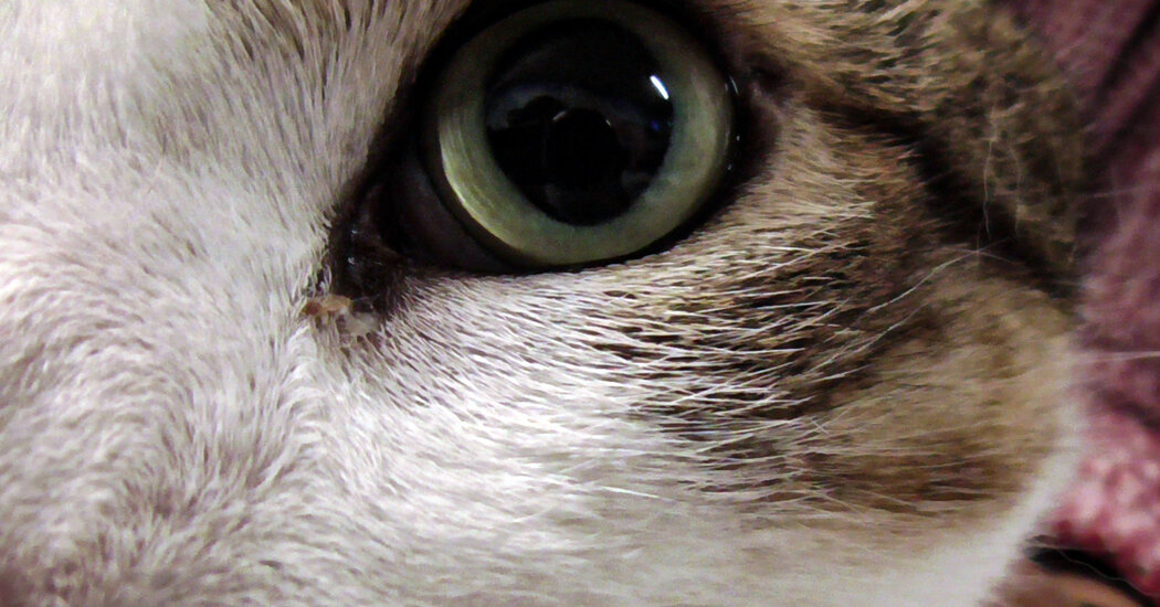
Veterinary telemedicine could help more pet owners access much-needed care and put anxious animals at ease, but challenges remain.Milkshake and Pickles are reluctant travelers. So when Patience Warren needed to take the two elderly cats on a 12-hour drive in February, she was hoping to get some pharmaceutical assistance, especially for Pickles, a petite gray tabby with a history of severe motion sickness.The dilemma: Taking Pickles to the vet typically triggered the very distress Ms. Warren was hoping to avoid.“Within like a minute of being put into her carrier and put into the car, she would usually vomit or lose her bowels,” said Ms. Warren, a political researcher in Missouri. “She would also just cower and be really scared and meow. And I didn’t want to put her through that stress.”Over the course of the pandemic, Ms. Warren had grown accustomed to having her own health needs met virtually, seeing a doctor, therapist and nutritionist online. She wondered whether there were veterinarians who might prescribe anti-anxiety and motion sickness medications over a video call. When she searched online, she was surprised to discover numerous options.“I honestly didn’t realize that virtual vets existed,” she said.While many people have embraced virtual visits with their own doctors, use of veterinary telemedicine by pet owners has lagged. In one new survey of more than 1,200 American cat owners, 72 percent reported using telemedicine for themselves, compared to just 3 percent who had used it for their felines.“But things are changing, and things are changing fast,” said Carly Moody, an animal welfare researcher at the University of California, Davis, who conducted the survey, which has not yet been published, as part of an ongoing project studying telemedicine for cats.A televisit at the Texas A&M University School of Veterinary Medicine in College Station, Texas, in 2020.Lori TellerPickles, Patience Warren’s agoraphobic cat, with some of her leftover medications.Patience WarrenDuring the pandemic, numerous states temporarily loosened restrictions on veterinary telemedicine and many clinics as well as pet owners tried remote appointments for the first time. Some states are now considering permanently expanding their use.Although hurdles remain, and it’s not appropriate for all pet care scenarios, scaling up telemedicine could bring a variety of benefits, experts said, like improving access to veterinary care and reducing stress for vet-averse pets like Pickles.For veterinary medicine, Covid-19 “served as a catalyst for change that was necessary,” said Dr. Christina Tran, a veterinarian at the University of Arizona who is on the board of directors of the Veterinary Virtual Care Association and is a paid adviser to a veterinary telehealth company.Pandemic practicesSome forms of telehealth are decades old; veterinarians have long fielded frantic calls from pet owners or consulted with colleagues over email.But remote video appointments are newer. “Before the pandemic, it was not very common to utilize telemedicine in that way,” said Dr. Lori Teller, the president of the American Veterinary Medical Association, who is also on the faculty at Texas A&M University, where she has developed a veterinary telehealth program. She is also a compensated adviser for another veterinary telehealth company.In part, that stemmed from restrictive state laws, many of which required veterinarians to have a pre-existing relationship with an animal — including having given a prior hands-on exam — before treating them remotely.But when the pandemic began, some states temporarily eased their requirements. Veterinary practices turned to telemedicine to conserve personal protective equipment and flatten the coronavirus curve. The share of vets offering remote video appointments rose to 30 percent from 4 percent, according to one survey of American and Canadian clinicians.Pet owners who had never considered virtual care suddenly had few other options. One morning in May 2020, Kristyn Booth, an educator who then lived in Austin, Texas, discovered that her dog’s eye, which had been injured nearly a decade prior, was bulging and red. The veterinarian would only offer a virtual appointment.Kristyn Booth of Cedar Creek, Texas, with her redbone coonhound, Lily, who had an injured eye.Montinique Monroe for The New York TimesInitially, Ms. Booth was nervous. “How can they do this?” she recalled thinking. “It’s her eye.”But the veterinarian suggested that Ms. Booth drive Lily, a redbone coonhound, to the office and take the virtual appointment in the parking lot. If the situation looked serious, they could rush Lily inside.So Ms. Booth sat inside her car and showed the doctor Lily’s eye over a video call. She followed the vet’s instructions to gently press around the dog’s eye socket and look under her eyelid for blood. “I felt like I was a vet that day,” she said. “They were right there in case it was going to go awry.”The doctor prescribed eye drops and sent Ms. Booth and Lily on their way.Last year, when Lily’s eye got worse, Ms. Booth used a video call again; this time, the doctor took one look and knew it was time for the injured eye to come out, Ms. Booth said. Lily has coped well, she added: “She’s great. She’s old now, and we do everything we can to keep her happy and healthy.”Scaredy catsSome organizations are pushing for a more permanent expansion of virtual vet care.“The pandemic really did open up our eyes to the utilization of telemedicine,” said Kevin O’Neill, the vice president of state affairs for the American Society for the Prevention of Cruelty to Animals, which is urging states to loosen their rules around telemedicine. “We see it as a real key component to establishing a broader ability for patients and pet owners to be able to access that vet care that’s so badly needed.”In June 2022, 26 percent of American pet owners reported that there had been times over the previous two years when they had been unable to access veterinary care, according to a survey of 5,000 people conducted by the A.S.P.C.A., which provided the data to The Times. Two-thirds of them said that their pets would probably be “seen by a veterinarian more often” if they could use telemedicine.Although not all medical care can be provided through a screen, routine appointments like post-surgical follow-ups or behavioral consultations work fine from a distance, experts said.Telemedicine could be especially useful for rural pet owners, who may live hours from an animal clinic, as well as those who cannot afford to take time off from work or lack reliable transportation, experts said. Virtual triage services could help people determine whether their pets’ symptoms require in-person care or can be monitored at home.“And then we open up those appointments for the brick and mortar so that they can in fact see things that need to be seen in person,” Dr. Tran said.Grace Boone, an animal welfare researcher in Carly Moody’s lab at the University of California, Davis.Rozette for The New York TimesCarly Moody of the University of California, Davis. “Things are changing, and things are changing fast,” she said of pet telemedicine.Rozette for The New York TimesVirtual care could also be a boon for cats, many of which are intensely stressed by trips to the vet, Dr. Moody said. In a small unpublished study funded by the A.S.P.C.A. and Maddie’s Fund, an animal welfare group, Dr. Moody and her colleagues found that cats displayed fewer signs of physiological stress — with smaller pupils, slower breathing rates and more relaxed ear postures — during remote appointments than during in-person ones. When given clear instructions, cat owners could even do some basic physiological assessments usually performed by a vet. “I think we only had one cat that the owner wasn’t able to get a respiration rate at home because the cat was very wiggly,” said Grace Boone, a researcher in Dr. Moody’s lab who ran the study.The virtual vet turned out to be perfect for motion-sickness-prone Pickles, who seemed to hate the vet just as much as she loved crashing Ms. Warren’s remote work meetings. “She loves being on camera during my work Zoom, so as soon as I turned it on, she was up on my lap,” Ms. Warren said.The vet prescribed medication for motion sickness and anxiety, which were shipped directly to Ms. Warren’s home. “Overall, it was wonderful,” she said. “I’ve been telling everybody.”Tele-troublesTelemedicine is not a panacea, and some pet owners and veterinarians have run into its limitations.When Alisa Crane’s 16-year-old cat developed an infection on his ears and face last November, the only appointment she could find was a virtual one. The appointment felt cursory, she said, lasting “maybe six minutes.”The virtual veterinarian prescribed painkillers and antibiotics, and the infection eventually cleared, but Ms. Crane, who lives in Sudbury, Ontario, still doesn’t know what caused it or whether it was a sign of a deeper health issue. If she were in the same situation again, she said, she’d probably opt for an in-person trip to the expensive emergency vet. “I don’t think they were able to properly diagnose him through the video call,” she said.After Friendship Hospital for Animals, in Washington, D.C., began offering telemedicine appointments for some patients in April 2020, some veterinarians soon found them to be more trouble than they were worth. “Because our patients can’t tell us their symptoms like a person can, I am relying on a pet owner to interpret their symptoms,” said Dr. Christine Klippen, an emergency veterinarian at the hospital. “And that can sometimes be very, very wrong.”A cat named Alfie who was not loving his in-person visit.Grace BooneFor instance, a pet owner who noticed their cat straining in the litter box might book an online appointment for constipation, typically a relatively minor ailment, when the cat in fact might have a urinary blockage, a potentially life-threatening emergency, Dr. Klippen said.“We would get a lot of folks that would not find the animal in time,” she added. “You’d have a scheduled appointment, and then they’re trying to take video of pets in the closet. It didn’t turn out to be an efficient use of the veterinarian’s time.”The hospital discontinued the appointments after less than six months, she said, although the hospital does occasional remote consultations. Virtual care is a “great idea,” she added, but vets and pet owners need clearer guidelines about how and when to use virtual visits.The legal landscape can be confusing too, with a patchwork of state and federal laws. But some states are embracing telemedicine. New Jersey, for instance, now allows vets to treat new patients virtually without a prior physical examination, and Arizona lawmakers are considering similar legislation.The American Veterinary Medical Association favors a more restrictive approach, largely limiting telemedicine to pet owners and veterinarians with a pre-existing relationship, with limited exceptions for emergencies and specialists. But Dr. Teller said that she expected a growing number of practices to offer virtual options.“Now that pandemic craziness overall is slowing down,” she said, “it’ll be easier for veterinarians and their teams to take a breath and figure out how to better integrate telemedicine.”

Read more →

Covid origins: Chinese scientists publish long-awaited data

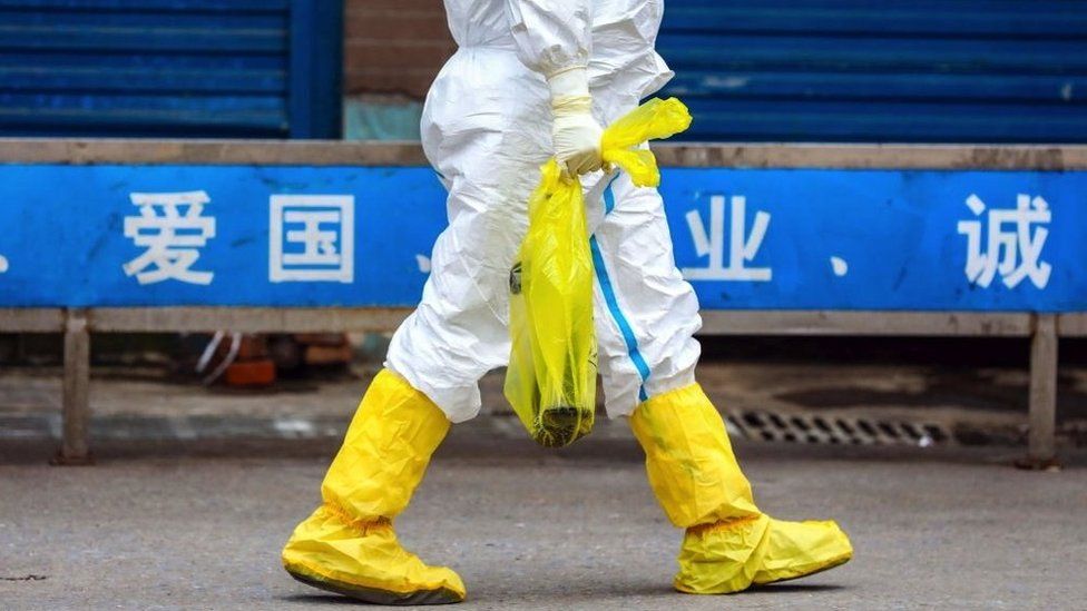
Published36 minutes agoShareclose panelShare pageCopy linkAbout sharingImage source, Getty ImagesBy Victoria GillScience correspondent, BBC NewsA research team in China has published analysis of samples taken more than three years ago from the market linked to the outbreak of Covid-19.The Huanan seafood and wildlife market has been a focal point in the search for the origin of the coronavirus. But this is the first peer-reviewed study of biological evidence gathered from the market back in 2020.By linking the virus with animals sold in the market, it could open new lines of inquiry into how the outbreak began.The research reveals swabs that tested positive for the virus also contained genetic material from wild animals. Some scientists say this is further evidence that the disease was initially transmitted from an infected animal to a human. But others have urged caution in interpreting the findings and it remains unclear why it took three years for the genetic content of the samples to be made public. Another theory has centred on the suggestion that the virus accidentally leaked from a laboratory in Wuhan. No definitive proofThe Chinese research team posted an early version of their study online in February, but they did not publish the full genetic information that was contained in the samples gathered from the market. Another international group of researchers later shared their own assessment of what those crucial market swabs had revealed, after spotting that the genetic sequences had been posted on a scientific data-sharing website. This new analysis, which has been validated by other scientists before being published in the journal Nature, includes more important detail about the content of those samples, which were collected from stalls, surfaces, cages and machinery inside the market. Image source, E HolmesThe Chinese research team’s paper showed that some samples – collected from areas where wildlife was being sold – had tested positive for the virus. Their analysis also showed that animals now known to be susceptible to the virus, particularly raccoon dogs, were being sold alive in those locations. But the Chinese researchers have pointed out that their discoveries fall short of definitive proof of how the outbreak started. “These environmental samples cannot prove that the animals were infected,” the paper explains. The possibility remains, it adds, that the virus was brought into the market by an infected person, rather than an animal. Prof David Robertson, from the University of Glasgow, is a virologist who has been involved in the genetic investigation into the origin of SARS-CoV-2 since it emerged in 2020. He told BBC News: “The most important thing is that this very important dataset is now published and available for others to work on.” But he added that the contents of the samples were “compelling evidence that animals there were probably infected with the virus”. “It’s the whole body of evidence that’s important,” he said. “When you bring this together with the fact that the early Covid-19 cases in Wuhan are linked to the market, it’s strong evidence that this is where a spillover from an animal in the market occurred.”Have we found the ‘animal origin’ of Covid?Covid origin likely China lab incident – FBI chiefCovid origin studies say evidence points to marketThe published findings come amid signs that the lab leak theory is gaining ground among authorities in the US.The Chinese government has strenuously denied suggestions that the virus originated in a scientific facility, but the FBI said it now believes that scenario is the “most likely”, as does the US Department of Energy.Various US departments and agencies have investigated the mystery and produced differing conclusions, but on 1 March the FBI’s director accused Beijing of “doing its best to try to thwart and obfuscate”, and disclosed the bureau had been convinced of the lab leak theory “for quite some time now”. The FBI has not made their findings public, which has frustrated some scientists.The lead researcher of the new report, from the Chinese Center for Disease Control and Prevention (China CDC) in Beijing, has been contacted by the BBC for comment. More on this storyHave we found the ‘animal origin’ of Covid?25 MarchCovid origin studies say evidence points to market26 July 2022Raccoon dogs: Are they pet or pest?1 June 2019

Read more →

Physicians should be on alert for group A strep as cases experience historic rise, study finds

The U.S. experienced an unprecedented number of group A streptococcal infections in children from October to December of 2022, which should alert physicians to check for the potentially deadly infectious disease as the country moves out of the pandemic, according to research published by UTHealth Houston.
The study, led by senior author Anthony R. Flores, MD, PhD, MPH, associate professor and chief of pediatric infectious diseases at McGovern Medical School at UTHealth Houston, was published this month in Clinical Infectious Diseases.
During the COVID-19 pandemic, the rate of other infectious diseases dropped due to safety measures such as social distancing and mask-wearing, according to researchers. But as those safety measures began to lift, diseases such as respiratory syncytial virus, the flu, and group A strep all experienced a resurgence in cases.
Infectious disease experts first noticed the rise in cases in the UK, now with 355 deaths of which 40 were children dying from severe group A strep infections. Researchers such as Flores then turned to what was occurring in the U.S.
“In 2020 and 2021, the overall number of infections that we saw due to group A strep were far lower than what we had seen before the pandemic,” Flores said. “When we look at the number of infections by quarter, historically, pre-pandemic cases of group A strep were pretty consistent from quarter to quarter with a little variation and more infections in the winter months. But during the last quarter of 2022, the number of infections we saw, including invasive infections, were far greater than what we’d ever seen before.”
In 2022, a total of 318 individual group A strep cases were identified in young children in Houston. Researchers looked at three group A strep disease types: invasive group A strep (iGAS), skin and soft tissue infections (SSTI), and pharyngeal, or throat, infections (PHG). The study found that group A strep strains derived from iGAS accounted for 31.4%, SSTI for 17.6%, and pharyngeal for 50.9%.
“Proportionately speaking, if we just looked at the percentage of all infections that were invasive, it was the same as we had seen in the past,” Flores said. “It wasn’t like, all of a sudden, 75% of our cases are invasive diseases. What we’re seeing is an increase in the total number of infections.”
The study also found that emm12 group A strep strains were disproportionately represented, compared to emm1 group A strep, which was the dominant emm type pre-pandemic. Emm type is a marker that differentiates different strains of group A strep.
“By looking at the molecular epidemiology, we can look at specific things about the bacteria that give us clues as to whether or not something is changing,” Flores said. “What we will be observing as we go forward is whether it will shift back to what it was pre-pandemic, and if it doesn’t, then we hope to have some work underway that is going to tell us why.”
As cases of group A strep continued to be high in the first quarter of 2023 in Houston, the disease is a cause for concern for pediatricians who see children with symptoms of group A strep.
“The reason why this is important is we’re seeing group A strep more frequently than what we have seen in the past, and therefore, if a physician has a child coming in with a sore throat or with a skin infection, we should have a high index of suspicion for group A strep,” Flores said.
Co-authors on the paper from McGovern Medical Schools department of pediatrics infectious diseases division included Aya Aboulhosn, MD; Misu A. Sanson-Iglesias, MD, PhD; Luis Alberto Vega, PhD; and Maria G. Segura, MD.

Read more →

Case for Candida auris wastewater surveillance

A rapid spike in cases of a potentially deadly, drug-resistant fungus has concerned public health officials across the nation. But a team of Southern Nevada researchers hope their new study applying wastewater surveillance can help health officials get a step ahead of this emerging global public health threat.
The Pathogen Problem
Candida auris is a fungus that can cause serious infections, particularly in patients who are immunocompromised, have pre-existing health conditions, are in long-term healthcare settings, or are undergoing treatment with invasive medical devices such as a catheter. Infection prevention and control is challenging because the fungus can grow on both dry and moist surfaces such as furniture, door handles, clothing, and medical equipment in healthcare facilities. It’s also shown resistance to many commonly used surface disinfectants and all three types of antifungal medicines. More than 1 in 3 patients with invasive C. auris infections — which can affect the blood, heart, or brain — dies.
What’s more, Nevada — one of six states with recently high burdens of C. auris — last year experienced outbreaks across multiple healthcare facilities and logged the most U.S. cases of the fungal infection. The Silver State experienced a 16-fold increase from just 24 cases in 2021 to 384 cases in 2022, according to the Centers for Disease Control and Prevention (CDC). Cases have also been reported in dozens of other countries.
What They Found
A research team led by Casey Barber, a UNLV School of Public Health doctoral student and Southern Nevada Water Authority (SNWA) graduate intern, recently published a study in the journal Environmental Science & Technology that analyzed 10 weeks’ worth of wastewater samples from seven Southern Nevada sewersheds.

The scientists detected the genetic material of C. auris in at least one untreated sewage sample from each Southern Nevada wastewater treatment facility and nearly 80% of all untreated sewage samples in the study. The sewersheds serving healthcare facilities involved in the outbreak also showed higher detection frequencies for the fungus. Researchers noted that no fungus was detected in untreated sewage samples from a wastewater treatment facility in Utah, an area with no known C. auris cases at the time. The fungus was not detected in the Las Vegas Wash, which contains treated wastewater effluent, nor in Lake Mead, indicating that there is no sign that C. auris poses a risk to drinking water.
“These results show that wastewater surveillance may help monitor the spread of C. auris and could serve as an early warning system for public health action,” Barber said.
Other Takeaways
The first human case of C. auris was reported in 2009, but it’s become more prevalent in recent years. The fungus is often spread via contaminated surfaces or skin-to-skin contact with infected individuals, including with those who are asymptomatic.
Scientists called the Southern Nevada fungus flare-up — which erupted in August 2021 and has now affected over 30 healthcare facilities — one of the largest recent outbreaks of healthcare-associated C. auris in the U.S. The research team formally launched C. auris-specific monitoring and data collection in late June 2022, as part of a larger ongoing UNLV wastewater surveillance collaboration with SNWA.
In addition to implications for large-scale C. auris detection and prevention, researchers said the study is groundbreaking in its progress towards helping establish new procedures for sewage sample processing, preparation, and analysis to look for C. auris.
Wastewater surveillance, they said, may provide a more accurate estimate of C. auris prevalence than traditional public health surveillance methods, in part because traditional methods may not accurately identify C. auris, leading to delays in targeted intervention measures. The team also anticipates that their previously established approach to monitoring COVID-19 levels in wastewater could be applied to watching for mutations and new strains of C. auris.
“Detection of Candida auris through wastewater surveillance has already prompted expanded screenings in Southern Nevada healthcare facilities in an effort to prevent larger outbreaks,” said SNWA principal research microbiologist Daniel Gerrity. “This demonstrates how wastewater surveillance can be applied to emerging public health threats beyond COVID-19.”

Read more →

Researchers clear the way for well-rounded view of cellular defects

Amrinder Nain is an associate professor in the Department of Mechanical Engineering, but he doesn’t build cars or robots. The mechanics he champions are the tiny building blocks of life and how they behave and move.
Cellular dynamics research studies living cells and their life, death, division, and multiplication. Over the past several years, Nain has taken many journeys down the microscopic roads where cells live. His past work has analyzed how cells move and even included projects with colleagues to measure cell forces and nucleus shapes and to electrify cells and observe how they heal.
A cell divided is how we stand
His latest collaboration investigates how cells divide, particularly in the fibrous environment of living tissue. Cells are typically studied in a flat environment, and the difference between flat and fibrous landscapes opens new windows into the behavior of cells and the diseases that impact them. The findings were published in the Proceedings of the National Academy of Sciences on Feb. 27. The work received funding from the National Science Foundation and support from both the Virginia Tech Institute for Critical Technology and Applied Science and the Virginia Tech Macromolecules Innovation Institute.
Cell division, called mitosis, is essential for developmental, repair, and disease biology. A cell, at its most fundamental level, duplicates its chromosomes, which are then separated and distributed equally between two daughter cells, each with its own complete set of genetic information. As new cells perform the same function over and over, they form organs, heal wounds, and replace dead cells, sustaining the cycle of healthy tissues and organs.
But cell division doesn’t always happen this smoothly. Sometimes, cells divide unevenly, or chromosomes can become unevenly split. When those misfires occur, the resulting cell will continue to duplicate copies of its faulty self, creating genetic defects that could cause widespread problems in a living body. These abnormalities account for many prenatal birth defects and can contribute to the origins of cancer.

Better understanding cellular mitosis increases our chances of diagnosing, treating, and preventing those mitotic defects. Nain’s discovery puts valuable information in the hands of researchers by painting a complete picture of what’s going on at the cellular level within the body’s fibrous environment.
Movement, multiplication, and division
At the microscopic level, cells move by way of an extracellular matrix (ECM), a three-dimensional lattice of organic material that provides the framework for cells to form organs by underlaying a strong foundation that holds them together.
Nain’sfoundational research focuses on re-creating and studying that lattice, and his team’s past studies on cellular motion have shown how cells travel along it. For a single fiber, a cell pulls itself along at each end, walking the fiber like a tightrope. Two fibers running parallel allow the cell to double those connections.
A dividing cell also makes use of the fibers around it. For a single fiber, each end of the cell adheres and pulls to create the division. If a cell is in an environment with multiple fibers, it will likely attach to those as well. The ECM may cross above and below the cell, providing a three-dimensional web onto which cells connect.

The number of fibers available for cells to attach to affects the timing of cell division and the types of defects a cell may produce. Cells take longer to divide on single fibers, and mitotic errors change with more attachments, creating a complex picture of the myriad ways in which a cell might fail.
This discovery affects future research because the complex view of cell division errors has not been previously investigated in fibrous environments.
A new dimension for research
“Cellular biology has predominantly been studied on a Petri dish, which is a flat, two-dimensional surface,” said Nain. “Flat 2D is limited in physiological output because there are very few places in the body where the environment can be considered two-dimensional.”
The team found that observing cells in the 3D environment of an ECM yielded new results beyond the capability of 2D Petri dishes. In this work, the team asked a central question: how does the shape of a cell affect its dividing behavior?
Cell shape depends on how a cell adheres to underlying substrates. For example, on a flat, two-dimensional Petri dish, a cell resembles a pancake. In a fibrous environment such as an ECM, shapes range from elongated aerofoils to kites, depending on the number of fibers and their architecture. While a cell might adhere above and below the fiber plane on suspended fibers, a flat surface causes the cell to flatten out and form connections outward. That flattening causes the cell to behave differently when it balls up and undergoes division.
Schematic of a rounded cell body attached to a single fiber and held by actin retraction fiber cables (red) connecting adhesion clusters (green) with the cell cortex (blue). Image courtesy of Amrinder Nain.
As a rounded cell body divides, it’s held in place by organic cables that attach the cell body, or cortex, to the fibers. On single fibers, near-perfect spherical cell bodies are held in place by two sets of cables, giving maximum freedom for the rounded cell body to move in 3D. As the number of fibers in the lattice increases, so does the number of places to which a cell can adhere. This results in multiple cable complexes that limit 3D movement of the rounded cell body.
This simple mechanical effect highlights the significant difference between the Petri dish and the ECM. On a Petri dish, monopolar spindle defects, which represent incomplete spindle pole (or centrosome) separation, do not often occur. However, when a cell is in a single-fiber environment with two cable attachment sites, monopolar spindle defects increase.
These results turn cell study quite literally on its head: in the environment of a Petri dish, some defects that occur during cellular mitosis cannot happen in the same way as they do in a living body.
“While bipolar division, the most common and error-free division mode, dominates division outcomes in fibrous environments, our work shows a switch in monopolar and multipolar defects by changing the number of fibers cells attach to,” said Nain. “It offers a glimpse into how cell division might occur in actual living tissues.”
Nain hopes that the fresh perspective provided by this foundational experimental-computational work will yield insights on how to treat disease and genetic disorders.
“With fiber networks, we provide more detail on a comprehensive in vivo picture, filling in some missing information and using our multi-disciplinary approach, we would like to ask some precise questions in mitotic biology as we move forward,” he said.
The multi-disciplinary team assembled for this project comprises leading experts, including cell division biologist Jennifer DeLuca, a professor at Colorado State University; theoretician and biophysicist Nir Gov, a professor at the Weizmann Institute of Science, Israel; and theoretician and computational expert Raja Paul, a professor at the Indian Association for the Cultivation of Science (IACS), India. The first author for the publication was Aniket Jana, now a postdoctoral fellow at the University of Maryland, College Park. Other student members involved in this study include Hoanan Zhang and Atharva Agashe from the Virginia Tech Department of Mechanical Engineering, Ji Wang from the Virginia Tech Department of Biomedical Engineering and Mechanics, and Apurba Sarkar from IACS, India.

Read more →