Broccoli consumption protects gut lining, reduces disease, in mice

Broccoli is known to be beneficial to our health. For example, research has shown that increased consumption of the cruciferous vegetable decreases incidence of cancer and type 2 diabetes. In a recent study, researchers at Penn State found that broccoli contains certain molecules that bind to a receptor within mice and help to protect the lining of the small intestine, thereby inhibiting the development of disease. The findings lend support to the idea that broccoli truly is a ‘superfood.’
“We all know that broccoli is good for us, but why? What happens in the body when we eat broccoli?” said Gary Perdew, H. Thomas and Dorothy Willits Hallowell Chair in Agricultural Sciences, Penn State. “Our research is helping to uncover the mechanisms for how broccoli and other foods benefit health in mice and likely humans, as well. It provides strong evidence that cruciferous vegetables, such as broccoli, cabbage, and Brussels sprouts should be part of a normal healthy diet.”
According to Perdew, the wall of the small intestine allows beneficial water and nutrients to pass into the body but prevents food particles and bacteria that could cause harm. Certain cells that line the intestine — including enterocytes, which absorb water and nutrients; goblet cells, which secrete a protective layer of mucus on the intestinal wall; and Paneth cells, which secrete lysosomes that contain digestive enzymes — help to modulate this activity and keep a healthy balance.
In their study, which published in the journal Laboratory Investigation, Perdew and his colleagues found that molecules in broccoli, called aryl hydrocarbon receptor ligands, bind to aryl hydrocarbon receptor (AHR), which is a type of protein called a transcription factor. This binding, they found, initiates a variety of activities that affect the functions of intestinal cells.
To conduct their study, the researchers fed an experimental group of mice a diet containing 15% broccoli — equivalent to about 3.5 cups per day for humans — and fed a control group of mice a typical lab diet that did not contain broccoli. They then analyzed the animals’ tissues to determine the extent to which AHR was activated, as well as the quantities of various cell types and mucus concentrations, among other factors, in the two groups.
The team found that mice that were not fed broccoli lacked AHR activity, which resulted in altered intestinal barrier function, reduced transit time of food in the small intestine, decreased number of goblet cells and protective mucus, decreased Paneth cells and lysosome production, and decreased number of enterocyte cells.
“The gut health of the mice that were not fed broccoli was compromised in a variety of ways that are known to be associated with disease,” said Perdew. “Our research suggests that broccoli and likely other foods can be used as natural sources of AHR ligands, and that diets rich in these ligands contribute to resilience of the small intestine.”
More broadly, added Andrew Patterson, John T. and Paige S. Smith Professor of Molecular Toxicology and of Biochemistry and Molecular Biology, “these data suggest that dietary cues, relayed through the activity of AHR, can reshape the cellular and metabolic repertoire of the gastrointestinal tract.”
Other authors on the paper include Xiaoliang Zhou, Debopriya Chakraborty, Iain A. Murray, Denise Coslo, Zoe Kehs, Anitha Vijay, Carolyn Ton, Dhimant Desai and Shantu G. Amin.
The National Institutes of Health Grants, U.S. Department of Agriculture and Penn State Cancer Institute supported this research.
Read more →

Tripping in the Bronze Age

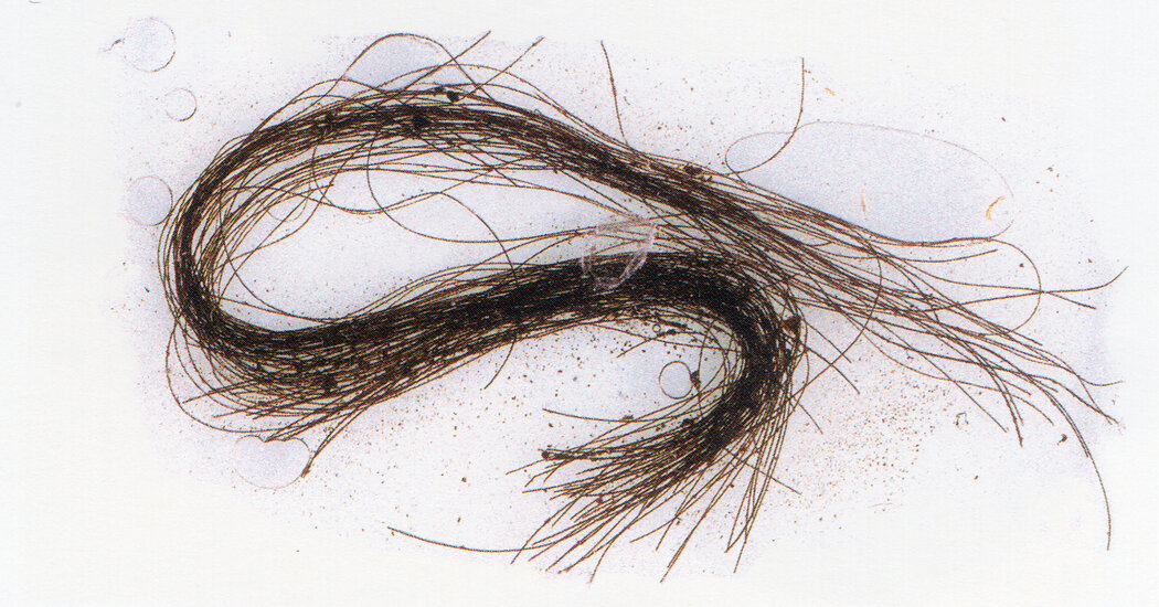
A new study based on strands of hair found in a Spanish burial cave reveals that humans living about 3,000 years ago used hallucinogens, likely derived from local plants, as part of their rituals.Bronze Age humans have been credited with a number of civilizational advancements: the invention of irrigation, the wheel, writing systems and the ability to forge weapons and tools from the durable metal that lends the era its name.Now, strands of human hair discovered in an ancient burial cave in Spain suggest another novelty: a proclivity for consuming psychoactive drugs.The hair, found inside wooden containers hidden deep within a sealed grotto on Minorca, an island off the coast of eastern Spain, tested positive for a number of mind-altering compounds, suggesting that the people who lived there 3,000 years ago incorporated hallucinogenic experiences into the rituals of their lives.The findings, published Thursday in Scientific Reports of the journal Nature, provide the first direct evidence that ancient Europeans consumed psychoactive drugs much like their pre-Columbian brethren in Mesoamerica, the researchers said.Elisa Guerra-Doce, the lead author of the study, said researchers were stunned by the results, especially because the cave interiors yielded no detectable signs of the drugs’ presence. A chemical analysis of the hair revealed evidence of three alkaloid substances known to produce altered states of consciousness: ephedrine, atropine and scopolamine.The compounds themselves are produced by flora native to Minorca. Atropine and scopolamine, powerful hallucinogens, can be found in plants in the nightshade family, among them mandrake, henbane and thorn apple. Ephedrine, a stimulant, can be extracted from joint pine.“These findings are so singular,” said Ms. Guerra-Doce, an expert in the anthropology of intoxication at the University of Valladolid in Spain. “Sometimes when people think about drugs, they think it’s a modern practice. These results tell a different story.”Ms. Guerra-Doce said the way the compounds were distributed through each hair strand suggests the drugs were consumed over the period of a year, and well before death.A thorn apple plant in Spain. The powerful hallucinogens atropine and scopolamine are found in plants in the nightshade family such as thorn apple, mandrake and henbane, which are native to Minorca.J.M. Barres/agefotostock, via AlamyThe cave, Es Càrritx, was discovered by spelunkers in 1995 and held the remains of more than 200 people who had been laid to rest over the course of six centuries, with the latest burial in roughly 800 B.C.E. Many were related across multiple generations. Curiously, the cave did not contain the bodies of pregnant women or babies.For anthropologists, the cave’s most significant treasure were the tubular boxes, mostly wooden but some made of antler, that held tufts of hair dyed red. The boxes and their contents survived in large part because the cave’s opening, more than 80 feet beneath the upper ledge of a 300-foot-tall gorge, had been sealed off by rubble that had collapsed long ago.Although there is no way to know why these ancient people were consuming such powerful drugs, Ms. Guerra-Doce noted that the boxes featured patterns that present-day humans might interpret as psychedelic inspired — a series of concentric circles suggesting the hypnotic bull’s-eye drawings of yore.Ancient humans are thought to have used drug plants for both medicinal purposes and religious ceremonies, but until now, much of the scholarship has been based on indirect evidence such as pottery vessels, smoking pipes or plant residue from opium poppies or cannabis found at archaeological sites across Eurasia.Giorgio Samorini, an Italian ethnobotanist who specializes in the archaeology of psychoactive plants and who was not involved in the study, said he was exhilarated by the findings. He said they added to a growing body of evidence suggesting that hallucinogens were an integral part of ancient societies worldwide.He said the context of the findings suggested the drugs were consumed as part of a religious ritual. “This was not a profane purpose of ‘searching for a high’ but more generally the search for existential meaning that has been largely lost to time,” he said in an email.Because the strands lacked hair bulbs, scientists were unable to do a DNA analysis that would allow them to determine the sex of those who had consumed the compounds.The three compounds have a long history of human use. Ephedrine is a stimulant that provides bursts of energy and mental clarity, and it can stave off sleepiness. Atropine and scopolamine are powerful deliriants that can produce hallucinations and out-of-body experiences. In higher concentrations, atropine can lead to respiratory failure, paralysis and death.Although impossible to know for sure, Ms. Guerra-Doce said the presence of these drugs suggested that the people who used them were guided by someone, perhaps a shaman, who understood their powers. “There is so much more we need to learn,” she said.

Read more →

New test could help identify type 2 diabetes risk

Analysing changes to DNA in the blood can improve the ability to predict a person’s risk of developing type 2 diabetes within a decade.
Scientists looked at the influence of these changes — known as DNA methylation — alongside other risk factors in almost 15,000 people to predict the likelihood of developing the condition years in advance of any symptoms developing.
The findings could lead to preventative measures being put in place earlier, reducing the economic and health burden caused by type 2 diabetes.
Methylation is a chemical process in the body in which a small molecule called a methyl group is added to DNA.
Current risk prediction tools for type 2 diabetes use information such as age, sex, BMI and family history of the disease.
Researchers from the University of Edinburgh found that the inclusion of DNA methylation data alongside these risk factors provided a more accurate prediction.

The scientists used their results to estimate the predictive performance using a hypothetical screening scenario of 10,000 people, where one in three individuals develop type 2 diabetes over a 10-year period.
The model that used DNA methylation correctly classed an extra 449 individuals compared with traditional risk factors alone.
The addition or removal of these methyl groups can affect how some molecules act in the body. These methylation patterns can help to track ageing processes and development of disease.
Data came from 14,613 volunteers in the Generation Scotland study — a large study designed to help scientists investigate the causes of disease, understand the country’s healthcare priorities, and inform future medical treatments and health policies.
The team also repeated the analyses in 1,451 individuals from a study based in Germany to ensure their findings could be replicated in people from different backgrounds.
Type 2 diabetes is a serious condition where the insulin a pancreas makes cannot work properly, or a pancreas cannot make enough insulin. This can lead to high blood sugar levels and, in turn, a range of health issues such as heart diseases and stroke, nerve damage and foot problems.
More than 4.9 million people live with diabetes in the UK, with 90 per cent of those with type 2.
Yipeng Cheng, a PhD student from the University of Edinburgh’s Centre for Genomic and Experimental Medicine, said: “It is promising that our findings were observed in the Scottish and German studies with both showing an improvement in prediction above and beyond commonly used risk factors. Delaying onset is important as diabetes is a risk factor for other common diseases, including dementias.”
The study’s principal investigator, Professor Riccardo Marioni, also from the University of Edinburgh’s Centre for Genomic and Experimental Medicine, said: “Similar approaches could be taken for other common diseases to generate broad health predictors from a single blood or saliva sample. We are incredibly grateful for our study volunteers who make this research possible — the more people that join our study, the more precisely we can identify signals that will help delay or reduce the onset of diseases as we age.”

Read more →

Researchers create embryo-like structures from monkey embryonic stem cells

Human embryo development and early organ formation remain largely unexplored due to ethical issues surrounding the use of embryos for research as well as limited availability of materials to study. In a paper published April 6 in the journal Cell Stem Cell, a team of investigators from China report for the first time the creation of embryo-like structures from monkey embryonic stem cells. The investigators also transferred these embryo-like structures into the uteruses of female monkeys and determined that the structures were able to implant and elicit a hormonal response similar to pregnancy.
“The molecular mechanisms of human embryogenesis and organogenesis are largely unclear,” says co-corresponding author Zhen Liu of the Chinese Academy of Sciences (CAS) in Shanghai. “Because monkeys are closely related to humans evolutionarily, we hope the study of these models will deepen our understanding of human embryonic development, including shedding light on some of the causes of early miscarriages.”
“This research has created an embryo-like system that can be induced and cultured indefinitely,” says co-corresponding author Qiang Sun, also of CAS. “It provides new tools and perspectives for the subsequent exploration of primate embryos and reproductive medical health.”
The investigators started with macaque embryonic stem cells, which they exposed to a number of growth factors in cell culture. These factors induced the stem cells to form embryo-like structures for the first time using non-human primate cells.
When studied under a microscope, the embryo-like structures, also called blastoids, were found to have similar morphology to natural blastocysts. As they further developed in vitro, they formed arrangements that looked like the amnion and yolk sac. The blastoids also started to form the types of cells that eventually make up the three germ layers of the body. Single-cell RNA sequencing revealed that the different types of cells found within the structures had similar gene expression patterns to cells found in natural blastocysts or post-implantation embryos.
The blastoids were then transferred into the uteruses of 8 female monkeys; in 3 of the 8, the structures implanted. This implantation resulted in the release of progesterone and chorionic gonadotropin, hormones normally associated with pregnancy. The blastoids also formed early gestation sacs, fluid-filled structures that develop early in pregnancy to enclose an embryo and amniotic fluid. However, they did not form fetuses and the structures disappeared after about a week.
In future work, the investigators plan to focus on further developing the system of culturing embryo-like structures from monkey cells. “This will provide us with a useful model for future study,” says co-corresponding author Fan Zhou of Tsinghua University. “Further application of monkey blastoids can help to dissect the molecular mechanisms of primate embryonic development.”
The researchers acknowledge the ethical concerns surrounding this type of research but emphasize that there are still many differences between these embryo-like structures and natural blastocysts. Importantly, the embryo-like structures do not have full developmental potential. They note that for this field to advance it’s important to have discussions between the scientific community and the public.

Read more →

Activating adult-born neurons through deep brain stimulation alleviates Alzheimer's symptoms in rodent models

People with Alzheimer’s disease develop defects in cognitive functions like memory as well as problems with noncognitive functions that can lead to anxiety and depression. In a paper published April 6 in the journal Cell Stem Cell, investigators used mice to study a process through which new neurons are generated in adulthood, called adult hippocampus neurogenesis (AHN). The research showed that deep brain stimulation of new neurons helped restore both cognitive and noncognitive functions in mouse models of Alzheimer’s disease.
“We were surprised to find that activating only a small population of adult-born new neurons was enough to make a significant contribution to these brain functions,” says senior author Juan Song, an associate professor at the University of North Carolina at Chapel Hill. The neurons were modified by deep brain stimulation of the suprammamillary nucleus (SuM), which is located in the hypothalamus. “We are eager to find out the mechanisms that underlie these beneficial effects,” Song says.
This research used two distinct mouse models of Alzheimer’s. The investigators used optogenetics to stimulate the SuM and enhance AHN in Alzheimer’s mice. Their earlier research had shown that stimulation of the SuM could increase the production of new neurons and improve their qualities in normal adult mice. In the new study, the investigators showed that this strategy was also effective in the Alzheimer’s mice, leading to the generation of new neurons that made better connections with other parts of the brain.
However, having more improved new neurons is not enough to improve memory and mood. Behavioral improvement in Alzheimer’s mice were seen only when these improved new neurons were activated by chemogenetics. The researchers used memory tests as well as established assessments to look for anxiety-like and depression-like behavior to confirm these improvements. The results suggested that multi-level enhancement of new neurons — enhancement in number, properties, and activity — is required for behavioral restoration in Alzheimer’s brains.
To further understand the mechanism, they also analyzed the protein changes in the hippocampus of Alzheimer’s mice in response to activation of SuM-modified adult-born new neurons. They found several well-known protein pathways activated inside cells, including those known to be important for improved memory performance, as well as those that allow clearance of the plaques related to Alzheimer’s.
“It was striking that multilevel enhancement of such a small number of adult-born new neurons made such a profound functional contribution to the animals’ diseased brains,” Song says. “We were also surprised to find that activation of SuM-enhanced neurons promoted the process that can potentially remove plaques.”
Future efforts of the team will focus on developing potential therapeutics that mimic the beneficial effects mediated by activation of SuM-modified new neurons. “We are hoping these drugs could exert therapeutic effects in patients with low or no hippocampal neurogenesis,” Song says. “Ultimately, the hope is to develop first-in-class, highly targeted therapies to treat Alzheimer’s and related dementia.”

Read more →

Super-sized nanocage could deliver bigger drug cargoes

Think about how frustrating it is to try to fit a gift into a box that is too small. Sometimes you just need a bigger box.
Building a bigger box
Nanocages are tiny artificial containers that can be used to deliver therapeutics to a target destination in the body. But some drug molecules are like gifts that are too big for a standard-sized nanocage ‘box’. Now, in an article published today in Nature Synthesis, researchers from the University of Cambridge describe how they have built a super-sized nanocage that could be used to deliver larger drug cargoes. They have built a bigger box.
Simple building blocks
Rational control over the self-assembly of these types of cages generally poses considerable challenges. So instead of following traditional self-assembly methods, the team decided to use a simple building block process inspired by natural biological systems. Using the new method, they were able to build progressively larger artificial nanocages, with the largest cage having an enclosed volume greater than 92 cubic nanometres — the largest ligand-enclosed inner cavity volume ever made.
Larger cages have been reported, but they have more open ligand frameworks, which are not as useful because these cages have not been able to bind cargoes. Prospective ‘guest’ molecules slip out between the widely-spaced bars, unless they are covalently tethered to the ‘host’ framework.
First author Dr Kai Wu, a postdoctoral researcher in the Nitschke lab in the Yusuf Hamied Department of Chemistry, said: “The findings of this study are important because they demonstrate how we are able to create ever-larger complex, functional structures using simple building blocks.”
Larger cargoes
The super-sized nanocages have potential applications in fields such as drug delivery and biotechnology, where they could be used to deliver larger therapeutic biomolecules to specific parts of the body.
The researchers also note that the large internal cavities of the nanocages could serve as a platform for the binding of large biomolecules, such as hydrophobic membrane proteins or proteases, which could be useful in drug discovery and development.
Wu said: “Overall, this research expands our understanding of how to create nanoscale structures and may have practical implications in a variety of fields.”
Professor Jonathan Nitschke, who led the research, said: “This work, sponsored in part by Astex Pharmaceuticals under its Sustaining Innovation Postdoctoral Programme, aims to have real-world impact in the field of new drug development.”

Read more →

Fasting diet reduces risk markers of type 2 diabetes

A fasting diet which focuses on eating early in the day could be the key to reducing the risk of developing type 2 diabetes.
Researchers from the University of Adelaide and South Australian Health and Medical Research Institute (SAHMRI) compared two different diets: a time restricted, intermittent fasting diet and a reduced calorie diet to see which one was more beneficial for people who were prone to developing type 2 diabetes.
“Following a time restricted, intermittent fasting diet could help lower the chances of developing type 2 diabetes,” said senior author the University of Adelaide’s Professor Leonie Heilbronn, Adelaide Medical School.
“People who fasted for three days during the week, only eating between 8am and 12pm on those days, showed a greater tolerance to glucose after 6 months than those on a daily, low-calorie diet.
“Participants who followed the intermittent fasting diet were more sensitive to insulin and also experienced a greater reduction in blood lipids than those on the low-calorie diet.”
Type 2 diabetes occurs when the body’s cells don’t respond effectively to insulin and it loses its ability to produce the hormone, which is responsible for controlling glucose in blood.

It’s estimated that nearly 60 per cent of type 2 diabetes cases could be delayed or prevented with changes to diet and lifestyle.
Almost 1.3 million Australians are currently living with the condition, for which there is no cure.
There were more than 200 participants recruited from South Australia in the 18-month study, which was published in scientific journal, Nature Medicine.
Participants on both the time restricted, intermittent fasting diet and the low-calorie diet experienced similar amounts of weight loss.
“This is the largest study in the world to date and the first powered to assess how the body processes and uses glucose after eating a meal, which is a better indicator of diabetes risk than a fasting test,” said first author Xiao Tong Teong, a PhD student at the University of Adelaide.
“The results of this study add to the growing body of evidence to indicate that meal timing and fasting advice extends the health benefits of a restricted calorie diet, independently from weight loss, and this may be influential in clinical practice.”
Further research is needed to investigate if the same benefits are experienced with a slightly longer eating window, which could make the diet more sustainable in the long term.

Read more →

Optimizing sepsis treatment timing with a machine learning model

A new machine learning model that estimates optimal treatment timing for sepsis could pave the way for support tools that help physicians personalize treatment decisions at the patient bedside, researchers say.
In a paper published today (April 6, 2023) in Nature Machine Intelligence, scientists from The Ohio State University describe the new model, which uses artificial intelligence to take on the complex question of when to administer antibiotics to patients with a suspected case of sepsis.
Time is of the essence because sepsis, the body’s overwhelming response to an infection, can rapidly lead to organ failure. And yet, its symptoms — fever, low blood pressure, increasing heart rate and breathing problems — can look like a lot of other conditions. Federal guidelines call for quick treatment with broad-spectrum antibiotics as the first line of defense — a strategy that typically requires action before cultures confirming a bacterial infection can be obtained from a lab.
The model was designed to take into account these uncertainties and time pressures.
Researchers tested the model’s performance using critical-care patient information from a U.S. database and a European database, comparing outcomes in patients whose actual treatment matched the model’s recommended treatment timeline to outcomes for patients whose actual treatment had differed from what the model would have recommended based on their vital signs, lab results and risk-related demographic data. The measure representing the outcome was patient survival 30 and 60 days after sepsis treatment.
“We showed that when the actual treatment and artificial intelligence agree, we have a lower mortality rate. If they don’t agree, the mortality rate can be as high as 25%,” said senior author Ping Zhang, PhD, assistant professor of computer science and engineering and biomedical informatics at Ohio State.

The model was trained and validated on a dataset obtained from a publicly available database, called MIMIC-III. The model was tested on different portions of MIMIC-III and a new external dataset from AmsterdamUMCdb. Key measures from almost 14,000 individuals with sepsis included changes to patient vital signs and lab test results as time passed — serving as indicators of illness severity and type of infection — and an innovative method devised to compare outcomes for patients who did and did not receive antibiotics at a specific time.
“We want the modeling to predict whether it’s beneficial to use antibiotics at a given time — yes or no. But we’ll never know what happens if we don’t give the antibiotic. So we applied a clinical trial concept to this model: For every patient who had taken the drug, we included a matched, clinically similar patient who didn’t take antibiotics at that time,” said Zhang, who leads the Artificial Intelligence in Medicine Lab and is also a core faculty member in Ohio State’s Translational Data Analytics Institute. “That way, we can predict the counterfactual outcome, and train the counterfactual treatment model to find whether treatment for sepsis works or not.”
Sepsis contributes to more than one-third of in-hospital deaths, and is seen most often in intensive care units and emergency departments, “where we’re often making decisions without the gold standard — results from a culture,” said study co-author Katherine Buck, MD, assistant professor of emergency medicine in the College of Medicine and director of the Geriatric Emergency Department at Ohio State Wexner Medical Center. “Not every patient that meets sepsis criteria goes on to have proof of a bacterial infection.”
Antibiotics don’t come without risks — they can be toxic to kidneys, prompt an allergic reaction or lead to C. difficile, an infection that causes severe diarrhea and inflammation of the colon.
“What this paper starts to get at is, can we use information available to the clinicians, sometimes at the forefront and sometimes not, to say: Things are changing in a way that suggests the patient will benefit from antibiotics,” Buck said. “A decision-support tool could tell clinicians if it matches what we’re already thinking or prompt us to ask ourselves what we’re missing. Hopefully, with time, all the electronic health record data we have will reveal signals — and from there it’s a matter of figuring out how to use them and how to get that to clinicians.”
Those insights — and availability of electronic health record data — were important to feeding the model with the right kind of data and designing it to take into account multiple considerations that come with changing medical circumstances, Zhang said.

“We modeled the patient record like it’s language,” he said. “And for machine learning, we always train the model batch by batch — you need the model to analyze the pattern of data, set parameters, and based on these parameters, add another training dataset to make improvements. And then the machine always finds better parameters to fit the model.”
A key measure used to guide how the model arrives at a recommendation is the Sequential Organ Failure Assessment (SOFA) score, which is used to regularly assess how an ICU patient’s organ systems are performing based on results from six lab tests. The researchers ran example case studies to demonstrate what an interface developed for the clinical setting might look like, showing how SOFA scores change when the model adjusts the recommended treatment timeline based on changes to personalized patient data.
“Our paper is the first to use AI to pursue an antibiotic recommendation for sepsis, using real-world data to help clinical decision making,” Zhang said. “Any research like this needs clinical validation — this is phase one for retrospective data analysis, and phase two will involve human-AI collaboration for better patient care.”
This work was supported by the National Science Foundation and the National Institutes of Health. Additional co-authors, both from Ohio State, were first author Ruoqi Liu, a PhD student in computer science and engineering, and Jeffrey Caterino, MD, professor and chair of emergency medicine and chief of emergency medical services.

Read more →

Study reveals how diet and probiotics boost melanoma immunotherapy response

In mice with melanoma, probiotic bacteria travel from the gut and establish in tumors, where they directly stimulate immune cells to make cancer immunotherapy more effective, according to a new study led by University of Pittsburgh researchers.
Published today in Cell, the study showed that Lactobacillus reuteri stimulates cancer-killing T cells by secreting a compound called indole-3-aldehyde, or I3A. When the researchers gave mice a diet rich in the amino acid tryptophan — which the bacteria convert to I3A — immunotherapy drugs had a stronger effect on restraining tumor size and prolonging survival. The findings lay the groundwork for clinical trials to test whether I3A treatments or combining probiotics and diet could improve outcomes in melanoma patients undergoing immunotherapy.
“We knew that gut microbes influence immunotherapy response, but there were still big questions about how they do this and whether they act from the gut or if they have to be at the tumor site,” said senior author Marlies Meisel, Ph.D., assistant professor in the Department of Immunology at Pitt’s School of Medicine and member of the Cancer Immunology and Immunotherapy Program (CIIP) at UPMC Hillman Cancer Center. “Our study is the first to show that orally administered bacteria increase efficacy of cancer immunotherapy by moving to tumors outside of the gut where they directly impact immune cells in the tumor.”
The gut microbiome is an important factor in why immunotherapy — which helps the body’s immune system recognize and kill cancer cells — is effective for some patients but not others. Several recent studies have also found a link between probiotic supplements and immunotherapy response in melanoma patients.
To learn more, Meisel and her colleagues fed L. reuteri, a bacterium that is often part of commercially available probiotics, to germ-free mice with melanoma. They showed that the bacteria moved from the gut to tumors, where they established and persisted over time.
Compared to control mice that did not receive bacteria, those given L. reuteri had greater quantities of more potent CD8, or “killer,” T cells at the tumor site, the tumors shrank more, and the mice lived longer.

And the effects of L. reuteri weren’t limited to melanoma. In mouse models of adenocarcinoma, fibrosarcoma and breast cancer, the bacterium similarly moved to tumors beyond the gut and suppressed cancer growth.
Delving deeper, the researchers showed that L. reuteri stimulates immunity in tumors by producing I3A, which activates a receptor in CD8 cells. Although the receptor is found in almost every cell in the body, I3A acts specifically on CD8 cells to enhance their cancer-killing abilities. When the researchers removed the receptor within these cells, the bacteria no longer induced anti-tumor immunity, showing that the effect is dependent on this receptor in CD8 T cells. Using a genetically modified strain of L. reuteri that can’t produce I3A, they demonstrated that this compound is essential for the bacterium’s effect on enhancing anti-tumor immunity and tumor suppression.
“While the microbiome of tumors beyond the gut, including melanoma, had been described, the concept that tumor microbes play an active role in mediating cancer immunotherapy efficacy had not been demonstrated,” said Meisel. “So, we were surprised to find that I3A released by L. reuteri within the tumor enhanced immunotherapy response whereas the presence of L. reuteri in the gut was insufficient to have an anti-tumor effect.”
To see whether I3A could play a role in human response to immunotherapy, Meisel teamed up with Diwakar Davar, M.D., assistant professor of medicine at Pitt and member of the CIIP, and Hassane Zarour, M.D., professor of medicine at Pitt and co-leader of the CIIP. Previously, they found that modifying gut bacteria via fecal transplants improved immunotherapy response in melanoma patients.
Analyzing blood samples from melanoma patients undergoing immune checkpoint inhibitor treatment, the researchers found that patients who responded well to immunotherapy had elevated I3A levels. Higher levels of I3A before treatment were also associated with a better chance of survival.

“Building on these findings in the future, it might be possible to use I3A levels as a biomarker to predict which patients are likely to respond to immunotherapy,” said Meisel. “Another direction I’m excited about is developing clinical trials to evaluate whether combining I3A with immunotherapy could improve outcomes for patients.”
L. reuteri requires tryptophan — an amino acid found in foods such as chicken, soybeans, oatmeal, nuts and seeds — to make I3A. When mice with melanoma ate a tryptophan-rich diet, tumors grew more slowly, and the mice lived longer than control mice fed an otherwise identical diet that was low in tryptophan. A diet high in tryptophan also enhanced the effect of immunotherapy on shrinking tumors.
Meisel cautioned that more research, including well-controlled clinical trials, is needed to understand whether a tryptophan-rich diet could influence outcomes in cancer patients.
“This paper shouldn’t serve as a recommendation, but as an initiation: We hope it will spark other studies that investigate how diet affects immunity and cancer outcomes,” she said. “My lab is interested in understanding whether holistic approaches, such as diet or lifestyle changes, could enhance the efficacy of immunotherapy and other cancer treatments. I think it’s empowering for patients that they could make these changes themselves — of course, after careful clinical consideration — and have some control over their treatment journey, rather than being entirely at the mercy of the health care system.”
Other authors on the study were Mackenzie J. Bender, Alex C. McPherson, Catherine M. Phelps, Surya P. Pandey, Ph.D., Colin R. Laughlin, Jake H. Shapira, Luzmariel Medina Sanchez, Mohit Rana, Angela M. Gocher-Demske, Ph.D., Steven J. Mullett, Stacy L. Gelhaus, Ph.D., Tullia C. Bruno, Ph.D., Dario A. A. Vignali, Ph.D., Reinhard Hinterleitner, Ph.D., and Alok V. Joglekar, Ph.D., all of Pitt and UPMC; Tanner G. Richie and Sonny T.M. Lee, Ph.D., both of Kansas State University; Tahliyah S. Mims and Joseph F. Pierre, Ph.D., both of the University of Wisconsin-Madison; Luisa Cervantes-Barragan, Ph.D., of Emory University; and Nikki Cannon and John A. McCulloch, Ph.D., both of the National Cancer Institute.
This research was supported by the University of Pittsburgh Department of Immunology, the National Institutes of Health (R01 DK130897, R21 CA259636, T32 CA082084, T32 AI089443, F32 CA247004-01, T32 CA082084, P01 AI108545, R01 CA203689, R56AI168478, R21AI163721, R01 DK129950, P20 GM103418, R01CA253329, U01CA272541, RO1CA222203, P50CA254865, U01CA268806, NIHS10OD023402 and P30CA047904) and the Melanoma Research Alliance (820677).

Read more →

New pesticide exposure test developed to protect inexperienced cannabis farmers

A chemical analyst and expert in micro-extraction at The University of Toledo created a more reliable, robust and efficient way to monitor pesticide exposure and help protect the health and safety of agricultural workers, especially for emerging sectors like the cannabis industry.
Dr. Emanuela Gionfriddo, an assistant professor of analytical chemistry, and Nipunika H. Godage, a Ph.D. candidate in UToledo’s Dr. Nina McClelland Laboratory for Water Chemistry and Environmental Analysis, published research in the journal Analytical and Bioanalytical Chemistry outlining their groundbreaking method that is able to detect 79 pesticide residues in human blood plasma at “ultra-trace” levels, or parts per trillion.
“This has the potential to be applied to human exposure studies for the general public such as exposure through food or contaminated water but, most importantly, agricultural workers who have a higher potential for acute exposure to these toxic chemicals, which typically occurs through the skin, with pesticides then passing into the bloodstream and circulating through the body,” Gionfriddo said.
Pesticides are widely used in farming to prevent or reduce produce losses caused by pests and improve the quality of fruits and vegetables, but human exposure during mixing or application has been reported to cause neurological disorders, poisoning, cancer, reproductive disruptions, respiratory problems and chronic kidney diseases among farm workers.
Though pesticides are regulated by the U.S. Environmental Protection Agency, Gionfriddo said the legalization of cannabis recently in several states has led to “inexperienced” farmers exposing themselves to the harmful chemicals since those workers are less familiar with pesticide safety equipment and procedures as well as proper pesticide storage and handling.
The pesticides selected for her study are the most commonly used pesticides during cannabis cultivation.
Gionfriddo’s new testing method uses what’s called bio solid-phase microextraction with liquid chromatography-tandem mass spectrometry.
“To meet the growing demands of regulatory agencies and routine analysis laboratories, sample throughput and method tunability is critical,” Gionfriddo said. “Using automated samplers, the preparation time per sample is 1.7 minutes.”
And as occupational exposure to pesticides can occur at varying concentration levels, it is important for any method to quantify pesticides at low concentrations. The new testing method demonstrated higher sensitivity, precision and accuracy and a drastic reduction in abnormalities compared to the commonly used approach, known as QuEChERS, which stands for Quick, Easy, Cheap, Effective, Rugged and Safe but can be labor intensive with prolonged workflows.
Last week during National Farmworker Awareness Week, the U.S. EPA said pesticide exposure doesn’t only happen when working in the fields. The federal agency said pesticide take-home exposure can occur when farm workers go home bearing pesticide residues that may cling to their skin, clothing, hats, boots, tools, lunch coolers or other items in their work environment. Their children may then be exposed to these pesticide residues.
“Assessing pesticide exposure quickly and thoroughly is crucial for the health and safety of workers and their families, to correct malpractices in pesticide storage and application, and to prevent further exposure,” Godage said. “Our new method can extract and analyze simultaneously a wide variety of pesticides from human plasma.”
To learn more about the U.S. EPA’s Occupational Pesticide Safety and Health program, visit the agency’s website (https://www.epa.gov/pesticide-worker-safety).

Read more →