Gene found in monkeys and mice could work as a new type of antiviral to block HIV, Ebola, and other deadly viruses in humans

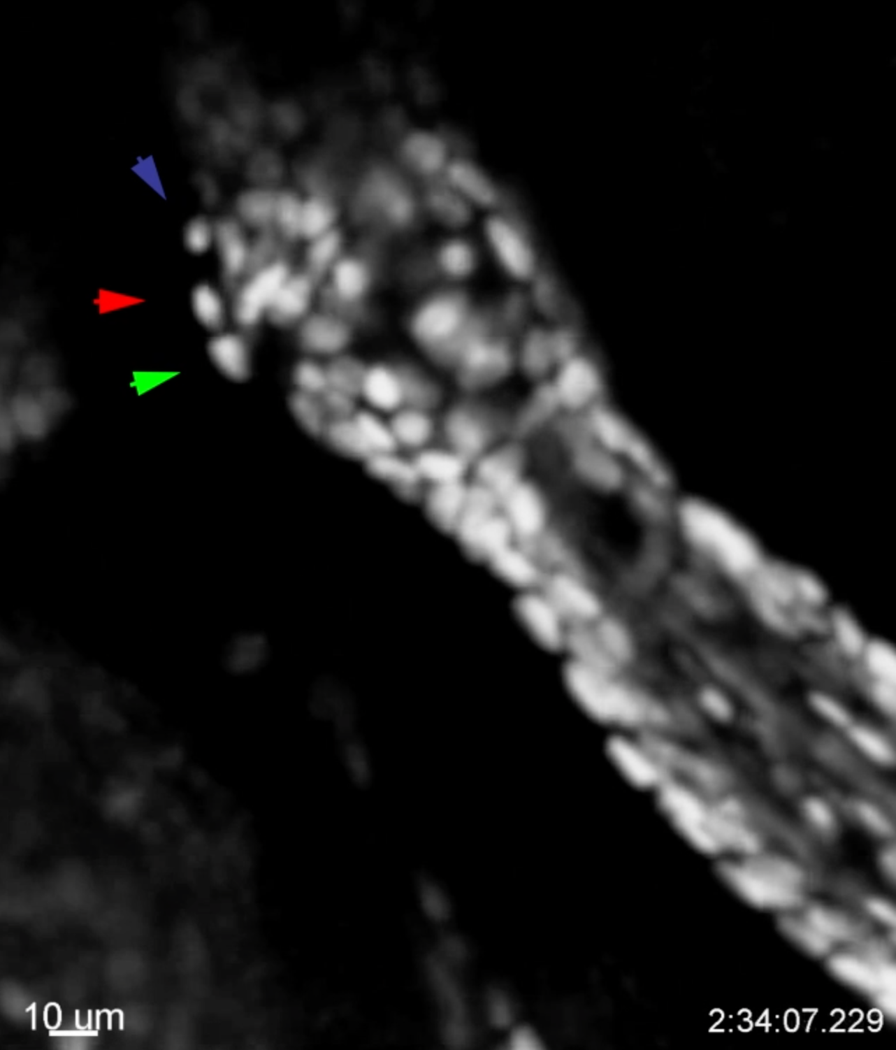
A nationwide team of researchers, led by scientists at University of Utah Health and The Rockefeller University, has determined how a genetic mutation found in mice and monkeys interferes with viruses such as HIV and Ebola. They say the finding could eventually lead to the development of medical interventions in humans.
The gene, called retroCHMP3, encodes an altered protein that disrupts the ability of certain viruses to exit an infected cell and prevents it from going on to infect other cells.
Normally, some viruses encase themselves in cell membranes and then make an exit by budding off from the host cell. RetroCHMP3 delays that process long enough that the virus can no longer escape.
“This was an unexpected discovery,” says Nels Elde, Ph.D., senior author of the study and an evolutionary geneticist in the Department of Human Genetics at U of U Health. “We were surprised that slowing down our cell biology just a little bit throws virus replication off its game.”
The study appears online Sept. 30 in advance of the Oct. 14 issue of Cell.
RetroCHMP3 originated as a duplicated copy of a gene called charged multivesicular body protein 3, or CHMP3. While some monkeys, mice, and other animals have retroCHMP3 or other variants, humans only have the original CHMP3.

Read more →

Bioengineers develop new class of human-powered bioelectronics

A team of bioengineers at the UCLA Samueli School of Engineering has invented a novel soft and flexible self-powered bioelectronic device. The technology converts human body motions — from bending an elbow to subtle movements such as a pulse on one’s wrist — into electricity that could be used to power wearable and implantable diagnostic sensors.
The researchers discovered that the magnetoelastic effect, which is the change of how much a material is magnetized when tiny magnets are constantly pushed together and pulled apart by mechanical pressure, can exist in a soft and flexible system — not just one that is rigid. To prove their concept, the team used microscopic magnets dispersed in a paper-thin silicone matrix to generate a magnetic field that changes in strength as the matrix undulated. As the magnetic field’s strength shifts, electricity is generated.
Nature Materials published today a research study detailing the discovery, the theoretical model behind the breakthrough and the demonstration. The research is also highlighted by Nature.
“Our finding opens up a new avenue for practical energy, sensing and therapeutic technologies that are human-body-centric and can be connected to the Internet of Things,” said study leader Jun Chen, an assistant professor of bioengineering at UCLA Samueli. “What makes this technology unique is that it allows people to stretch and move with comfort when the device is pressed against human skin, and because it relies on magnetism rather than electricity, humidity and our own sweat do not compromise its effectiveness.”
Chen and his team built a small, flexible magnetoelastic generator (about the size of a U.S. quarter) made of a platinum-catalyzed silicone polymer matrix and neodymium-iron-boron nanomagnets. They then affixed it to a subject’s elbow with a soft, stretchy silicone band. The magnetoelastic effect they observed was four times greater than similarly sized setups with rigid metal alloys. As a result, the device generated electrical currents of 4.27 milliamperes per square centimeter, which is 10,000 times better than the next best comparable technology.
In fact, the flexible magnetoelastic generator is so sensitive that it could convert human pulse waves into electrical signals and act as a self-powered, waterproof heart-rate monitor. The electricity generated can also be used to sustainably power other wearable devices, such as a sweat sensor or a thermometer.
There have been ongoing efforts to make wearable generators that harvest energy from human body movements to power sensors and other devices, but the lack of practicality has hindered such progress. For example, rigid metal alloys with magnetoelastic effect do not bend sufficiently to compress against the skin and generate meaningful levels of power for viable applications.
Other devices that rely on static electricity tend not to generate enough energy. Their performance can also suffer in humid conditions, or when there is sweat on the skin. Some have tried to encapsulate such devices in order to keep water out, but that cuts down their effectiveness. The UCLA team’s novel wearable magnetoelastic generators, however, tested well even after being soaked in artificial perspiration for a week.
A patent on the technology has been filed by the UCLA Technology Development Group.
Story Source:
Materials provided by University of California – Los Angeles. Note: Content may be edited for style and length.

Read more →

New treatment uses reverse vaccination to teach immune system not to attack life-saving drugs

For nearly a third of patients with hemophilia A and almost all patients with Pompe disease, their own immune system is their greatest obstacle to treatment. When given essential proteins and enzymes, their body perceives the treatments as a threat and attacks.
However, University at Buffalo researchers have developed a new treatment that uses reverse vaccination to pre-expose the body to medications and build immune tolerance. The novel treatment pairs essential proteins and enzymes with lysophosphatidylserine (Lyso-PS), a fatty acid that helps the immune system tolerate foreign substances, reducing adverse reactions to the drugs.
Unlike traditional vaccination, which uses pre-exposure to teach the immune system to attack potential threats, reverse vaccination uses exposure to teach the immune system to ignore foreign substances. The treatment could be applied to a broad range of drug therapies, autoimmune disorders and allergies, says lead investigator Sathy Balu-Iyer, PhD, professor of pharmaceutical sciences and associate dean for research in the UB School of Pharmacy and Pharmaceutical Sciences.
The results were published this month in Scientific Reports. Balu-Iyer recently received funding to continue preclinical research from the Empire Discovery Institute, which will license the technology and advance the treatment to the market.
“The safety and effectiveness of several life-saving therapeutic drugs are compromised by anti-drug antibodies. Once antibodies develop, clinical options available for patients become expensive and, in several cases, ineffective,” said Balu-Iyer.
“Instead of attempting to reverse the anti-drug antibodies, which is highly challenging, clinical treatments that prevent antibody development may be a more effective strategy,” says Nhan Hanh Nguyen, first author and pharmaceutical sciences graduate student at UB. “Our approach is based on the rationale that pre-exposure of a protein in the presence of Lyso-PS teaches the immune system not to mount a response.”
Hemophilia A is a genetic bleeding disorder caused by the lack of blood-clotting protein Factor VIII. Patients with the disorder are at severe risk for bleeding after injury or surgery. Recombinant Factor VIII is the first line of defense in treatment, however, the body may associate Factor VIII with other threats and produces antibodies that destroy it. A third of patients experience these adverse reactions, and once antibodies develop, the cost of clinical treatments may rise above $700,000 per year.

Read more →

Relieving pain by mapping its biological signatures

Many people are confronted with chronic pain that can last for months or even years. How to best treat chronic pain? First, pain must be categorized for the right treatment to be prescribed. However, is that it is very challenging for patients to define their pain, its intensity or even its location using questionnaires. To overcome this difficulty, scientists from the University of Geneva (UNIGE) have joined forces with the research department of the Clinique romande de réadaptation (CRR) in Sion to carry out a complete epigenomic analysis of patients, making it possible to find the epigenetic signatures specific to each pain category. Thus, a simple blood test would make it possible to define which pain the person suffers from and, in the future, to prescribe treatment accordingly and to observe whether the biomarkers modified by the pain return to normal. These results can be read in the Journal of Pain.
Chronic pain is classified into two main categories: nociceptive pain — defined by the activation of receptors at the end of nerve fibres and found in osteoarthritis, burns or infections — and neuropathic pain, which is caused by damage to nerve structures, such as pain caused by shingles. In order to classify which pain the patient suffers from, they fill in several questionnaires and quantify pain intensity of using assessment scales. However, this is very subjective and time-consuming.
Blind genome analysis
“At the CRR, we treat many people suffering from chronic diseases,” explains Bertrand Léger, a researcher at the CRR and last author of the study. “We joined forces with UNIGE scientists to carry out a complete epigenomic study and define specific biomarkers for each type of pain, in order to be able to categorise the various types of pain quickly and reliably.”
To do this, the Geneva team carried out an analysis of the entire genomes of 57 patients: 20 with no pain, 18 with nociceptive pain and 19 with neuropathic pain. “The aim was to start without any prior hypothesis to probe the genome as a whole and identify all the biomarkers involved in pain,” explains Ariane Giacobino, the study’s coauthor and a professor in the Department of Genetic Medicine and Development at UNIGE Faculty of Medicine.
Specific and potentially reversible biomarkers
Unexpectedly, not only did the scientists identify very striking epigenetic signatures of pain, but there was no overlap between nociceptive and neuropathic pain. “This total absence of similarities between the two categories of pain is very surprising, because intuitively, we might think that the difficulty in defining one’s pain comes from a similarity in the epigenetic signature. We could prove that itis absolutely not the case,” notes Ariane Giacobino.
Indeed, the biomarkers specific to nociceptive pain are expressed by the genes of the opioid system — involved in emotion, reward and pain — as well as by the genes of inflammation, specific to irritation. Conversely, the biomarkers for neuropathic pain are linked only to genes of the GABA system, the neurotransmitters of the central nervous system.
“Now that these epigenetic signatures are clearly defined, a simple blood test will make it possible to define the type of pain the person is suffering from and prescribe the appropriate treatment,” says Bertrand Léger. The treatment will thus no longer target the symptoms, but the very root of the problem. And finally, since epigenetics is characterised by the fact that the expression of a gene is durably modified, the right treatment may return it to normal. “We could imagine monitoring the reversion of pain by observing, from an epigenetic point of view, whether the biomarkers return to normal, and adapt the treatment accordingly,” concludes Ariane Giacobino.
Story Source:
Materials provided by Université de Genève. Note: Content may be edited for style and length.

Read more →

Cell labelling method from microscopy adapted for use in whole-body imaging

Processes and structures within the body that are normally hidden from the eye can be made visible through medical imaging. Scientists use imaging to investigate the complex functions of cells and organs and search for ways to better detect and treat diseases. In everyday medical practice, images from the body help physicians diagnose diseases and monitor whether therapies are working. To be able to depict specific processes in the body, researchers are developing new techniques for labelling cells or molecules so that they emit signals that can be detected outside the body and converted into meaningful images. A research team at the University of Münster has now adapted a cell labelling strategy currently used in microscopy — the so-called SNAP-tag technology — for use in whole-body imaging with positron emission tomography (PET) for the first time.
This method labels cells in two steps that work for completely different cell types such as tumour and inflammatory cells. First, the cells are genetically modified to produce a so-called SNAP-tag enzyme on their surface that is unique to the targeted cells. The enzyme is then brought into contact with a suitable SNAP-tag substrate. The substrate is labelled with a signal emitter and chemically structured so that it is recognised and split by the enzyme allowing the signal emitter to be transferred to the enzyme. In the process, the enzyme is modified so that it is no longer active and, as a result, the signal emitter remains tightly coupled to it. “Through its biological activity, the SNAP-tag enzyme labels itself, so to speak — this happens very quickly and without disturbing the natural processes in the organism,” explains Dominic Depke, a biology doctoral student and one of the lead authors of the new study.
In microscopy, fluorescent dyes are used to label cells, but they are mostly not suitable for whole-body imaging because their signals are scattered by thicker tissue layers with the result that they can no longer be measured. To solve this problem, the scientists synthesised a new SNAP-tag substrate using the radioactive signal emitter fluorine-18. The team have successfully labelled tumour cells in mice by injecting this substrate into the organism via the bloodstream and were then able to visualise the tumours using PET imaging. “The exciting thing for us about SNAP-tag technology is that it opens up the prospect of visualizing genetically encoded cells in the body with different imaging modalities and at different temporal stages — we call it multiscale imaging,” explains nuclear medicine specialist Prof Michael Schäfers. “Radioactive signals from fluorine-18 remain stable for only a short time,” adds radiochemist Dr Christian Paul Konken, “but as we can repeat the second labelling step, we can potentially visualise the same cells again and again over days and weeks.” The high level of detail provided by microscopy makes it possible to study how individual cells communicate with each other. The big picture view provided by whole-body imaging enables scientists to assess how these cells function as part of whole organ systems. Time may reveal what role individual cell types play in inflammation, for example, as it begins, continues and resolves. “Only by combining all this information can we understand how everything is connected in the body,” says Michael Schäfers.
A small beginning with great potential
“Our investigations are a very first step, in which we have shown that labelling cells with SNAP-tags works, in principle, in living organisms,” emphasises biochemist Prof Andrea Rentmeister. “What matters here is that the substrate is distributed rapidly in the organism and that it binds exclusively to the cells to be studied.” The next crucial steps will be to test how many cells are needed to obtain a sufficiently strong signal and whether the method can also be used to visualise cells that move within the organism — in particular immune system cells. If the approach continues to prove successful, the technique may become important for future research into immunotherapies in which the body’s own immune cells are genetically modified in the laboratory so that they can combat a specific disease. Such therapies are already being used for cancer treatment and have the potential to help treat inflammatory diseases as well. Imaging could help develop and improve such treatments.
When the scientists presented their results for the first time at a scientific symposium, they were in for a surprise — colleagues from Tübingen presented a similar study there at the same time. Independently of each other, both research teams had the same fundamental idea, a SNAP-tag substrate labelled with fluorine-18. Chemically speaking, they implemented the idea differently but they tested the resulting substrates using the same biological model system and arrived at similar findings. “This shows how topical our question is and that our results are reproducible and really promising,” says Michael Schäfers. He adds that the Tübingen team is developing new labelling methods to study immune cells in cancer, while the team in Münster is focusing on inflammatory diseases, so the research complements each other very well. The research team from Münster published their study in the scientific journal “Chemical Communications,” only a few days later the publication from Tübingen was released in “Pharmaceuticals.”
Creating a new substrate for the SNAP-tag
Like all SNAP-tag substrates, the newly developed molecule is based on benzylguanine to which the scientists attached the radioactive isotope fluorine-18, which is, in turn, ideally suited for PET imaging. “Our goal was to design the synthesis in a few quick steps so that we get as strong a signal as possible — because fluorine-18 has a short half-life, its radioactivity is reduced by half after every 110 minutes,” explains Christian Paul Konken. Initially, the scientists found that the fluorine-18 did not attach to the desired position on the molecule. “The benzylguanine was apparently too sensitive to be labelled directly with fluorine-18,” says Lukas Rösner, a biochemistry doctoral student, “so we first labelled a small molecule that is insensitive to the necessary chemical reactions — the fluoroethylazide — and then attached it to the benzylguanine using a click reaction, which is very fast and selective.”
Tests in test tube, cell cultures and the organism
The scientists first checked whether the synthesised substrate remained stable when in contact with blood in the test tube and then examined how the cells interacted with the substrate in the first practical tests in cell cultures. In doing so, they compared human tumour cells into which they had genetically incorporated the SNAP-tag enzyme with those that did not produce the enzyme. “We could see very clearly that the radioactivity was only taken up by the cells that produced the SNAP-tag enzyme,” says Dominic Depke. Finally, the team conducted targeted studies on individual mice. “This step was decisive once again,” explains Michael Schäfers, “because how a molecule behaves in the complex biological environment in a living organism cannot be fully simulated in cell culture or with artificially produced organs.” The scientists were able to show that once the substrate is injected into the bloodstream it is distributed through the body very quickly. Additionally, they identified the pathways through which it is excreted. They then compared how tumour cells with and without the SNAP-tag enzyme reacted to the substrate in living organisms. For this purpose, the tumour cells were injected under the skin of mice and removed again after the examination in order to confirm the results with autoradiography.
Story Source:
Materials provided by University of Münster. Original written by Doris Niederhoff. Note: Content may be edited for style and length.

Read more →

Novel fluorescence imaging assay gives new insight to develop biomimetic nanoparticles for targeted cancer therapy

Cell membrane coated biomimetic nanoparticles (NPs) have been widely studied in nanomedicine because of their unique properties such as immune escaping, long blood circulation, specific molecular recognition and efficient cancer targeting, indicating a great potential in targeted cancer therapy. However, the integrity of the cell membrane coating on NPs, a key metrics related to the quality of these biomimetic-systems and to the resulting biomedical function, has remained largely unexplored. In the present study, the researchers developed a fluorescence quenching assay to probe the integrity of the cell membrane coating.
The study published in Nature Communications shows that the great majority of the cell membrane coated NPs were only partially coated when the traditional coating techniques were applied. The information is essential as the coating degree impacts the biological fate of NPs. The research was carried out at the University of Eastern Finland, Department of Applied Physics, in Professor Vesa-Pekka Lehto’s research group.
“The present methods for characterizing the cell membrane coating are only qualitative and fail to statistically evaluate the degree and variability of the coating,” says Lizhi Liu, the first author of the publication. “When we applied the developed quantification method to evaluate the success of the commonly used protocols to produce fully coated NPs, we found that the fraction never exceeded 20%.”
“Our discovery is a big surprise to whole scientific community in nanomedicine because it has been generally accepted that the cell membrane coating is perfect. Despite of the partial coating, biomimetic NPs could still be internalized by the target cells via different pathways,” says Dr Wujun Xu, one of the corresponding authors of the paper. To explain this, the authors proposed a new endocytic entry mechanism for these partially coated NPs by computational simulations. Specifically, the NPs with a high coating degree (? 50%) entered the cells individually, whereas the NPs with a low coating degree (< 50%) needed to aggregate together before internalization. "The present study highlights some of the limitations of the current cell membrane coating protocols and motivates the efforts to improve the protocols. The developed quantification method is a practical tool to assess the success of these efforts and establish a standard for comparing the different coating designs," summarises Professor Vesa-Pekka Lehto. Story Source: Materials provided by University of Eastern Finland. Note: Content may be edited for style and length.

Read more →

Toxic DNA buildup in eyes may drive blinding macular degeneration

Damaging DNA builds up in the eyes of patients with geographic atrophy, an untreatable, poorly understood form of age-related macular degeneration that causes blindness, new research from the University of Virginia School of Medicine reveals. Based on the discovery, the researchers think it may be possible to treat the disease with common HIV drugs or an even safer alternative.
The harmful DNA, known as Alu cDNA, was previously discovered to be manufactured in the cytoplasm by UVA’s Jayakrishna Ambati, MD, and his collaborators. The new findings are believed to be the first time toxic Alu cDNA accumulation has been confirmed in patients in any disease.
The new findings offer insights into how geographic atrophy progresses over time. “Although we’ve known that geographic atrophy expands over time, we didn’t know how or why,” said Ambati, of UVA’s Department of Ophthalmology and Center for Advanced Vision Science. “Our finding in human eyes that the levels of toxic Alu cDNA are highest at the leading edge of the geographic atrophy lesion provides strong evidence that it is responsible for this expansion over time that leads to vision loss.”
About Age-Related Macular Degeneration
Geographic atrophy is an advanced form of age-related macular degeneration, a potentially blinding disease estimated to affect 200 million people around the world. The disease ultimately destroys vital cells in the retina, the light-sensing portion of the eye.
Ambati, a top expert in macular degeneration, and his colleagues found that this destruction is caused by the buildup of Alu DNA, which the researchers discovered floating in the cytoplasm of cells. That Alu DNA was being manufactured in cytoplasm came as a surprise, as DNA is typically thought to be contained within the cell nucleus.
As Alu DNA accumulates in the eye, it triggers harmful inflammation via a part of the immune system called the inflammasome. The researchers identified how this happens, discovering a previously unknown structural facet of Alu that triggers the immune mechanism that leads to the death of the vital retinal cells.
That’s where HIV drugs called nucleoside reverse transcriptase inhibitors, or NRTIs, could come in. The researchers’ new work in lab mice suggests these drugs, or safer derivatives known as Kamuvudines, could block the harmful inflammation and protect against retinal cell death.
“Over the last two decades, dozens of clinical trials for geographic atrophy that have targeted other pathways have failed,” Ambati said. “These findings from patient eyes provide a strong impetus for a new direction.”
Ambati says his latest findings offer further support for conducting clinical trials testing the drugs in patients with macular degeneration. A prior study of four different health insurance databases — encompassing more than 100 million patients over two decades — found that people taking NRTIs were almost 40% less likely to develop dry macular degeneration.
“Our findings from human eyes show that these toxic molecules, which activate the inflammasome, are most abundant precisely in the area of greatest disease activity,” Ambati said. “We are very hopeful that a clinical trial of Kamuvudines will be launched soon in geographic atrophy so that we can potentially offer a treatment for this devastating condition.”
Story Source:
Materials provided by University of Virginia Health System. Note: Content may be edited for style and length.

Read more →

Breastfeeding status and duration significantly impact postpartum depression risk

According to the United States Centers for Disease Control and Prevention, between 11 and 20 percent of women who give birth each year in the U.S. have postpartum depression symptoms, which is the greatest risk factor for maternal suicide and infanticide. Given that there are 4 million births annually, this equates to almost 800,000 women with postpartum depression each year.
Current biological and psychosocial models of breastfeeding suggest that breastfeeding could possibly reduce a woman’s risk for postpartum depression. However, prior studies only have looked at the initiation of breastfeeding and breastfeeding length. In addition, small and often homogenous samples have yielded ungeneralizable results lacking in statistical power with biased results due to higher levels of education, income, and proportions of white participants compared to the general population of the sampled country.
Researchers from Florida Atlantic University’s Christine E. Lynn College of Nursing and collaborators are the first to examine current breastfeeding status in association with postpartum depression risk using a large, national population-based dataset of 29,685 women living in 26 states.
Results of the study, published in the journal Public Health Nursing, demonstrate that postpartum depression is a significant health issue among American women with nearly 13 percent of the sample being at risk. Findings showed that women who were currently breastfeeding at the time of data collection had statistically significant lower risk of postpartum depression than women who were not breastfeeding. In addition, there is a statistically significant inverse relationship between breastfeeding length and risk of postpartum depression. As the number of weeks that women breastfed increased, their postpartum depression decreased. An unexpected finding was that there was no significant difference in postpartum depression risk among women with varying breastfeeding intent (yes, no, unsure).
“Women suffering from postpartum depression, which occurs within four weeks and up to 12 months after childbirth, endure feelings of sadness, anxiety and extreme fatigue that makes it difficult for them to function,” said Christine Toledo, Ph.D., senior author and an assistant professor in FAU’s Christine E. Lynn College of Nursing. “Women with postpartum depression who are not treated also may have negative outcomes, including difficulty bonding with and caring for their children, thoughts of harming themselves or their infant, and also are at an increased risk of substance misuse.”
Woman who have experienced postpartum depression have a 50 percent increased risk of suffering further episodes of postpartum depression in subsequent deliveries. In addition, they have a 25 percent increased risk of suffering further depressive disorders unrelated to childbirth up to 11 years later. Postpartum depression increases maternal morbidity and is associated with increased risks for cardiovascular disease, stroke and type-2 diabetes.
For the study, Toledo and collaborators from the University of Miami School of Nursing and Health Studies, University of North Carolina School of Nursing, Chapel Hill, Seattle University of Nursing, and The University of British Columbia School of Nursing, analyzed dataset from the 2016 Pregnancy Risk Assessment Monitoring System (PRAMS) questionnaire to investigate the association of breastfeeding practices taking into consideration significant covariates such as age, race, marital status, education, abuse before and during pregnancy, cigarette smoking, among others.
“Findings from this important study suggest that breastfeeding is a cost efficient and healthy behavior that can decrease a woman’s risk for postpartum depression,” said Safiya George, Ph.D., dean, FAU Christine E. Lynn College of Nursing. “Nurses in particular play an important role in educating and promoting both the maternal health benefits of breastfeeding and infant benefits such as providing necessary nutrients and protecting them against allergies, diseases and infections.”
Study co-authors are Rosina Cianelli, Ph.D.; Giovanna De Olivera, Ph.D.; and; Karina Gattamorta, Ph.D., all with the University of Miami School of Nursing and Health Studies; Natalia Villegas Rodgriguez, Ph.D., University of North Carolina School of Nursing, Chapel Hill; Danuta Wojnar, Ph.D., Seattle University of Nursing; and Emmanuela Ojukwu, Ph.D., The University of British Columbia School of Nursing.
The study was funded by the Ph.D. Scholarly Award by the Sigma Theta International, Beta Tau Chapter.
Story Source:
Materials provided by Florida Atlantic University. Original written by Gisele Galoustian. Note: Content may be edited for style and length.

Read more →

Dental care: The best, worst and unproven tools to care for your teeth

Do probiotics prevent gum disease? Is flossing necessary? Many patients are unable to confidently answer these questions and more due to the abundance of conflicting medical information. However, new research led by the University at Buffalo aims to separate fact from fiction in determining which oral hygiene tools actually prevent gum disease.
The paper, published in the October issue of the Journal of the International Academy of Periodontology, examines the effectiveness of various oral hygiene devices.
The result: Only a handful of self-administered interventions provide additional protection against gingivitis and periodontitis beyond brushing one’s teeth with a basic toothbrush. At the moment, all other oral hygiene interventions are only supported by insufficient evidence, says Frank Scannapieco, DMD, PhD, principal investigator and chair and professor of oral biology in the UB School of Dental Medicine.
The findings, he says, will help dental practitioners and the public identify best practices for preventing gum disease, which affects nearly half of adults 30 and older in the United States, according to the Centers for Disease Control and Prevention (CDC).
“Patients can be confident that the oral care tools and practices supported by research, as described in the paper, will prevent the initiation and progression of periodontal disease, if they are performed regularly and properly,” says Scannapieco.
Additional investigators include Eva Volman, DDS, first author, UB alumna and resident dentist at the Eastman Institute for Oral Health; and Elizabeth Stellrecht, interim head of health sciences library services at UB.

Read more →

Virtual care with remote monitoring catches drug errors and reduces patient pain

Patients using take-home technology following non-elective surgery resulted in significantly greater detection and correction of drug errors, and reduction in patients’ pain, says a national study led by Hamilton researchers.
The study looked at patient outcomes from virtual care and remote automated monitoring (RAM) — video calls with nurses and doctors, and self-monitoring of vital signs using wearable devices.
The research also raised the possibility of a reduction in acute-hospital care as the result of virtual care and RAM.
“We began the study in the first months of the pandemic, when hospitals were challenged to drastically reduce non-emergency care,” said P.J. Devereaux, co-principal investigator of the study.
He is a senior scientist at the Population Health Research Institute (PHRI), professor and director of the division of perioperative care at McMaster University, and a cardiologist and perioperative care physician at Hamilton Health Sciences.
“Our study provides proof of concept that virtual care with RAM can improve outcomes after discharge following non-elective surgery — outcomes that are important to patients,” he said.

Read more →