
White House Pledges $1 Billion for At-Home Covid Tests

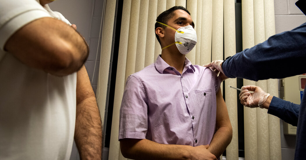
Males between 16 and 29 years of age have an increased risk of developing heart problems after receiving a second dose of coronavirus vaccines made by Pfizer-BioNtech or Moderna, according to a large new analysis published on Wednesday in the New England Journal of Medicine.The study, conducted in Israel, estimated that nearly 11 of every 100,000 males in that age group developed myocarditis, inflammation of the heart, a few days after having been fully vaccinated. That figure is higher than most earlier estimates.Boys between 16 and 19 years of age had the highest incidence of myocarditis after the second dose, according to a second study in the journal. The risk of heart problems in boys of that age was about nine times higher than in unvaccinated boys of the same age.The absolute risk is still very small, and the condition temporary. And studies have shown that Covid-19 is much more likely to cause heart problems compared with vaccination.Based on data available in June, advisers to the Centers for Disease Control and Prevention unanimously voted to recommend the vaccine, saying the benefits far outweighed the risk. For every million vaccinated boys ages 12 to 17, the shots might cause a maximum of 70 myocarditis cases, but would prevent 5,700 infections, 215 hospitalizations and two deaths, the agency has estimated.Myocarditis is among the concerns that may have led the Food and Drug Administration to ask Pfizer-BioNTech and Moderna to enroll more children in their vaccine trials. Potential side effects are likely to dominate the discussion when agency advisers meet next week to review the evidence for a vaccine in children aged 5 to 11 years.One of the new studies looked at data from 2.5 million vaccinated members of Israel’s largest health care network who were aged 16 years or above. The researchers identified 54 cases of myocarditis, and deemed 41 of them to be mild. About 70 percent of the myocarditis cases were observed after the second dose.One case was severe enough to require ventilation. One patient with a history of heart disease died of an unknown cause soon after being discharged from the hospital. Of the 14 patients who showed heart abnormalities when they were admitted to the hospital, 10 still had some signs of problems when they were discharged.But when the patients were examined again a few weeks later, all five of those for whom results were available appeared to have fully recovered.Across all age groups and sexes, the overall incidence was just over two cases per 100,000 people, the study found. But when the researchers analyzed the results by age and sex, they found the highest incidence among males 16 to 29 years. The risk in females of every age was negligible.
Read more →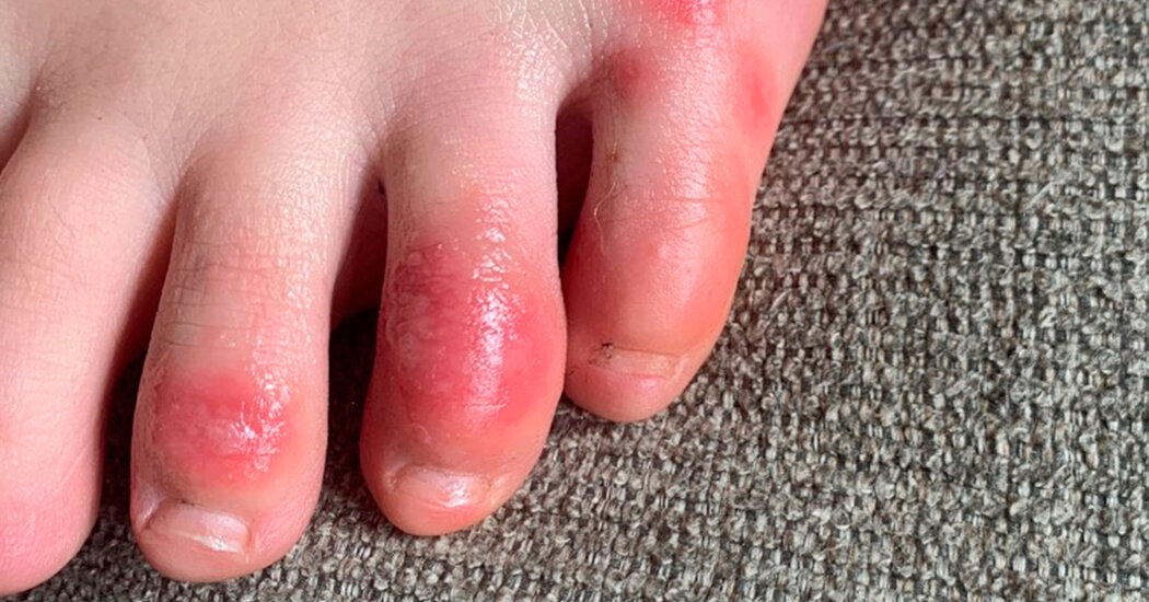
Shortly after the pandemic erupted last year, doctors were baffled by a surge of patients, mostly teenagers and young adults, who came in complaining of chilblains — painful lesions on their toes, and sometimes also on their fingers.The condition came to be called Covid toes. They were seen, like the loss of smell and taste, as yet another strange telltale sign of the disease, even though most of the patients tested negative for coronavirus. Physicians were hard-pressed to explain the association.The lesions are red or purple in white people and often purplish or brownish in people of color. They cause painful burning or itching sensations and sometimes make it difficult for people to wear shoes or walk.Now a new study from France, published in the British Journal of Dermatology, sheds some light on the causes of Covid toes. The research indicates that the lesions may be a side effect of the immune system’s shift into overdrive in response to exposure to the virus, which can damage cells and tissues in the process.The French researchers analyzed blood samples and skin biopsies from 50 patients who had chilblainlike lesions for the first time in April 2020, and who were referred to St.-Louis Hospital in Paris. Slightly more than half of the patients had other symptoms suggestive of Covid-19, like coughing, shortness of breath and loss of smell, but all of them tested negative for the virus on PCR tests.The samples showed high levels of Type 1 interferon, a protein that activates the body’s immune system to fight viruses, but which may also cause damage. The researchers also found high levels of an antibody that can inadvertently attack the body’s own cells.Abnormal changes in the linings of the blood vessels may also play a role in the lesions, the study suggests.Although the relationship between coronavirus infection and chilblainlike lesions “is still controversial,” the authors wrote, the “peaks of chilblainlike lesions concomitant with peaks of Covid-19 deaths in 2020 strongly suggest that this disorder is closely related to SARS-CoV-2 infection.”The explanation for Covid toes is not entirely surprising; one of the hallmark features of the disease is an immune system overreaction called a cytokine storm, which may ultimately cause more illness than the virus itself.German scientists published a paper last year saying they had found a strong localized interferon-driven response in three young men who came in with chilblains. That paper suggested that the men, who tested negative for the coronavirus, may have developed chilblains several weeks after an initial infection caused mild or asymptomatic disease, and that the interferon-driven immune response may have led to early control of the virus and prevented respiratory disease.Dermatologists say that people with Covid toes generally do well and are unlikely to develop severe Covid, and that the symptoms reflect a healthy immune response to the virus.The new study suggested treating Covid toes with local or systemic anti-inflammatory agents may be effective.
Read more →
Whether it’s reporting on conflicts abroad and political divisions at home, or covering the latest style trends and scientific developments, Times Video journalists provide a revealing and unforgettable view of the world.Whether it’s reporting on conflicts abroad and political divisions at home, or covering the latest style trends and scientific developments, Times Video journalists provide a revealing and unforgettable view of the world.
Read more →
Researchers at the University of Saskatchewan (USask) are hopeful new understanding of cellular defects related to Cystic Fibrosis (CF) could help pave the way for treatment of the disease.
A team in the College of Medicine led by Drs. Juan Ianowski (PhD) and Julian Tam (MD) found that sodium transport is abnormal in lungs with CF. The researchers, affiliated with the Respiratory Research Centre, studied the swine model of CF and used a specialized microelectrode technique that allowed them to perform experiments with very high resolution. They discovered there is excessive sodium absorption in the small airways, a previously unstudied site in the body.
“A precise understanding of the cellular basis of CF lung disease is a prerequisite for the development of treatments such as gene therapy that have the potential to cure CF,” said Tam. “CFTR modulators, such as Trikafta, can improve life for about 90 per cent of patients. Our work is especially relevant to that 10 per cent of people with CF who cannot benefit from these medications.”
Their findings were published in the journal Cell Reports on Oct. 5.
According to Cystic Fibrosis Canada, CF is the most common fatal genetic disease affecting Canadian children and young adults. There is presently no cure for the disorder that varies in symptoms from patient to patient, but mainly affects the lungs and digestive system. About one in every 3,600 children born in Canada has CF, which occurs when a person receives two copies of a defective gene — one from each parent.
Ianowski has been working with Tam for about six years, and has been able to meet people with CF in his clinic. He said this adds a sense of purpose and desire to create applicable outcomes through the research he performs in the lab.
“In this context I get to know the patients by name and see their struggles,” Ianowski said. “Working with Julian has created a meaningful partnership and we can inform and strengthen each other’s work.”
Tam and Ianowski point to the power of USask’s rich research environment, which gave them access to the expertise of veterinarians that supported their work. They also are grateful for the vital contributions of team members like post-doctorate fellow Dr. Xianojie Luan (PhD), who played a crucial role in developing the research protocols and collecting data.
The research was funded by the Canadian Institute for Health Research and Cystic Fibrosis Canada.
Story Source:
Materials provided by University of Saskatchewan. Note: Content may be edited for style and length.
Colorectal cancer is projected to claim 53,000 lives in the United States this year alone and, as with most cancers, the disease is deadliest when it metastasizes. It follows that the most effective way to control it would be a drug that targets metastasis itself — preventing cancer cells from breaking off the primary tumor, or reining in rogue cells before they spread throughout the body and seed secondary tumors.
Now, a new study identifies a small molecule that could, in the future, be administered alongside standard chemotherapies to stave off colorectal cancer metastasis. The research, published in Science Advances, demonstrated how the compound, named RGX-202, foils a key pathway that cancer cells rely upon to hoard energy, thereby killing them and shrinking tumors in mice.
The findings have already led to a clinical trial in humans and may eventually give rise to a novel therapy that increases survival rates for multiple gastrointestinal cancers.
“Colorectal cancer is one of the top causes of cancer-related mortality,” says Rockefeller’s Sohail Tavazoie, head of the Elizabeth and Vincent Meyer Laboratory of Systems Cancer Biology. “We’ve found a critical pathway that promotes colorectal cancer metastasis and a novel therapeutic that appears to inhibit it.”
Stockpiling phosphocreatine
Back in 2016, researchers in the Tavazoie lab noticed that colorectal cancer cells had developed an ingenious strategy for surviving under oxygen-poor conditions. The tumors were mass-producing a unique enzyme within their cells, only to pump the enzyme out into the extracellular space. There, the enzyme would convert the metabolite creatine (which is abundant outside of cells) into phosphocreatine, which the cancer cells would finally import back across their membranes.
Metformin, a drug commonly prescribed against diabetes, holds promise against a rare type of childhood brain tumor in laboratory studies, an international team of researchers led by the University of Michigan Health Rogel Cancer Center report in Science Translational Medicine.
Experiments that uncovered new understandings of group A posterior fossa ependymomas — or PFAs — led the research team to the potential new treatment approach. The universally devastating tumors are a subset of ependymomas, which account for about 5% of childhood brain cancers and affect about 240 children each year in the U.S., according to Cancer.net statistics.
“These types of tumors actually challenge our fundamental understanding of cancer,” said study senior author Sriram Venneti, M.D., Ph.D., the Al and Robert Glick Family Research Professor of Pediatrics in the Department of Pathology at Michigan Medicine.
That’s because most cancers are known to arise from genetic mutations or errors. Precision medicine approaches strive to identify these mutations and target them with specific drugs.
But majority of PFAs lack such cancer-driving genetic mutations.
“We discovered back in 2016 that epigenetic changes are actually the main drivers of these tumors,” Venneti added — referring not to permanent mutations to the DNA itself, but to changes in how cells access and read DNA sequences.
Pairing a newly developed gel with immunotherapy that was delivered to post-surgical mouse brains with glioblastoma, a highly malignant and deadly cancer, improved the immunotherapy’s effectiveness, report researchers from the University of North Carolina Lineberger Comprehensive Cancer Center and colleagues. The findings appeared Oct. 6, 2021, in Science Advances.
The researchers used CAR-T cell (chimeric antigen receptor-T cell) immunotherapy, which involves harvesting immune-system T cells from a patient and genetically re-engineering them in the lab to recognize targets on the surface of cancer cells. In this mouse study, the CAR-T cells and gel were placed to fill in the area where a glioblastoma tumor had just been surgically removed. Previous studies have shown that administering T cells alone has produced limited benefit.
Glioblastoma is an aggressive tumor that can form quickly in the brain and is diagnosed most often in people in their sixties. Only 40 percent of people live for one year after diagnosis; just 17 percent survive for two years. Surgery is the first treatment used but presents many challenges in removing the tumor while also allowing for wound healing.
“We developed a gel made of fibrin, a protein most often associated with helping blood to clot. Applying a gel substance to an area of the brain to aid CAR-T cell therapy is unique in glioblastoma treatment,” said Edikan Ogunnaike, PhD, a biomedical engineer at UNC and first author of the article. “The gel aided CAR-T cell distribution in the brain by acclimating the T cells to the post-surgical wound environment while also stopping the tumor from recurring.”
The researchers used concentrations of human fibrinogen, a protein produced by the liver, which was transformed to fibrin with enzymes to develop a porous gel that was mixed with CAR-T cells and placed in the post-surgical brain area. The gel created web-like fibrin scaffolds in the brain, in which the CAR-T cells uniformly enmeshed themselves into the pores of scaffolds. The scaffolds are biodegradable and do not cause inflammation, tissue death or scarring.
Nine of 14 (64%) mice that received the gel and T cells were tumor free 94 days after treatment, compared to two of 10 (20%) mice who only received T cells. The researchers said if these findings can be replicated in human studies — they caution that many early laboratory findings don’t lead to clinical studies or new therapies — it would result in a great improvement in current treatment rates.
“Our approach was beneficial in glioblastoma and we believe that it could also control growth or return of tumors in the brain, eye and other organs,” said Gianpietro Dotti, MD, professor in the UNC School of Medicine Department of Microbiology and Immunology, co-leader of the Immunology Program at UNC Lineberger and corresponding author of this article. “It should be noted that direct delivery of CAR-T cells into a post-surgical area must result in broad coverage of the surface of the surgical cavity to maximize the possibility that T cells come in contact with residual tumor cells, which is where our flexible scaffold has shown to be of great benefit.”
The investigators are currently looking at injecting CAR-T cells to other parts of the brain. The process is being tested in pilot clinical studies to assess safety as well as to see if it elicits a greater distribution of CAR-T cells.
“We hope that our gel will provide antitumor activity over an extended period of time after surgery in patients without inducing toxicity, which seems to be the case in mice,” Ogunnaike said. “The gel might also allow for local delivery of other biological agents that could sustain T cell growth and counter suppression of an immunotherapy.”
In addition to Ogunnaike and Dotti, the paper’s other authors at UNC include Alain Valdivia, Mostafa Yazdimamaghani, PhD, Ernesto Leon, Seema Nandi, PhD, Hannah Hudson, Hongwei Du, PhD, Simon Khagi, MD, Barbara Savoldo, MD, PhD, Frances S. Ligler, PhD, and Shawn Hingtgen, PhD. Zhen Gu, PhD, is at the University of California, Los Angeles and Zhejiang University, China.
Massage has been used to treat sore, injured muscles for more than 3,000 years, and today many athletes swear by massage guns to rehabilitate their bodies. But other than making people feel good, do these “mechanotherapies” actually improve healing after severe injury? According to a new study from researchers at Harvard’s Wyss Institute for Biologically Inspired Engineering and John A. Paulson School of Engineering and Applied Sciences (SEAS), the answer is “yes.”
Using a custom-designed robotic system to deliver consistent and tunable compressive forces to mice’s leg muscles, the team found that this mechanical loading (ML) rapidly clears immune cells called neutrophils out of severely injured muscle tissue. This process also removed inflammatory cytokines released by neutrophils from the muscles, enhancing the process of muscle fiber regeneration. The research is published in Science Translational Medicine.
“Lots of people have been trying to study the beneficial effects of massage and other mechanotherapies on the body, but up to this point it hadn’t been done in a systematic, reproducible way. Our work shows a very clear connection between mechanical stimulation and immune function. This has promise for regenerating a wide variety of tissues including bone, tendon, hair, and skin, and can also be used in patients with diseases that prevent the use of drug-based interventions,” said first author Bo Ri Seo, Ph.D., who is a Postdoctoral Fellow in the lab of Core Faculty member Dave Mooney, Ph.D. at the Wyss Institute and SEAS.
A more meticulous massage gun
Seo and her coauthors started exploring the effects of mechanotherapy on injured tissues in mice several years ago, and found that it doubled the rate of muscle regeneration and reduced tissue scarring over the course of two weeks. Excited by the idea that mechanical stimulation alone can foster regeneration and enhance muscle function, the team decided to probe more deeply into exactly how that process worked in the body, and to figure out what parameters would maximize healing.
They teamed up with soft robotics experts in the Harvard Biodesign Lab, led by Wyss Associate Faculty member Conor Walsh, Ph.D., to create a small device that used sensors and actuators to monitor and control the force applied to the limb of a mouse. ” The device we created allows us to precisely control parameters like the amount and frequency of force applied, enabling a much more systematic approach to understanding tissue healing than would be possible with a manual approach,” said co-second author Christopher Payne, Ph.D., a former Postdoctoral Fellow at the Wyss Institute and the Harvard Biodesign Lab who is now a Robotics Engineer at Viam, Inc.
The isolated Suba and Luo communities of Mfangano Island on Lake Victoria in Western Kenya experience some of the highest maternal and neonatal mortality rates in East Africa. To understand factors at the heart of this complex challenge, researchers from the University of Minnesota have partnered with one of the most remote and underserved populations in the world.
Published in Global Public Health, the Monitoring Maternal Emergency Navigation and Triage on Mfangano (MOMENTUM) Study uses an innovative adaptive design to measure delays and barriers faced by mothers and newborns seeking emergency care.
Since 2018, Charles Salmen, MD, director of the Mfangano Community Health Field Station and assistant professor in the Department of Family Medicine and Community Health at the Medical School, and Co-Principal Iinvestigator Louisa Ndunyu, PhD, of Maseno University in Kenya, have mentored a team of local and international investigators to launch the MOMENTUM Study. At the same time, the study was designed to provide pragmatic, actionable data to support a locally-directed health navigation program serving this remote population. Through a 12-month, mixed-methods cohort study, the group of local and international investigators evaluated barriers and delays among patients seeking care for pregnancy-related, obstetric, and neonatal emergencies at nine remote health centers within Mfangano Island Division.
“The study was nearly derailed by the start of the COVID-19 pandemic in the middle of our data collection period. Yet through all, and thanks to the ingenuity and dedication of our team on the ground, and the remote support from our team at the University of Minnesota, we persevered to ensure that the harrowing stories of these mothers and their families were preserved and understood,” said Salmen. “Because they were willing to share their experiences from beginning to end, this study now represents an important contribution towards addressing the vexing problem of maternal emergencies in sub-Saharan Africa.”
The MOMENTUM study points to an urgent need for community education efforts regarding awareness of emergency conditions, implementation of streamlined hand-off systems for emergency referrals, as well as policy level interventions to address the recurrent health worker strikes. Particularly, in light of frequent facility closures and staffing interruptions, the study suggests a critical role for community-based care coordinators. The study suggests that these recurrent gaps can, in fact, be bridged by local interventions that flexibly connect the dots for families and providers across dynamic care environments, syncing communication and facilitating timely transport to available resources, and above all, advocating for patients.
With the completion of the first round of quantitative data analysis, the team in Kenya is currently conducting focus group discussions and key informant interviews to understand how the COVID-19 pandemic has impacted maternal and neonatal emergency care on Lake Victoria. This research is led by Kenyan investigators from Maseno University, local research staff at the Ekialo Kiona Center, with remote support from a team of University of Minnesota medical and public health students.
Story Source:
Materials provided by University of Minnesota. Original written by Gao Vang. Note: Content may be edited for style and length.