Ancient DNA reveals leprosy hit the Americas long before colonization

Hansen’s Disease (leprosy) is thought to have originated in Eurasia: previous studies on Mycobacterium leprae, the dominant form of leprosy, suggest the disease originated in Eurasia. Ancient pathogen genomes from old bones: a team of scientists from Germany and Argentina have reconstructed two genomes of Mycobacterium lepromatosis in 4000-year-old human skeletons from Chile. This pathogen is regarded as a second, less common, cause of Hansen’s Disease. A new American chapter for Hansen’s Disease: two pathogens that are responsible for the same disease evolved separately on opposite sides of the world for thousands of years.Hansen’s Disease, more commonly known as leprosy, is a chronic disease that can lead to physical impairment. Today it exists in over 100 countries, and while the infection is treatable, access to treatment varies widely with socioeconomic conditions. Its mention in historical texts give us a glimpse into its past impact on population health in Europe and Asia. Prolonged untreated infection can result in characteristic changes in bone, and these have been documented in archaeological skeletons as early as 5000 years ago in Europe, Asia, and Oceania. So far, absence of these characteristic changes in the pre-contact American contexts suggests that leprosy was one of the many diseases introduced to the continent in the colonial period. Thereafter it afflicted humans and curiously also armadillos.From a genetic perspective, Hansen’s Disease is caused by either the globally dominant Mycobacterium leprae or the newly identified and rare Mycobacterium lepromatosis. The recovery of M. leprae from archaeological bone in Europe suggests the disease originated in Eurasia sometime during the Neolithic transition about 7000 years ago. Similar emergence estimates have been proposed for other notorious diseases such as plague, tuberculosis, and typhoid fever. Ancient genomes for M. lepromatosis have remained elusive, and these may hold important clues on the history of Hansen’s Disease.
Past disease in the American continent
We know comparatively little about the infectious disease experience of the diverse communities of people living in the Americas before the colonial period. This accounts for almost 20,000 years of human history, and the diverse ecosystems into which humans integrated across the continent would have presented challenges to the immune system not otherwise encountered in other parts of the world. We know very little of these diseases, as they were smothered by the onslaught of pathogens that Europeans later brought with them. Archaeological study of human bones from the pre-contact Americas confirm that the time was far from disease-free, but often the traces seen on the bones aren’t specific enough to assign to a known disease.
“Ancient DNA has become a great tool that allows us to dig deeper into diseases that have had a long history in the Americas,” says Kirsten Bos, group leader for Molecular Paleopathology at the Max Planck Institute for Evolutionary Anthropology. She and her team have been studying pathological bone from American context for over a decade. Some diseases that the group has found were expected – just last year they found evidence that the family of diseases closely related to syphilis had its roots in the Americas, which many had suspected. “The advanced techniques now used to study ancient pathogen DNA allows us to look beyond the suspects and into other diseases that might not be expected from the context,” she adds.
Re-writing the history of Hansen’s Disease
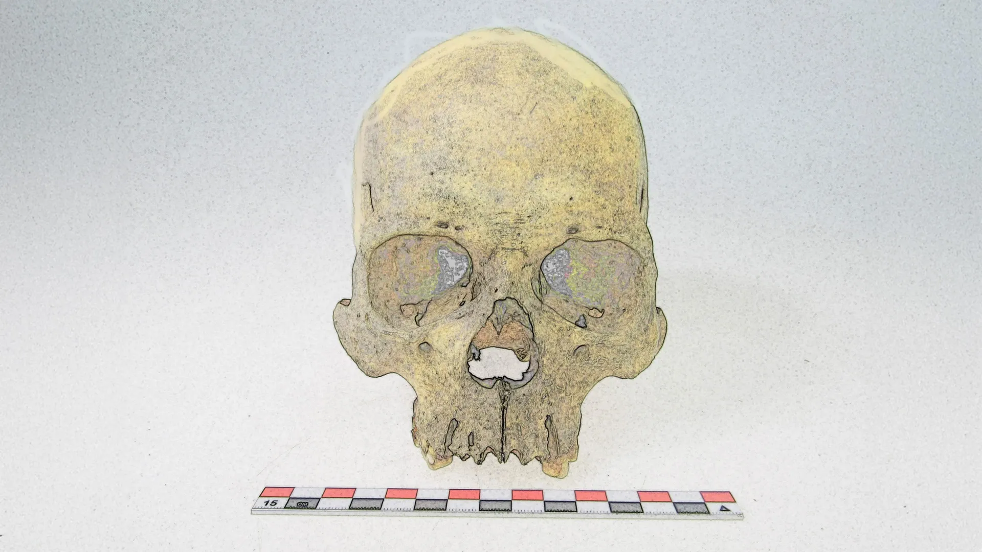
Bos’s team worked closely with researchers from Argentina and Chile to both identify bones suitable for analysis and to carry out the meticulous work of isolating the DNA of ancient pathogens. Doctoral candidate Darío Ramirez of the University of Córdoba, Argentina, worked extensively with such material, and was the first to identify a genetic signature related to leprosy in some 4000-year-old skeletons from Chile. “We were initially suspicious, since leprosy is regarded a colonial-era disease, but more careful evaluation of the DNA revealed the pathogen to be of the lepromatosis form.” This provided the first clue that M. lepromatosis and M. leprae, though nominally both pathogens that cause Hansen’s Disease, might have very different histories. Reconstruction of the genome was key in looking into this. While putting the molecular puzzle of an ancient genome back together is never an easy task, these pathogens in particular had “amazing preservation, which is uncommon in ancient DNA, especially from specimens of that age,” comments Lesley Sitter, a postdoctoral researcher in Bos’s team who carried out the analysis.
The pathogen is related to all known modern forms of M. lepromatosis, but as there are so few genomes available for comparison, there is still much to be learned about it. This work has shown that a pathogen considered rare in a modern context caused disease for thousands of years in the Americas. Rodrigo Nores, professor of Anthropology at the University of Córdoba, Argentina is convinced that more cases, both ancient and modern, will be identified in the coming years: “this disease was present in Chile as early as 4000 years ago, and now that we know it was there, we can specifically look for it in other contexts.” Once more genomes surface, we’ll be able to piece out further details of its history and better understand its global distribution today. The pathogen has recently been discovered in squirrel populations from the United Kingdom and Ireland, but in the Americas it has yet to be found in any species other than humans. With such little data, mystery still surrounds its origin. “It remains to be determined if the disease originated in the Americas, or if it joined some of the first settlers from Eurasia,” adds Bos. “So far the evidence points in the direction of an American origin, but we’ll need more genomes from other time periods and contexts to be sure.”

Read more →

What should you look for when choosing sunscreen?

30 June 2025ShareSaveShareSaveGetty ImagesTemperatures are set to be well above average in many parts of the UK, and UV levels are expected to be high. How can you protect your skin from the sun’s rays and what should you do if you get burned?What do the SPF numbers mean on sunscreen?The most prominent number on sunscreen bottles is the sun protection factor or SPF.The higher the number, the greater the protection it offers.SPF tells you how much protection your sunscreen provides from UVB radiation. The number refers to how much UVB it allows in, not how much it blocks.For example, a sunscreen with SPF 15 allows one-fifteenth of the sun’s rays to reach your skin, or about 7%.So it filters out about 93% percent of UVB rays while SPF 30 filters about 97%.This means if you could stay in the sun for 10 minutes unprotected without burning, SPF 15 would in theory give you 15 times that protection, or two-and-a-half hours before you would burn.The British Association of Dermatologists says sunscreen with SPF 30 is a “satisfactory form of sun protection in addition to protective shade and clothing”.It says sunscreen should be reapplied at least every two hours, regardless of its SPF.What are UVA and UVB and what do the star ratings mean?Many brands also carry a star rating from one to five.This tells you the percentage of UVA radiation that is absorbed by the sunscreen in comparison to how much UVB is absorbed. The more stars the better.UVA and UVB refer to different wavelengths of radiation from the sun that enter the Earth’s atmosphere.UVA is associated with ageing of the skin and pigmentation as well as skin cancer. It can affect human skin even through glass.UVB causes sunburn, and is linked to particular types of skin cancer – basal cell carcinoma (the most common type) and malignant melanoma.A low SPF sunscreen could have a high star rating if the ratio of UVA to UVB protection is the same as in a higher SPF product.Ideally, you want a sunscreen with a high SPF and a high star rating.How effective is the best sunscreen?These levels of protection assume sunscreen has been applied in ideal conditions.In reality, most people do not apply sunscreen perfectly, and it can rub off with sweat or while in water. Experts think most people only apply half the recommended quantity.You should not use sunscreen which is past its expiry date as it may be less effective.Getty ImagesEU guidance states that sunscreen should only be marketed as having sun protection of “50+” and not the ratings of 80 or 100 which can be found in some countries.It thinks these could be misleading about how much extra protection they provide: SPF 50 provides about 98% protection, while SPF 100 would provide less than 100%.No product provides 100% protection from the sun’s rays so the advice is that everyone should cover up and seek shade when the sun is strongest.What about ‘once-a-day’ sunscreens?There are lots of “extended wear” sunscreens on the market that advertise themselves as being for use “once a day”. Many claim to offer protection for up to eight hours – if applied correctly.But some dermatologists recommend that these products should still be applied at least every two hours, like any other sunscreen, since the risk that you may have missed a spot – or that it will rub or wash off in that time – are too high.A Which? report in 2016 criticised four of these products for not providing the protection promised. It found that after six to eight hours, the average protection offered decreased by 74%.But this claim was rejected by some of the manufacturers, who said their own testing had produced different results.What should you do if you get sunburned?Sunburn results in hot and sore skin which may peel after a few days. In extreme cases, skin can blister.It usually gets better within a week or so, but there are things you can do to ease the symptoms.The NHS recommends getting out of the sun as soon as possible, and keeping burned skin covered to protect it from the sun until it has fully healed.A cool shower or a damp towel can help, as can moisturising aftersun products, painkillers and drinking plenty of water. Ask your pharmacist for advice.You should not put petroleum jelly or ice on sunburned skin. You should resist scratching the burned areas and avoid tight-fitting clothes.Contact your GP or NHS 111 if your skin is blistered or swollen or you have other symptoms of heat exhaustion or heatstroke.Regularly getting sunburned can increase the risk of skin cancer.

Read more →

Scientists discover ‘off switch’ enzyme that could stop heart disease and diabetes

Scientists at The University of Texas at Arlington have identified a new enzyme that can be “turned off” to help the body maintain healthy cholesterol levels — a significant development that could lead to new treatments for diseases that affect millions of Americans.
“We found that by blocking the enzyme IDO1, we are able to control the inflammation in immune cells called macrophages,” said Subhrangsu S. Mandal, lead author of a new peer-reviewed study and professor of chemistry at UT Arlington. “Inflammation is linked to so many conditions — everything from heart disease to cancer to diabetes to dementia. By better understanding IDO1 and how to block it, we have the potential to better control inflammation and restore proper cholesterol processing, stopping many of these diseases in their tracks.”
Inflammation plays a crucial role in the immune system, helping the body fight infections and heal injuries. But when inflammation becomes abnormal — due to stress, injury or infection — it can damage cells, disrupt normal functions and increase the risk of serious diseases. During these periods, macrophages can’t absorb cholesterol properly, which can lead to chronic disease.
The team — Dr. Mandal, postdoctoral researcher Avisankar Chini; doctoral students Prarthana Guha, Ashcharya Rishi and Nagashree Bhat; master’s student Angel Covarrubias; and undergraduate researchers Valeria Martinez, Lucine Devejian and Bao Nhi Nguyen — found that the enzyme IDO1 becomes activated during inflammation, producing a substance called kynurenine that interferes with how macrophages process cholesterol.
When IDO1 is blocked, however, macrophages regain their ability to absorb cholesterol. This suggests that reducing IDO1 activity could offer a new way to help prevent heart disease by keeping cholesterol levels in check.
The researchers also found that nitric oxide synthase (NOS), another enzyme linked in inflammation, worsens the effects of IDO1. They believe that inhibiting NOS could provide another potential therapy for managing cholesterol problems driven by inflammation.
“These findings are important because we know too much cholesterol buildup in macrophages can lead to clogged arteries, heart disease and a host of other illnesses,” Mandal said. “Understanding how to prevent the inflammation affecting cholesterol regulation could lead to new treatments for conditions like heart disease, diabetes, cancer and others.”
Next, the research team plans to dig deeper into how IDO1 interacts with cholesterol regulation and whether other enzymes play a role. If they can find a safe way to block IDO1, it could open the door for more effective drugs to prevent inflammation-related diseases.
This work is supported by the National Institutes of Health (1 R15 997 HL170257-01), National Science Foundation (NSF AGEP 998 Award — 2243017), and the Schwartzberg Companies.

Read more →

Fire smoke exposure leaves toxic metals and lasting immune changes

Healthy adults exposed to fire smoke — which can be composed of gases, particulate matter, and toxins — showed alterations to their immune systems, including changes in the regulation of genes associated with asthma and allergies, and in immune cells that play a key role in fighting pathogens, compared to adults who had no exposure to fire smoke. The study is the first to examine the specific immune mechanisms associated with fire smoke exposure, showing cellular-level changes related to smoke’s known negative impacts on respiratory, cardiac, neurological, and pregnancy outcomes. According to the researchers, the findings may pave the way for new therapeutics to mitigate, or prevent altogether, the health effects of smoke exposure and environmental contaminants.Exposure to fire smoke — which can be composed of particulate matter, gases, materials from buildings such as perfluoroalkyl and polyfluoroalkyl substances (PFAS), toxic metals, and carcinogenic compounds — may alter the immune system on a cellular level, according to a new study led by researchers at Harvard T.H. Chan School of Public Health. The study is the first to examine the specific cellular changes associated with fire smoke exposure, documenting how smoke can damage the body through the immune system.”We’ve known that smoke exposure causes poor respiratory, cardiac, neurological, and pregnancy outcomes, but we haven’t understood how,” said corresponding author Kari Nadeau, John Rock Professor of Climate and Population Studies and chair of the Department of Environmental Health. “Our study fills in this knowledge gap, so that clinicians and public health leaders are better equipped to respond to the growing threat of difficult to contain, toxic wildfires.”
The study was published pn June 26 in Nature Medicine.
The researchers collected blood from two cohorts matched by age, sex, and socioeconomic status: 31 smoke-exposed adults, both firefighters and civilians, and 29 non-smoke-exposed adults. None of the participants had an acute or chronic condition, and none were taking immunomodulatory drugs at or before the time of the blood draw, which took place within one month after participants had been exposed to fire smoke.
Using cutting-edge single-cell -omic techniques — epigenetic assays and mass cytometry — and bioinformatic analytical tools, the researchers examined and analyzed individual cells within each blood sample.
The study found several cellular-level changes in the smoke-exposed individuals compared to the non-smoke-exposed individuals. Smoke-exposed individuals showed an increase in memory CD8+ T cells (a type of immune cell critical to long-term immunity against pathogens) and elevated activation and chemokine receptor biomarkers (indicators of inflammation and immune activity) within multiple cell types. Additionally, those who had been exposed to smoke showed changes in133 genes related to allergies and asthma, and more of their immune cells were bound with toxic metals, including mercury and cadmium.

“Our findings demonstrate that the immune system is extremely sensitive to environmental exposures like fire smoke, even in healthy individuals,” said lead author Mary Johnson, principal research scientist in the Department of Environmental Health. “Knowing exactly how may help us detect immune dysfunction from smoke exposure earlier and could pave the way for new therapeutics to mitigate, or prevent altogether, the health effects of smoke exposure and environmental contaminants.”
The researchers also noted that the study could help inform environmental and public health policies and investments. “Knowing more about exactly how smoke exposure is harming the body, we may increase public health campaigns about the dangers of smoke exposure and the importance of following evacuation procedures during wildfires,” Nadeau said. “We may also reconsider what levels of smoke exposure we consider toxic.”
Other Harvard Chan co-authors included Abhinav Kaushik, Olivia Kline, Xiaoying Zhou, and Elisabeth Simonin.
The study was funded by the National Institute of Environmental Health Sciences (R01 ES032253), the National Heart, Lung, and Blood Institute (P01 HL152953, T32HL007118), the National Institute of Allergy and Infectious Diseases (U19AI167903), the San Francisco Cancer Prevention Foundation, the Asthma and Allergic Diseases Cooperative Research Center, and the Keck Foundation.

Read more →

Covid inquiry to look at impact on care services

4 days agoShareSaveAlison HoltJames MelleySenior Social Affairs ProducerShareSaveBBCThe Covid inquiry will start examining the impact of the pandemic on care services for elderly and disabled people on Monday. Bereaved families say they have been waiting for this moment for years, describing the way Covid swept through care homes as one of the clearest and most devastating failures of the pandemic. Nearly 46,000 care home residents died with Covid in England and Wales between March 2020 and January 2022, many of them in the early weeks of the pandemic. The government says it supports the inquiry and is committed to learning lessons from it. There are key questions families and care staff want answering, including why the decision was made in March 2020 to rapidly discharge some hospital patients into care homes.They blame this, in part, for seeding the virus into care homes in the early stages of the pandemic.There are also questions about blanket “do not resuscitate” notices being placed on some care home residents by medical services, and about visiting policies which prevented families seeing their loved ones for months.”It was an awful, awful time,” says Maureen Lewis, manager of St Ives Lodge care home in northeast London.The home cares for up to 35 people, many of whom have dementia. When we visited earlier this month, some of the residents were sitting round a table in the dining area, making brightly coloured collages of flower baskets. Staff and residents sat cheek by jowl chatting to each other.This was a huge contrast to our first visit on 15 April 2020. Then, staff were dressed head to toe in protective suits, their faces covered by masks. It was three weeks after the UK locked down to try to stop Covid spreading. Despite this, St Ives Lodge had just lost six residents in a week.”That was the hardest. And that was right at the very beginning of Covid,” Maureen remembers. On 17 April 2020 the number of deaths in care homes reached its peak, with 540 people dying in England and Wales in one day.St Ives Lodge had locked down in mid March, but a resident who had returned from hospital developed Covid symptoms. Those who sat at the same dining table as him quickly showed the same signs. They died within seven days of each other. The home later lost another resident to the virus.At the time, Maureen described how doctors and district nurses refused to come in, how care homes struggled to get protective equipment (PPE) and there was no helpful guidance from government.”We were like a mini hospital,” she told BBC News, “dealing with end-of-life care… Googling what to do”. It was an experience described by many other care home managers who felt abandoned by the government and NHS in the early months of the pandemic. Looking back, Maureen says: “There were no protocols, no testing and [the NHS] was basically getting rid of any residents back to care homes. They were discharged as normal, but we were in a pandemic.”She wants the inquiry to find out why such decisions were made, and she wants those in positions of responsibility held to account.She remains angry with the former Health and Social Care Secretary, Matt Hancock, for his statement on 15 May 2020, that the government had “thrown a protective ring around care homes”. “There was no ring of protection for care homes at all,” she says. “He needs to take accountability for the decisions he made.”Mr Hancock is due to give evidence within the next few days. It will be his seventh, and likely final appearance.For Jean Adamson, the evidence which will be heard at the inquiry over the coming weeks will be the most important yet.Her father, Aldrick, died with Covid on 15 April 2020. The last time she saw him was when, whilst she stood outside, she glimpsed him through the window of his care home a few days before he passed away. She was devastated she could not be at his side.”We weren’t allowed to say goodbye to him, to hold his hand,” she says. “It’s an experience that will stay with me forever. There is no grief like it.”She too has questions about the policy of discharging patients to care homes. On 17 March 2020 the NHS sent a letter to all hospitals telling them to free up beds. Over the next four weeks, an estimated 25,000 patients were discharged into care homes many were not tested for Covid. On 2 April 2020, the government advised “negative [Covid] tests are not required” before patients were discharged into care homes.This was changed on 15 April 2020, the day Aldrick Adamson died. New government guidance said anyone discharged from hospital to a care setting would be tested first. Jean Adamson believes patients who arrived at her father’s care home from hospital could have seeded the virus there. Other factors such as staff moving between care homes and the spread of the virus in the general community will have played a part but, she says, the rapid discharge of patients to care homes without them being tested or isolated was a choice. “It was a reckless decision,” she says. “The way that my father and tens of thousands of other care home residents were sacrificed. It really gets me because I think it smacks of ageism and disability discrimination.”In the five years since her father’s death she has become an active member of the campaign group, Covid-19 Bereaved Families for Justice UK.She says the group wants the inquiry to look at the ban on care home visits that kept families apart for months.They also want answers to why some people were “routinely placed on do not attempt resuscitation orders [DNARs]”, without discussion with relatives or an assessment.”There was an abuse of the do not attempt resuscitation policy. We need to understand how that came about,” she says.The inquiry will also examine the difficulties faced by care workers who supported people in their own homes.This phase is expected to last five weeks, with the report not due until next year.For both Maureen Lewis and Jean Adamson the memories of the pandemic remain traumatic and both say what they want now is the truth.”We need to understand so that we can learn lessons going forward for when we have the next pandemic and what happened here should never happen again,” says Jean.Maureen would like more recognition of how care services survived without much help. In future, she says: “There needs to be more investment” and better planning for emergencies.

Read more →

The gene that hijacks fear: How PTEN rewires the brain’s anxiety circuit

PTEN Connection to Autism: Up to 25% of those with brain overgrowth and autistic spectrum disorder (ASD) carry variations in a gene called PTEN; PTEN-deficient mouse models exhibit ASD-like characteristics Cell-type Specific Model: PTEN loss in specific neurons leads to circuit imbalance and altered behavior Excitation-Inhibition Imbalance: Strengthened excitatory drive and loss of local inhibitory connections in an amygdala circuit Behavioral effects: This circuit imbalance results in increased fear learning and anxiety in mice — core traits seen in ASD. Researchers at the Max Planck Florida Institute for Neuroscience have discovered how loss of a gene strongly associated with autism and macrocephaly (large head size) rewires circuits and alters behavior. Their findings, published in Frontiers in Cellular Neuroscience, reveal specific circuit changes in the amygdala resulting from PTEN loss in inhibitory neurons, providing new insights into the underlying circuit alterations that contribute to heightened fear and anxiety.
PTEN has emerged as one of the most significant autism risk genes. Variations in this gene are found in a significant proportion of people with autism who also exhibit brain overgrowth, making it a key player in understanding differences in brain function. To investigate the impact of PTEN misregulation, researchers have turned to animal models, where global reduction of PTEN results in altered sociability, repetitive behaviors, and increased anxiety that are often associated with ASD in humans.
But understanding how PTEN dysfunction results in specific circuit and behavioral changes has been difficult in animal models that disrupt PTEN throughout the nervous system. Therefore, MPFI research group leader Dr. McLean Bolton and her team have focused on the changes in the central lateral amygdala driven by loss of PTEN in a critical neuronal population — somatostatin-expressing inhibitory neurons.
Alterations in the function of inhibitory neurons in the development of ASD have been seen through both human tissue studies and genetic mouse models. Moreover, the PTEN gene is known to regulate the development of inhibitory neurons. Therefore, a cell-type-specific disruption of PTEN in inhibitory neurons was a valuable target for understanding specific circuit changes associated with ASD.
“Although a cell-type specific disruption does not replicate the genome-wide changes seen in humans, it is essential to examine how genetic risk factors operate within distinct neural circuits,” explained Dr. Bolton. “Understanding these mechanisms is a crucial step toward targeted interventions for specific traits such as severe anxiety.”
The team, led by Dr. Tim Holford, combined a genetic model that disrupted PTEN only in somatostatin-containing inhibitory neurons with a unique circuit mapping approach previously developed in the lab. This approach measured the electrical responses of individual neurons to the sequential optogenetic activation of hundreds of nearby neurons, allowing rapid mapping of connectivity and strength with the precision of electrical recordings and the scale of imaging approaches.
“This is a powerful method that we can use to determine changes in local neuron connectivity and strength resulting from genetic variations. We were interested in uncovering how the disruption of PTEN signaling in a single cell type would change the way the brain processes information and contribute to the broad ASD phenotype,” described Dr. Holford.
The scientists focused on the circuits in the central amygdala (CeL) – a brain region known to serve as an inhibitory gate on the downstream expression of fear responses – and found striking results. Deleting PTEN specifically in somatostatin-containing interneurons disrupted local inhibitory connectivity in the CeL by roughly 50% and reduced the strength of the inhibitory connections that remained. This diminished connectivity between inhibitory connections within the CeL was contrasted by an increase in the strength of excitatory inputs received from the basolateral amygdala (BLA), a nearby brain region that relays emotionally-relevant sensory information to the CeL.
Behavioral analysis of the genetic model demonstrated that this imbalance in neural signaling was linked to heightened anxiety and increased fear learning, but not alterations in social behavior or repetitive behavior traits commonly observed in ASD. The results not only confirm that PTEN loss in this specific cell type is sufficient to induce specific ASD-like behaviors, but also provide one of the most detailed maps to date of how local inhibitory networks in the amygdala are affected by genetic variations associated with neurological disorders. Importantly, the altered circuitry did not affect all ASD-relevant behaviors — social interactions remained largely intact — suggesting that PTEN-related anxiety and fear behaviors may stem from specific microcircuit changes.
As Dr. Holford explains, “By teasing out the local circuitry underlying specific traits, we hope to differentiate the roles of specific microcircuits within the umbrella of neurological disorders, which may one day help in developing targeted therapeutics for specific cognitive and behavioral characteristics. In future studies, we hope to evaluate these circuits in different genetic models to determine if these microcircuit alterations are convergent changes that underlie heightened fear and anxiety expression across diverse genetic profiles.”

Read more →

Brain scan breakthrough reveals why Parkinson’s drugs don’t always work

Simon Fraser University researchers are using a new approach to brain imaging that could improve how drugs are prescribed to treat Parkinson’s disease.
The new study, published in the journal Movement Disorders, looks at why levodopa – the main drug used in dopamine replacement therapy – is sometimes less effective in patients.
The drug is typically prescribed to help reduce the movement symptoms associated with the neurodegenerative disorder.
While it is effective in improving symptoms for the vast majority of patients, not everyone experiences the same level of benefit.
In order to find out why this is the case, an SFU collaboration with researchers in Sweden has used magnetoencephalography (MEG) technology to determine how the drug affects signals in the brain.
“Parkinson’s is the second most prevalent neurodegenerative disease worldwide and it is the most rapidly increasing, in terms of incidence,” says Alex Wiesman, assistant professor in biomedical physiology and kinesiology at SFU.
“Treating this disease, both in terms of helping people with their symptoms, but also trying to find ways to reverse the effects, is becoming more and more important.

“If clinicians can see how levodopa activates certain parts of the brain in a patient, it can help to inform a more personalised approach to treatment.”
The study was a collaboration with researchers at Karolinska Institute in Sweden, who used MEG to collect data from 17 patients with Parkinson’s disease – a relatively small sample size.
Researchers mapped participants’ brain signals before and after taking the drug, in order to see how and where the drug impacted brain activity.
MEG is an advanced non-invasive technology that measures the magnetic fields produced by the brain’s electrical signals.
It can help clinicians and researchers to study brain disorders and diseases, including brain injuries, tumors, epilepsy, autism, mental illness and more.
Using this rare brain imaging technology, Wiesman and team developed a new analysis that lets them “search” the brain for off-target drug effects.

“With this new way of analyzing brain imaging data, we can track in real time whether or not the drug is affecting the right brain regions and helping patients to manage their symptoms,” says Wiesman.
“What we found was that there’s sometimes ‘off target’ effects of the drug. In other words, we could see the drug activating brain regions we don’t want to be activating and that’s getting in the way of the helpful effects.
“We found that those people who showed ‘off target’ effects are still being helped by the drug, but not to the same extent as others.”
Parkinson’s disease is a neurodegenerative disorder, meaning parts of the brain become progressively damaged over time. It affects predominately the dopamine-producing neurons in a specific area of the brain called the substantia nigra.
People with Parkinson’s disease may experience a range of movement-related symptoms, such as tremors, slow movement, stiffness and balance problems.
Wiesman hopes that a better understanding of how levodopa affects an individual’s brain signals could improve how drugs are prescribed to treat Parkinson’s.
“This might be really helpful for tracking individualized responses to these types of drugs and helping with prescribing and therapeutics,” he says.
“So maybe we try different medications, maybe we adjust dosages differently. And this helps clinicians get at that question of how we prescribe personalized medicine in a way that really helps the patient.
“The more we can personalize that approach, make it more expedient, make it a bit more specific to that person, the better.”
This new type of brain imaging analysis is not only for studying Parkinson’s disease; any medications that affect brain signaling can be studied using the method developed by Wiesman and colleagues.
SFU’s ImageTech Lab, at the Surrey Memorial Hospital, is home to the only MEG in western Canada.
“We have this really fantastic technology right here at SFU, and combined with the new analysis approaches that we’re developing, it gives us a really unprecedented look into what’s happening in the brain,” says Wiesman.
“We can use this technology moving forward to study Parkinson’s disease in ways that no one has ever done before worldwide.
“Our next step is to take our new approach and apply it to a larger patient group. We also need to translate this research to more accessible brain imaging methods, like electroencephalogram (EEG).
“Ultimately, we want to make sure this technology is useful for a diverse population and more widely accessible to patients with Parkinson’s disease.”

Read more →

This brain scan sees Alzheimer’s coming—but only in some brains

A team of researchers from the Keck School of Medicine of USC’s Mark and Mary Stevens Neuroimaging and Informatics Institute (Stevens INI) has identified a new brain imaging benchmark that may improve how researchers classify biologically meaningful changes associated with Alzheimer’s disease, especially in Hispanic and non-Hispanic White populations. The new study, published in Imaging Neuroscience, is part of the Health and Aging Brain Study-Health Disparities (HABS-HD), a multi-university collaboration led by the University of North Texas Health Science Center and supported by the National Institute on Aging.
Using an advanced brain imaging scan called tau PET, the research team studied over 675 older adults from HABS-HD, aiming to identify the optimal brain signal that distinguishes individuals with clinically-relevant biological markers of AD from those who are aging normally.
Tau PET enables researchers to visualize abnormal proteins in the brain which contribute to Alzheimer’s disease, known as tau, by using a small amount of a special radioactive tracer that highlights areas where tau has accumulated. With these scans, researchers can establish tau cut-points, a new type of biomarker used to determine whether a scan shows an amount of tau protein in the brain high enough to suggest possible early signs of Alzheimer’s disease or related conditions. This new benchmark could eventually inform the way clinicians interpret tau PET scans and better identify who may be at risk for AD.
In this study, researchers compared tau PET scans of study participants who were cognitively impaired with those who were not impaired based on cognitive tests to establish a tau cut-point that would indicate a higher risk for Alzheimer’s disease. They found one — but it was only effective in certain circumstances.
“Our tau cut-point was able to distinguish whether study participants had cognitive impairment – but only when another abnormal protein, amyloid, was also present in those with cognitive impairment, and only in Hispanic and non-Hispanic White participants,” said senior author Meredith N. Braskie, PhD, assistant professor of neurology. “In non-Hispanic Black participants, the tau cut-point did not perform as expected. This suggests that other pathologies or conditions may be driving cognitive decline in this group. Our study is an important step toward better understanding how tau relates to cognition in diverse populations and has important implications for future clinical trials that aim to target tau.”
The team used a new imaging tracer called 18F-PI-2620, to measure tau protein buildup in the brain. They found that when tau levels in the medial temporal lobe­ — a region deep in the brain — exceeded a certain threshold, it strongly indicated cognitive impairment related to AD.
“While our findings support prior research linking medial temporal lobe tau to cognitive impairment, establishing a cut-point in this region using 18F-PI-2620, marks an important step toward defining tau positivity for both research and clinical applications. At the same time, the limited reliability of tau as an indicator of cognitive impairment in non-Hispanic Black participants highlights the need for more diverse populations in research and for future studies to examine both biological and social determinants of Alzheimer’s disease,” said lead author Victoria R. Tennant, a PhD candidate in USC’s Neuroscience Graduate Program.

The findings reflect a growing focus in AD research on making sure diagnostic tools work for everyone — not just in narrow clinical trial populations. Alzheimer’s disease is known to affect the brain in stages. While amyloid plaques often build up early, tau tangles are more closely tied to memory loss and other symptoms.
“This type of imaging is critical for understanding who is at risk and how the disease develops,” said Stevens INI director Arthur W. Toga, PhD. “These findings are just the latest to come from HABS-HD, which is the most comprehensive study of Alzheimer’s disease and related dementias in diverse communities. HABS-HD has already produced key findings related to ethnic variations in AD biomarkers, the influences of social determinants on cognitive health, and vascular contributions to dementia, just to name a few. We hope this work will lead to more personalized care and better outcomes for all communities.”
In addition to Tennant and Braskie, the study’s other authors are Koral V. Wheeler, Noelle N. Lee, Jamie A. Terner, Maxwell W. Hand, Suchita Ganesan, Patrick Walsh, Aisha Greene, Tyler Berkness, Tiantian Lei, Arthur W. Toga from the Mark and Mary Stevens Neuroimaging and Informatics Institute, Keck School of Medicine of USC, University of Southern California; Rema Raman and Robert A. Rissman from the Alzheimer’s Therapeutic Research Institute, Keck School of Medicine of USC, University of Southern California; Bradley T. Christian from the Waisman Center, University of Wisconsin-Madison; Melissa Petersen, Ann D. Cohen, Karin L. Meeker, Zhengyang Zhou, Rajesh R. Nandy and Sid E. O’Bryant from the University of North Texas Health Science Center at Fort Worth; Beau M. Ances from the Washington University School of Medicine in St. Louis; and Kristine Yaffe from the Department of Psychiatry, Neurology, and Epidemiology/Biostatistics, University of California, San Francisco.
This research was supported by the National Institute on Aging of the National Institutes of Health [R01AG054073, R01AG058533, R01AG070862, P41EB015922, and U19AG078109], and by the Office of the Director, the National Institutes of Health, [S10OD032285].

Read more →

Make healthy food more appealing, government tells supermarkets

3 hours agoShareSaveNick TriggleShareSaveGetty ImagesFood businesses must make it easier for customers to buy healthy food, under new government plans announced on Sunday. Supermarkets and food manufacturers in England will partner with the government to tackle obesity rates by encouraging people to make their weekly shop healthier.Ministers say it will be up to food retailers to decide how they do that, but it could involve offering promotions on healthy food, tweaking loyalty points to incentivise healthy options, or changing shop layouts.It may also involve changing products to make them healthier.It is hoped the plans will help to make healthy eating more accessible to customers and relieve pressure on the NHS. The policy will see major food retailers report on healthy food sales in a bid to increase accountability, but it does not impose specific targets. The plan is part of a raft of measures aimed at improving the health of the nation that will be included in the 10-year plan for the NHS in England, which is expected to be published next week.”Unless we curb the rising tide of cost and demand, the NHS risks becoming unsustainable,” Health Secretary Wes Streeting said. “This government’s ambition for kids today is for them to be part of the healthiest generation of children ever. That is within our grasp.”The upcoming report will show that more than one in five children are living with obesity by the time they leave primary school, rising to almost one in three in the most deprived areas. A recent report found a basket of healthy food costs more than double that of less healthy options.The Food Foundation reported that 1,000 calories of healthy food such as fruit and veg costs £8.80, compared to £4.30 for the equivalent amount of less healthy food, such as ready meals and processed meats.Andrew Opie of the British Retail Consortium said the news is “really positive” but said “all food businesses” must work together to drive down obesity. “Engaging all food businesses makes a difference,” Opie told the BBC.”It can’t just be about supermarkets- we consume about a quarter of our calories outside the home, so unless we get all supermarkets, food retailers, and restaurants on board, we won’t move the dial on obesity.”Opie said that he agreed with the government’s flexible approach on the standards, as businesses have “a lot of insights and data” into how we shop for food and what would be best for their customers. Katharine Jenner, director of the Obesity Health Alliance said: “The government has rightly identified the root cause of obesity-related ill health: a food system that makes healthy eating difficult.”She said it was encouraging to see the government putting the spotlight on businesses “rather than placing the burden on individuals who are already struggling to get by.”Alongside the new partnership, the government said it will also: Offer shopping vouchers to customers in return for being active and eating healthily, via a new appDouble the number of spaces on the NHS Digital Weight Management Programme, which supports obese individuals with either diabetes or high blood pressureIntroduce stricter rules on advertising alcohol to in line with current regulation on promoting junk foodIt is hoped the new guidance will reduce people’s sugar and calorie intake overall. The government cited research showing that cutting just 50 calories a day would lift 340,000 children and two million adults out of obesity.If everyone who is overweight reduced their calorie intake by just 216 calories a day, which is equivalent to a single bottle of fizzy drink, obesity would be halved, researchers said. This is not the first time the government has sought voluntary partnerships with industry. Over the last 20 years there has been numerous food reformulation programmes aimed at reducing salt, sugar and calories in certain foods.Success has been mixed with a push to reduce sugar in certain foods by 20% between 2015 and 2020 falling well short.Sarah Woolnough, from The King’s Fund think tank, said while welcome, the impact of the scheme may be limited.“A lot of less healthy food and drink is purchased from local convenience shops and takeaways. “The stark fact remains that unhealthy food is far cheaper and more readily available and so unless this change is part of a wider, comprehensive strategy it will not be enough.”Additional reporting by Rachel Muller-Heyndyk

Read more →

School sun safety lessons trialled to tackle skin cancer

5 hours agoShareSaveSophie van BrugenBBC NewsReporting fromLondonShareSaveBBCChildren as young as five are being taught how to check UV levels and apply sunscreen, as part of a new pilot aimed at reducing future skin cancer cases.The initiative, currently being trialled in primary schools across Kent, is planned to be introduced into Personal, Social, Health and Economic (PSHE) lessons nationwide from 2026.Melanoma, the most serious form of skin cancer, is linked to UV exposure – and experts say sunburn in childhood significantly increases the risk later in life.In 2021, around 18,300 people were diagnosed with melanoma in the UK, according to Cancer Research. That figure is projected to rise to 21,300 by 2026, the charity says.Michelle Baker, from the charity Melanoma Fund, which is behind the education project, said changing children’s habits early is key. “People think melanoma is an older person’s disease,” she says, “but it’s often seeded in childhood.”She says the project aims to “grow skin cancer out of the next generation”. Giving children a sense of control and responsibility for their sun protection is central to this. “We’re saying this is your superpower,” she adds.At Platt Primary school in Maidstone, pupils are learning to read UV indexes, apply sunscreen properly, and understand when they need protection.Headteacher Emma Smith said the pupils have been “really receptive”.”If we educate them early, they’re more likely to keep that knowledge as they get older – especially when social media starts to influence their choices.”No ‘safe tan’ The childhood sun safety drive comes as Cancer Research UK says the rise in melanoma cases among adults is a particular cause for concern.A recent study from the charity found that cancer deaths cost the UK economy £10.3bn a year, more than any other health condition – underlining the importance of preventative measures.Consultant dermatologist Dr Katie Lacy says that educating children about how to look after their skin is key to reducing melanoma rates.Research shows that nine out of 10 cases are caused by UV exposure from the sun and sunbeds.Getting sunburnt regularly increases the chances of skin cancer. Dr Lacy stresses there is no such thing as a “safe tan”, explaining that tanning is a response to skin damage.”Most melanomas don’t come from existing moles – so if you notice something new, get it checked,” she adds.She also highlights the growing role of AI in screening suspicious moles within the NHS – which could help streamline referrals to specialist services.The ABCDE checklist can help identify if a mole is abnormal:A – asymmetrical (does the mole have an uneven shape?)B – border (are the edges blurry or jagged?)C – colour (is it an uneven colour with different shades and tones?)D – diameter (is the mole bigger than your other ones?)E – evolving (is it changing, such as starting to itch, bleed or become crusty?)Source: Cancer Research UKFor Kara Leece, diagnosed with melanoma at 29, the message is personal.”If I’d had that education at primary school, I think I could’ve prevented it,” she told BBC News.”Now I have a scar that reminds me of what I’ve been through. When children ask about it, I tell them my story – because I don’t want this to happen to anyone else.”Tips for keeping kids sun safeTry to keep children in the shade between 11am and 3pm, when the sun is at its strongestKit them out with wide-brimmed hats, sunglasses and clothing to cover their skin. Sunscreen is also very important – apply it regularly and generously, and make sure it’s at least SPF 30 and four or five starsSource: Cancer Research UK

Read more →