Multivitamin improves memory in older adults, study finds

Taking a daily multivitamin supplement can slow age-related memory decline, finds a large study led by researchers at Columbia University and Brigham and Women’s Hospital/Harvard.
“Cognitive aging is a top health concern for older adults, and this study suggests that there may be a simple, inexpensive way to help older adults slow down memory decline,” says study leader Adam M. Brickman, PhD, professor of neuropsychology at Columbia University Vagelos College of Physicians and Surgeons.
Many older people take vitamins or dietary supplements under the assumption that they will help maintain general health. But studies that have tested whether they improve memory and brain function have been mixed, and very few large-scale, randomized trials have been done.
Study methods
In the current study, more than 3,500 adults (mostly non-Hispanic white) over age 60 were randomly assigned to take a daily multivitamin supplement or placebo for three years. At the end of each year, participants performed a series of online cognitive assessments at home designed to test memory function of the hippocampus, an area of the brain that is affected by normal aging. The COSMOS-Web study is part of a large clinical trial led by Brigham & Women’s Hospital and Harvard called the COcoa Supplement and Multivitamin Outcomes Study (COSMOS).
By the end of the first year, memory improved for people taking a daily multivitamin, compared with those taking a placebo. The researchers estimate the improvement, which was sustained over the three-year study period, was equivalent to about three years of age-related memory decline. The effect was more pronounced in participants with underlying cardiovascular disease.

The results of the new study are consistent with another recent COSMOS study of more than 2,200 older adults that found that taking a daily multivitamin improved overall cognition, memory recall, and attention, effects that were also more pronounced in those with underlying cardiovascular disease.
“There is evidence that people with cardiovascular disease may have lower micronutrient levels that multivitamins may correct, but we don’t really know right now why the effect is stronger in this group,” says Brickman.
Good nutrition important for aging brain
Though the researchers did not look at whether any specific component of the multivitamin supplement was linked to the improvement in memory, the findings support growing evidence that nutrition is important for optimizing brain health as we age.
“Our study shows that the aging brain may be more sensitive to nutrition than we realized, though it may not be so important to find out which specific nutrient helps slow age-related cognitive decline,” says Lok-Kin Yeung, PhD, a postdoctoral researcher in Columbia’s Taub Institute for Research on Alzheimer’s Disease and the Aging Brain and first author of the study.

“The finding that a daily multivitamin improved memory in two separate cognition studies in the COSMOS randomized trial is remarkable, suggesting that multivitamin supplementation holds promise as a safe, accessible, and affordable approach to protecting cognitive health in older adults,” says co-author JoAnn Manson, MD, chief of the Division of Preventive Medicine at Brigham and Women’s Hospital.
“Supplementation of any kind shouldn’t take the place of more holistic ways of getting the same micronutrients,” adds Brickman. “Though multivitamins are generally safe, people should always consult a physician before taking them.”
More information
The study, titled “Multivitamin supplementation improves memory in older adults: A randomized clinical trial,” was published in the American Journal of Clinical Nutrition.
All authors: Lok-Kin Yeung (Columbia), Daniel M. Alschuler (New York State Psychiatric Institute), Melanie Wall (Columbia), Heike Luttman-Gibson (Brigham and Women’s Hospital/Harvard), Trisha Copeland (Brigham and Women’s/Harvard), Richard P. Sloan (Columbia), Howard D. Sesso (Brigham and Women’s/Harvard), JoAnn E. Manson (Brigham and Women’s/Harvard), and Adam M. Brickman (Columbia).
Dr. Manson and Dr. Sesso are co-leaders of the parent COSMOS trial.
The study was supported by grants from Mars Edge, a segment of Mars Inc., and the National Institutes of Health (AG050657, AG071611, EY025623, and HL157665).
Multivitamins were supplied by Pfizer. Dr. Sesso reported receiving investigator-initiated grants from Pure Encapsulations and Pfizer and/or travel funds for lectures from the Council for Responsible Nutrition, BASF, NIH, and the American Society of Nutrition during the study.

Read more →

New chemical compound demonstrates potential in nerve regeneration

Research led by UCL, in partnership with the MRC Laboratory of Molecular Biology (MRC LMB) and AstraZeneca, has identified a new compound that can stimulate nerve regeneration after injury, as well as protect cardiac tissue from the sort of damage seen in heart attack.
The study, published in Nature, identified a chemical compound, named ‘1938’, that activates the PI3K signalling pathway, and is involved in cell growth. Results from this early research showed the compound increased neuron growth in nerve cells, and in animal models, it reduced heart tissue damage after major trauma and regenerated lost motor function in a model of nerve injury.
Though further research is needed to translate these findings into the clinic, 1938 is one of just a few compounds in development that can promote nerve regeneration, for which there are currently no approved medicines.
Phosphoinositide 3-kinase (PI3K) is a type of enzyme that helps to control cell growth. It is active in various situations, such as initiating wound healing, but its functions can also be hijacked by cancer cells to allow them to proliferate. As a result, cancer drugs have been developed that inhibit PI3K to restrict tumour growth. But the clinical potential of activating the PI3K pathway remains underexplored.
Dr Roger Williams, a senior author of the study from the MRC Laboratory of Molecular Biology, said: “Kinases are ‘molecular machines’ that are key to controlling the activities of our cells, and they are targets for a wide range of drugs. Our aim was to find activators of one of these molecular machines, with the goal of making the machine work better. We found that we can directly activate a kinase with a small molecule to achieve therapeutic benefits in protecting hearts from injury and stimulating neural regeneration in animal studies.”
In this study, researchers from UCL and MRC LMB worked with researchers from AstraZeneca to screen thousands of molecules from its chemical compound library to create one that could activate the PI3K signalling pathway. They found that the compound named 1938 was able to activate PI3K reliably and its biological effect were assessed through experiments on cardiac tissue and nerve cells.

Researchers at UCL’s Hatter Cardiovascular Institute found that administering 1938 during the first 15 minutes of blood flow restoration following a heart attack provided substantial tissue protection in a preclinical model. Ordinarily, areas of dead tissue form when blood flow is restored that can lead to heart problems later in life.
When 1938 was added to lab-grown nerve cells, neuron growth was significantly increased. A rat model with a sciatic nerve injury was also tested, with delivery of 1938 to the injured nerve resulting in increased recovery in the hind leg muscle, indicative of nerve regeneration.
Professor James Phillips (UCL School of Pharmacy), a senior author of the study, said: “There are currently no approved medicines to regenerate nerves, which can be damaged as a result of injury or disease, so there’s a huge unmet need. Our results show that there’s potential for drugs that activate PI3K to accelerate nerve regeneration and, crucially, localised delivery methods could avoid issues with off-target effects that have seen other compounds fail.”
Given the positive findings, the group is now working to develop new therapies for peripheral nerve damage, such as those sustained in serious hand and arm injuries. They are also exploring whether PI3K activators could be used to help treat damage in the central nervous system, for example due to spinal cord injury, stroke or neurodegenerative disease.
Professor Bart Vanhaesebroeck (UCL Cancer Institute), a senior author of the study, said: “This is a prime example of interdisciplinary research, in which people with expertise ranging from basic science, drug development and clinical studies join forces around an innovative idea, whilst also crossing boundaries between academia and industry. ‘Blue sky’ research of this kind is difficult to get funding for in a world of increasing specialisation, but hopefully this project can provide something of a model for future ambitious research.”
An important factor in the overall success of the study was UCL’s Drug Discovery Group from the Translational Research Office supporting the drug discovery programme and participation in AstraZeneca’s ‘Open Innovation’ programme, which sees the company collaborating with academics that have innovative ideas to advance drug discovery and development.
Mike Snowden, Senior Vice President, Discovery Sciences at AstraZeneca, said: “Our Open Innovation programme aims to create an open research environment that connects our expertise and technologies with the innovative and ambitious research ideas of collaborators like UCL and MRC LMB, with the aim of uncovering novel biology and biological mechanisms.”
This research was funded by Wellcome, UKRI, MRC, NIHR UCLH Biomedical Research Centre, European Union Horizon 2020, the British Heart Foundation, the Rosetrees Trust and CRUK.

Read more →

Where do our limbs come from?

An international collaboration that includes scientists from the University of Colorado School of Medicine has uncovered new clues about the origin of paired appendages — a major evolutionary step that remains unresolved and highly debated.
The researchers describe their study in an article published today in the journal Nature.
“This has become a topic that comes with bit of controversy, but it’s really a very fundamental question in evolutionary biology: Where do our limbs come from?” says co-corresponding author Christian Mosimann, PhD, associate professor and Johnson Chair in the Department of Pediatrics, Section of Developmental Biology at CU School of Medicine.
That question — where do our limbs come from? — has been subject of debate for more than 100 years. In 1878, German scientist Carl Gegenbaur proposed that paired fins derived from a source called the gill arch, which are bony loops present in fish to support their gills. Other scientists favor the lateral fin fold hypothesis, concluding that lateral fins on the top and bottom of the fish are the source of paired fins.
“It is a highly active research topic because it’s been an intellectual challenge for such a long time,” Mosimann says. “Many big labs have studied the various aspects of how our limbs develop and have evolved.” Among those labs are Dr. Mosimann’s colleagues and co-authors, Tom Carney, PhD, and his team at the Lee Kong Chian School of Medicine at Nanyang Technological University in Singapore.
Chasing the odd cells
For Mosimann, the inquiry into where limbs come from is an offshoot of other research conducted by his laboratory on the CU Anschutz Medical Campus. In his laboratory, his team uses zebrafish as a model to understand the development from cells to organs. He and his team study how cells decide their fate, looking for explanations for how development can go awry leading to congenital anomalies, in particular cardiovascular and connective tissue diseases.

Along the way, Mosimann and his lab team observed how a peculiar cell type with features of connective tissue cells, so-called fibroblasts that share a developmental origin with the cardiovascular system, migrated into specific developing fins of the zebrafish. It turns out that these cells may support a connection between the competing theories of paired appendage evolution.
“We always knew these cells were odd,” he says. “There were these fibroblast-looking cells that went into the so-called ventral fin, the fin at the belly of the developing zebrafish. Similar fibroblast cells didn’t crawl into any other fin except the pectoral fin, which are the equivalent of our arms. So we kept noticing these peculiar fibroblasts, and we could never make sense of what these were for many years.”
The Mosimann lab has developed several techniques to track cell fates during development in pursuit of their main topic, which is an improved understanding of how the embryonic cell layer, called the lateral plate mesoderm, contributes to diverse organs. The lateral plate mesoderm is the developmental origin of the heart, blood vessels, kidneys, connective tissue, as well as major parts of limbs.
The paired fins that form the equivalent of our arms and legs are seeded by cells from the lateral plate mesoderm, while other fins are not. Understanding how these particular fins became more limb-like has been at the core of a long-standing debate.
Developing new theories
Hannah Moran, who is pursuing her PhD in the Cell Biology, Stem Cells and Development program in the Mosimann lab, adapted a method of tracking lateral plate mesoderm cells that contribute to heart development so that researchers could track the peculiar fibroblasts related to limb development.

“My primary research project focuses on the development of the heart rather than limb development,” Moran says, “but there was a genetic technique that I had adapted to map early heart cells, and so we were able to implement that into mapping where the mysterious cells of the ventral fin came from. And turns out, they are also from the lateral plate mesoderm.”
This crucial discovery provides a new puzzle piece to the big picture of how we evolved our arms and legs. Increasing evidence supports a hypothesis of paired appendage evolution called the dual origin theory.
“Our data fit nicely into this combined theory, but it can also stand on its own with the lateral fin theory,” says Robert Lalonde, PhD, postdoctoral fellow in the Mosimann lab. “While paired appendages arise from the lateral plate mesoderm, that does not rule out an ancient connection to unpaired, lateral fins.”
By observing the mechanisms of embryonic development and comparing the anatomy of existing species, research groups like Mosimann’s can develop theories on how embryonic structures may have evolved or have been modified over time.
“The embryo has features that are still ancient remnants that they have not lost yet, which provides insight into how animals have evolved,” Mosimann says. “We can use the embryo to learn more about features that just persist today, allowing us to kind of travel back in time,” Mosimann says. “We see that the body has a fundamental, inherent propensity to form bilateral, two-sided structures. Our study provides a molecular and genetic puzzle piece to resolve how we came to have limbs. It adds to this 100-plus year discussion, but now we have molecular insights.”
International collaboration
Collaborations with colleagues in laboratories across the country and around the world are another important part of the study. Those scientists bring additional specializations and contribute data from other models, including paddlefish, African clawed frogs, and a variant of split-tail goldfish called Ranchu, to study embryonic development.
“There are labs on this on this paper that work on musculoskeletal diseases, toxicology, fibrosis. We work on cardiovascular, congenital anomalies, cardiopulmonary anomalies, limb development, all related to our interest on the lateral plate mesoderm,” says Mosimann. “And then together, you get to make such fundamental discoveries. And that’s where team science enables us to do something that is more than just the sum of the parts.”
For all the considerable work and significance of the study, the Mosimann team recognizes that it is a key step, but not the end of the journey in the debate about paired appendages.
“I wouldn’t say we’ve solved the question, or even disproven either existing theory,” says Lalonde. “Rather, we’ve contributed meaningful data towards answering a major evolutionary question.”

Read more →

SWI/SNF complexes 'bookmark' cell identity during division

When a cell divides, it retains information about how to grow and instructions about what type of cell to become. Scientists at St. Jude Children’s Research Hospital have gained a new understanding of how these processes can work, revealing a previously unappreciated role for the SWI/SNF chromatin remodeling complex. The study was published today in Nature.
When a cell undergoes differentiation, stem cells (the earliest cells that develop) undergo changes that transform them into a different type of cell, typically one with a more specialized function (such as a skin or muscle cell). As cells divide, they must retain the “memory” of their current differentiation state to transfer the proper identity to the daughter cells.
Chromatin is a complex of DNA and protein tightly compacted inside cells. Chromatin must unwind to turn genes on and off in closely regulated processes. SWI/SNF complexes control a cell’s identity during differentiation by changing chromatin architecture to regulate gene expression. However, it was unknown whether SWI/SNF complexes contribute to the memory of cell identity during cell division.
Cancers often carry mutations that affect the SWI/SNF chromatin remodeling complex. One example is the loss of the core subunit SMARCB1. Other studies have also linked mutations in the complex to neurodevelopmental disorders. In this study, scientists at St. Jude have discovered how subunits of SWI/SNF act as “bookmarks” during mitosis to safeguard cell identity during division. The work points to the importance of SWI/SNF core subunits SMARCE1 and SMARCB1 and their roles in the process.
“This work provides an understanding of a new component of mitotic memory, as well as provides clues to why a mutation of this SWI/SNF complex subunit would disrupt memory of what a cell should normally be doing and allow it to go into a cancerous state,” said senior author Charles W.M. Roberts, M.D., Ph.D., Executive Vice President and St. Jude Comprehensive Cancer Center director.
SWI/SNF subunits help cells “remember”
Previously, researchers believed the enzymatically active subunit of the SWI/SNF complex was not bound to DNA in mitosis, so scientists have assumed that SWI/SNF complexes had no role during mitosis. In launching this study, Roberts wondered, “If a piece of this complex is mutated, how can a cancer cell remember to be a cancer cell coming out on the other side of mitosis? What is the memory mechanism sustaining the cancer cell?”

Surprisingly, Roberts’s team found that two individual subunits of SWI/SNF complexes, but not the rest of the complex, bind to mitotic DNA. They then showed that the binding of SMARCE1 and SMARCB1 is required for the appropriate reactivation of bound genes after mitosis.
Further experiments, which removed SMARCE1 during mitosis, found that loss of SMARCE1 disrupts gene expression, impairs the ability of some other “bookmarks” to bind to their targets and causes abnormal neural differentiation. These findings suggest that SMARCE1 plays a mitotic bookmarking role and is essential for the retention of appropriate differentiation programming during mitosis.
“In a normal cell, SMARCB1 would be bound in mitosis to bookmark the genes that should be turned on after the cell divides,” Roberts said. “But SMARCB1 is deleted in nearly all cases of a highly lethal type of cancer that strikes young children called rhabdoid tumors. Consequently cell identity genes fail to turn on immediately after a cell divides. This may be a key component that enables the cells to stay in a cancerous state, as they fail to activate the genes that normally help them differentiate.”
Implications beyond pediatric cancer
Previous work showed that SWI/SNF subunit abnormalities exist in approximately 20% of all cancers. Additionally, research has identified SWI/SNF subunit mutations in several types of neurodevelopmental diseases.

“History has shown that genes that mutate in the earliest onset cancers, in babies and toddlers, are often a bit of a ‘canary in a coal mine’ for broader principles in cancer.” Roberts said. “Indeed, while it was studies of rhabdoid tumors in young children that first linked SWI/SNF complexes to cancer, we now know that genes that encode subunits of SWI/SNF complexes are mutated in 20% of all cancers. Studying rhabdoid tumors, thus, not only provides insight into these often fatal cancers of young children, it is quite informative about many types of cancer.”
“Unlike adult cancers, where many genes are mutated, and it is harder to figure out what any single abnormality does, here we have a cancer driven by just this one mutation,” Roberts added. “It’s a beautiful model to understand how these processes work and then begin to leverage that understanding in adult cancers too.”
Authors and funding
The study’s co-first and co-corresponding author is Zhexin Zhu, formerly of the St. Jude Department of Oncology. The other co-first author is Xiaolong Chen, St. Jude Department of Computational Biology. Other authors are Ao Guo, Trishabelle Manzano, Patrick Walsh, Kendall Wills, Rebecca Halliburton, Sandi Radko-Juettner, Raymond Carter, Janet Partridge, Douglas Green and Jinghui Zhang of St. Jude.
The work was part of the St. Jude Collaborative Research Consortium on Chromatin Regulation in Pediatric Cancer. Through the collaborative, investigators at different institutions conduct research that requires the expertise of scientists with various specialties, streamlining and speeding up progress.
The study was also supported by the National Institutes of Health (R01CA216391, R01 AI123322), the National Cancer Institute (NCI) Cancer Center Support Grant (CCSG 2 P30 CA021765), NCI grants (R01CA113794 and R01CA172152), CURE AT/RT Now, Garret B. Smith Foundation, the Ruth L. Kirschstein National Research Service Award (F31 CA261150) and ALSAC, the fundraising and awareness organization of St. Jude.

Read more →

Discovery slows down muscular dystrophy

A team of researchers at the University of Houston College of Pharmacy is reporting that by manipulating TAK1, a signaling protein that plays an important role in development of the immune system, they can slow down disease progression and improve muscle function in Duchenne muscular dystrophy (DMD).
DMD, caused by mutations in dystrophin gene, is an inheritable neuromuscular disorder that occurs in one out of 3,600 male births. DMD patients undergo severe muscle wasting, inability to walk and eventually death in their early thirties due to respiratory failure. The disease is marked by an inflammatory response and death of muscle fibers. Eventually, the muscle fibers are replaced with fat and fibrotic tissue that causes severe muscle weakness.
“Our results suggest that TAK1 (transforming growth factor β-activated kinase1) is a regulator of skeletal muscle mass. By specifically targeting this protein, we can suppress the death of muscle fibers, known as myonecrosis, and slow down disease progression in DMD,” said Ashok Kumar, Else and Philip Hargrove Endowed Professor and chair, Department of Pharmacological and Pharmaceutical Sciences, whose results were published in JCI Insight. “Our research shows that activating TAK1 can stimulate myofiber growth in a model of DMD, with no negative impact on muscle health.”
In a previous breakthrough, Kumar’s team uncovered a surprising fact: TAK1 is essential for maintaining skeletal muscle mass and that activating TAK1 beyond normal levels can enhance skeletal muscle growth.
For this research, supported by the National Institutes of Health, the team designed experiments to reduce or augment the levels of TAK1 protein in skeletal muscle at different stages of disease progression.
“Our experiments demonstrate that depletion of TAK1 activity during peak necrotic phase followed by re-introduction of TAK1 at post-necrotic phase leads to substantial improvement in muscle pathology,” said Anirban Roy, research assistant professor.
The current standard of care for DMD is focused on reducing inflammation with corticosteroids, which modestly reduces disease progression, but has serious side effects.
“Accumulating evidence suggests that regulation of immune response, autophagy, and metabolism along with gene correction therapy can be promising approaches to slow down disease progression in DMD patients,” said Roy.

Read more →

Consistent link between the seaside and better health

Seaside residents and holidaymakers have felt it for centuries, but scientists have only recently started to investigate possible health benefits of the coast. Using data from 15 countries, new research led by Sandra Geiger from the Environmental Psychology Group at the University of Vienna confirms public intuition: Living near, but especially visiting, the seaside is associated with better health regardless of country or personal income.
The idea that being near the ocean may boost health is not new. As early as 1660, doctors in England began promoting sea bathing and coastal walks for health benefits. By the mid-1800s, taking ‘the waters’ or ‘sea air’ were widely promoted as health treatment among wealthier European citizens. Technological advances in medicine in the early 20th century led to the decline in such practices, which are only recently gaining popularity again among the medical profession.
As part of the EU-funded Horizon 2020 project Seas, Oceans, and Public Health In Europe led by Professor Lora Fleming, Geiger and colleagues from the Universities of Vienna, Exeter, and Birmingham, as well as Seascape Belgium and the European Marine Board, surveyed over 15,000 participants across 14 European countries (Belgium, Bulgaria, Czechia, France, Germany, Greece, Ireland, Italy, the Netherlands, Norway, Poland, Portugal, Spain, the United Kingdom) and Australia about their opinions on various marine-related activities and their own health.
The findings, published in the journal Communications Earth & Environment, surprised the team. Lead author Geiger said: “It is striking to see such consistent and clear patterns across all 15 countries. We also now demonstrate that everybody seems to benefit from being near the seaside, not just the wealthy. Although the associations are relatively small, living near and especially visiting the coast can still have substantial effects on population health.”
Understanding the potential benefits of coastal access for all members of society is key for policymaking. Dr. Paula Kellett from the European Marine Board said: “The substantial health benefits of equal and sustainable access to our coasts should be considered when countries develop their marine spatial plans, consider future housing needs, and develop public transportation links.”
But what does this mean for landlocked residents like Geiger and her colleagues in Austria? “Austrians and other central Europeans visit the coasts in their millions during the summer months, so they too get to experience some of these benefits. Besides, we are also starting to appreciate the similar health benefits offered by inland waters such as lakes and natural pools.”

Read more →

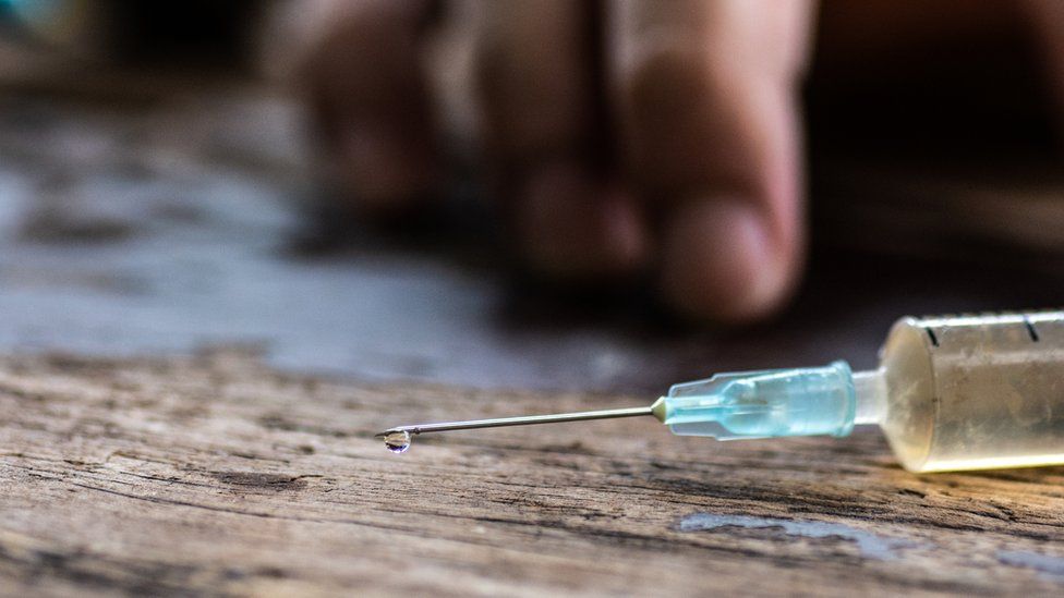
First UK death linked to 'zombie' drug xylazine

Published16 hours agoShareclose panelShare pageCopy linkAbout sharingImage source, Getty ImagesBy Philippa RoxbyHealth reporterThe death of a 43-year-old man is the first in the UK to be linked to the “zombie” drug xylazine, which is prompting overdose warnings in the US.Normally used as a large-animal tranquiliser but now being found in heroin, it can cause a dangerously low heart rate and large open skin wounds.UK experts are calling it “a really concerning drug”.They say drug users should be warned it is now present in the UK but there is no safe dose in humans.’Bought heroin’Karl Warburton, form Solihull, West Midlands, died in May 2022 at home and had a history of illicit drug use, according to the coroner’s report. He had been referred to addiction services on a number of occasions.An examination of his body detected heroin, fentanyl and cocaine in his system, as well as xylazine.A report on his death in the Journal of Forensic and Legal Medicine says he was “likely to have bought heroin and not known it was laced with xylazine and fentanyl”.”To the best of our knowledge, this is the first death associated with xylazine use reported in the UK, and even Europe, and indicates the entry of xylazine into the UK drug supply,” it adds.Skin ulcersXylazine is used by vets as a powerful sedative but it’s not approved for use in humans.Known as “tranq” or “tranq dope” when cut with heroin and fentanyl by drug dealers, xylazine has been causing huge problems in the US.If injected directly into someone’s bloodstream, it can cause large open skin ulcers to form. These can start to rot and lead to amputation.It also lowers breathing and heart rate to dangerously low levels, which has led to it being dubbed a “flesh-eating zombie drug”.Xylazine emerged on the illicit drug market in Puerto Rico in the early 2000s and has since been found in the US, mainly in the east, and in Canada.The US government has called it “an emerging threat” because of its growing role in fatal overdoses across the country – about 7% of the total.And in some states, the drug was found in more than a quarter of overdoses.But until now, there has been no sign of xylazine in the UK.The drug was detected only because the Birmingham lab that carried out tests after the man’s death noticed some strange results and identified xylaxine.Death certificate”The drug is not included in standard drug screens in the UK, so we don’t know how widespread the xylazine problem is,” said Dr Caroline Copeland, King’s College London lecturer and director of the National Programme on Substance Abuse Deaths.”We need to find out how that person ended up with it in his system.” Xylazine was listed on the man’s death certificate as contributing to his death but there was no way of recording it in the UK drug-deaths database.The report highlights the need to monitor changes in illicit-drug markets and in emerging drugs.”There is no safe dose to use”, it concludes, because there is an overlap between fatal and non-fatal doses of xylazine reported in people.

Read more →

Surgeon General Warns That Social Media May Harm Children and Adolescents

The report by Dr. Vivek Murthy cited a “profound risk of harm” to adolescent mental health and urged families to set limits and governments to set tougher standards for use.The nation’s top health official issued an extraordinary public warning on Tuesday about the risks of social media to young people, urging a push to fully understand the possible “harm to the mental health and well-being of children and adolescents.”In a 19-page advisory, the United States surgeon general, Dr. Vivek Murthy, noted that the effects of social media on adolescent mental health were not fully understood, and that social media can be beneficial to some users. Nonetheless, he wrote, “There are ample indicators that social media can also have a profound risk of harm to the mental health and well-being of children and adolescents.”The report included practical recommendations to help families guide children’s social media use. It recommended that families keep mealtimes and in-person gatherings free of devices to help build social bonds and promote conversation. It suggested creating a “family media plan” to set expectations for social media use, including boundaries around content and keeping personal information private.Dr. Murthy also called on tech companies to enforce minimum age limits and to create default settings for children with high safety and privacy standards. And he urged the government to create age-appropriate health and safety standards for technology platforms.Adolescents “are not just smaller adults,” Dr. Murthy said in an interview on Monday. “They’re in a different phase of development, and they’re in a critical phase of brain development.”The report, effectively elevating long-simmering concerns around social media in the national conversation, came as state and federal lawmakers, many of them raised in an era when social media barely existed or didn’t exist at all, have been struggling with how to set limits on its use.Montana’s governor recently signed a bill banning TikTok from operating in the state, prompting the Chinese-owned app to file a lawsuit and young TikTok users to lament what one called a “kick in the face.” In March, Utah became the first state to prohibit social media services from allowing users under 18 to have accounts without the explicit consent of a parent or guardian. That law could dramatically curtail young people’s access to apps like Instagram and Facebook.Survey results from Pew Research have found that up to 95 percent of teens reported using at least one social media platform, while more than one-third said they used social media “almost constantly.” As social media use has risen, so have self-reports and clinical diagnoses among adolescents of anxiety and depression, along with emergency room visits for self-harm and suicidal ideation.The report could help encourage further research to understand whether these two trends are related. It joins a growing number of calls for action around adolescents and social media. Earlier this month, the American Psychological Association issued its first-ever social media guidance, recommending that parents closely monitor teens’ usage and that tech companies reconsider features like endless scrolling and the “like” button.A large body of research has emerged in recent years on the potential connection between social media use and soaring rates of distress among adolescents. But the results have been consistent only in their nuance and complexity.An analysis published last year, examining research from 2019 to 2021 on social media use and mental health, found that “most reviews interpreted the associations between social media use and mental health as ‘weak’ or ‘inconsistent,’ whereas a few qualified the same associations as ‘substantial’ and ‘deleterious.’”At their clearest, the data indicate that social media can have both a positive and negative impact on the well-being of young people, and that heavy use of social media — and screen time generally — appears to displace activities like sleep and exercise that are considered vital to developing brains.On the positive side, social media can help many young people by giving them a forum to connect with others, find community and express themselves.At the same time, the surgeon general’s advisory noted, social media platforms brim with “extreme, inappropriate and harmful content,” including content that “can normalize” self-harm, eating disorders and other self-destructive behavior. Cyberbullying is rampant. Moreover, social media spaces can be fraught for young people especially, the advisory added: “In early adolescence, when identities and sense of self-worth are forming, brain development is especially susceptible to social pressures, peer opinions and peer comparison.”The advisory noted that technology companies have a vested interest in keeping users online, and that they use tactics that entice people to engage in addictive-like behaviors. “Our children have become unknowing participants in a decades-long experiment,” the advisory states.A spokesperson for Meta, the owner of Instagram and Facebook, said that the advisory included recommendations that “are reasonable and, in large part, Meta has already implemented.” Those measures include automatically making the accounts of people under 16 private when they join Instagram and limiting the types of content teens can see on the app.TikTok did not immediately respond to requests for comment on Tuesday afternoon.The advisory did not provide guidance on what a healthy use of social media might look like, nor did it condemn social-media use for all young people. Rather, it concluded, “We do not yet have enough evidence to determine if social media is sufficiently safe for children and adolescents.”The surgeon general’s position lacks any real power beyond its potential as a bully pulpit, and Dr. Murthy’s advisory does not carry the force of law or policy. It was intended, the report said, to call Americans’ attention to “an urgent public health issue” and to make recommendations for how it should be addressed.Similar reports from past surgeons general helped to shift the national conversation around smoking in the 1960s, drew attention to H.I.V. and AIDS in the 1980s and declared in the early 2000s that obesity had become a nationwide epidemic. Dr. Murthy has declared gun violence to be an epidemic and has decried what he has called a “public health crisis of loneliness, isolation, and the lack of connection in our country.”In the interview on Monday, Dr. Murthy acknowledged that the lack of clarity around social media was a heavy burden for users and families to bear.“That’s a lot to ask of parents, to take a new technology that’s rapidly evolving and that fundamentally changes how kids perceive themselves,” Dr. Murthy said. “So we’ve got to do what we do in other areas where we have product safety issues, which is to set in place safety standards that parents can rely on, that are actually enforced.”Remy Tumin

Read more →

Vaping: How do you quit e-cigarettes?

Published24 May 2023Shareclose panelShare pageCopy linkAbout sharingImage source, Getty ImagesQuitting ain’t easy. But vaping’s helped millions of people to ditch traditional cigarettes.It’s widely considered less harmful than smoking tobacco – and the UK government endorses vapes as a quitting aid.But what do you do when you want to take the next step and quit completely?Why is vaping so addictive?The main reason vaping gets you hooked is nicotine – the same ingredient that makes smokers crave cigarettes.It’s thought to be pretty harmless on its own, but is powerfully addictive. That’s why vaping isn’t recommended for non-smokers.Nicotine creates new receptors in the brain when it enters the body, and you quickly come to associate it with feeling good.So when you stop getting it you’ll feel cravings. These tend to be worse in the first three to four days after your last dose.According to the NHS, other withdrawal symptoms, like bad moods, are likely in the early stages of quitting.How long does it take to quit vaping?There’s no firm answer to this – it varies for each person – but there are some things we do know.It’s technically possible to go “cold turkey” and suddenly stop vaping. But most people find this very difficult.Experts, such as the National Centre for Smoking Cessation and Training (NCSCT), generally recommend some sort of gradual approach.One tip for vapers is to reduce the nicotine strength of their e-liquid in two to four-week stages, working down to 0% solutions.This can be trickier with disposable vapes, which tend to come in higher strengths.But zero-nicotine versions are available, and the NCSCT says using one alongside your regular vape can help.Failing that, a temporary switch to a tank-style vape, giving you access to different e-liquids, might be better. Image source, Getty ImagesOther reduction strategies include:Extending the time between vapesTaking shorter puffs (especially if you use a disposable)Setting rules for yourself, like only vaping outside the homeAnother option is Nicotine Replacement Therapy (NRT) – patches, gum and sprays used instead of a vape.Nicotine pouches that go between the gum and lip are starting to appear on some shop shelves but these aren’t currently recommended by the NHS.Pull the triggersLike smoking, if you’ve been vaping a long time you’re likely to have formed habits around it.Certain situations or times of day – triggers – will have got you used to reaching for your vape. This can be hard to snap out of, so it can help to figure out what they are.How easy is it to buy an illegal vape?High lead and nickel levels found in illegal vapesVaping – is it a risk-free option?It’s also a good idea to try and fill this time with something else to distract you, like exercise.If you are a former smoker, the NCSCT says it’s worth keeping a vape on hand for emergencies.Because, if you do relapse, it’s better than reaching for a cigarette.Follow Newsbeat on Twitter and YouTube.Listen to Newsbeat live at 12:45 and 17:45 weekdays – or listen back hereMore on this storyHigh lead and nickel levels found in illegal vapesPublished23 May 2023Free vapes to be handed out in anti-smoking drivePublished11 April 2023Vaping – is it a risk-free option?Published24 June 2022

Read more →