Diet tracking: How much is enough to lose weight?

Keeping track of everything you eat and drink in a day is a tedious task that is tough to keep up with over time. Unfortunately, dutiful tracking is a vital component for successful weight loss, however, a new study in Obesity finds that perfect tracking is not needed to achieve significant weight loss.
Researchers from UConn, the University of Florida, and the University of Pennsylvania tracked 153 weight loss program participants for six months where users self-reported their food intake using a commercial digital weight loss program. The researchers wanted to see what the optimal thresholds were for diet tracking to predict 3%, 5%, and 10% weight loss after six months.
“We partnered with WeightWatchers, who was planning on releasing a new Personal Points program, and they wanted to get empirical data via our clinical trial,” says co-author and Department of Allied Health Sciences Professor Sherry Pagoto.
Pagoto explains that the new program takes a personalized approach to assigning points including a list of zero-point foods to eliminate the need for calculating calories for everything,
“Dietary tracking is a cornerstone of all weight loss interventions, and it tends to be the biggest predictor of outcomes. This program lowers the burden of that task by allowing zero-point foods, which do not need to be tracked.”
Researchers and developers are seeking ways to make the tracking process less burdensome, because as Pagoto says, for a lot of programs, users may feel like they need to count calories for the rest of their lives: “That’s just not sustainable. Do users need to track everything every single day or not necessarily?”
With six months of data, Assistant Professor in the Department of Allied Health Sciences Ran Xu was interested to see if there was a way to predict outcomes based on how much diet tracking participants did. Ran Xu and Allied Health Sciences Ph.D. student Richard Bannor analyzed the data to see if there were patterns associated with weight loss success from a data science perspective. Using a method called receiver operating characteristics (ROC) curve analysis they found how many days people need to track their food to reach clinically significant weight loss.

“It turns out, you don’t need to track 100% each day to be successful,” says Xu. “Specifically in this trial, we find that people only need to track around 30% of the days to lose more than 3% weight and 40% of the days to lose more than 5% weight, or almost 70% of days to lose more than 10% weight. The key point here is that you don’t need to track every day to lose a clinically significant amount of weight.”
This is promising since Pagoto points out that the goal for a six-month weight loss program is typically 5% to 10%, a range where health benefits have been seen in clinical trials.
“A lot of times people feel like they need to lose 50 pounds to get healthier, but actually we start to see changes in things like blood pressure, lipids, cardiovascular disease risk, and diabetes risk when people lose about 5-to-10% of their weight,” says Pagoto. “That can be accomplished if participants lose about one to two pounds a week, which is considered a healthy pace of weight loss.”
Xu then looked at trajectories of diet tracking over the six months of the program.
The researchers found three distinct trajectories. One they call high trackers, or super users, who tracked food on most days of the week throughout six months, and on average lost around 10% of their weight.

However, many participants belonged to a second group that started tracking regularly, before their tracking gradually declined over time to, by the four-month mark, only about one day per week. They still lost about 5% of their weight.
A third group, called the low trackers, started tracking only three days a week, and dropped to zero by three months, where they stayed for the rest of the intervention. On average this group lost only 2% of their weight.
“One thing that is interesting about this data is, oftentimes in the literature, researchers just look at whether there is a correlation between tracking and overall weight loss outcomes. Ran took a data science approach to the data and found there is more to the story,” Pagoto says. “Now we’re seeing different patterns of tracking. This will help us identify when to provide extra assistance and who will need it the most.”
The patterns could help inform future programs which could be tailored to help improve user tracking based on which group they fall into. Future studies will dig deeper into these patterns to understand why they arise and hopefully develop interventions to improve outcomes.
“For me, what’s exciting about these digital programs is that we have a digital footprint of participant behavior,” says Xu. “We can drill down to the nitty-gritty of what people do during these programs. The data can inform precision medicine approaches, where we can take this data science perspective, identify patterns of behavior, and design a targeted approach.”
Digitally delivered health programs give researchers multitudes of data they never had before which can yield new insights, but this science requires a multidisciplinary approach.
“Before, it felt like we were flying in the dark or just going by anecdotes or self-reported measures, but it’s different now that we have so much user data. We need data science to make sense of all these data. This is where team science is so important because clinical and data scientists think about the problem from very different perspectives, but together, we can produce insights that neither of us could do on our own. This must be the future of this work,” says Pagoto.
Xu agrees: “From a data science perspective, machine learning is exciting but if we just have machine learning, we only know what people do, but we don’t know why or what to do with this information. That’s where we need clinical scientists like Sherry to make sense of these results. That’s why team science is so important.”
No longer flying in the dark, these multi-disciplinary teams of researchers now have the tools needed to start tailoring programs even further to help people achieve their desired outcomes. For now, users of these apps can be assured that they can still get significant results, even if they miss some entries.
Read more →

Study unravels the mysteries of actin filament polarity

Actin filaments — protein structures critical to living movement from single cells to animals — have long been known to have polarity associated with their physical characteristics, with growing “barbed” and shrinking “pointed” ends. The ends of the filament are also different in the way they interact with other proteins in cells. However, the mechanism that determines these differences has never been entirely clear to scientists. Now, researchers from the Perelman School of Medicine at the University of Pennsylvania have revealed key atomic structures of the ends of the actin filament through the use of a technique called cryo-electron microscopy (cryo-EM). The study, published in Science, provides fundamental insights that may help fill in details behind disorders affecting some muscle, bone, heart, neurological, and immune disorders that are the result of actin defects or deficiencies.
Actin is the most abundant protein inside the cells of higher organisms, such as animals. It serves as the building-block for long, thin structures called filaments, which provide key structural support as part of the cell “cytoskeleton,” the system that gives cells their shape and polarity. Rapid changes in actin filaments underlie key cellular events such as movement along surfaces, cell-to-cell contact, and cell division. Actin filaments also are major elements in muscle fibers.
“The results of our study provide a mechanistic understanding of a process we have known about for more than 40 years, referred to as filament treadmilling, and impacts how we view the cellular roles of actin in health and disease,” said the study senior author Roberto Dominguez, PhD, the William Maul Measey Presidential Professor of Physiology at Penn.
The dynamics of actin filaments are governed largely by the “treadmilling” process, through which individual actin proteins are shed from one filament end, known as the pointed end, and added at the other, barbed end. Actin filaments can be stabilized by distinct so-called “capping” proteins that bind to the filament ends to stop further addition or loss of individual actin proteins. Many other proteins also bind to the barbed and pointed ends of the actin filament. But the structural details determining the specificity of these interactions — the details that explain why these two ends function so differently — have been murky.
In their study, the researchers, including two Penn students — Peter Carman, PhD, a recent graduate student in Dominguez’s lab, and Kyle Barrie, PhD, a graduate student currently in the lab, who served as co-first authors — analyzed actin filaments using cryo-EM. With this high-resolution imaging technique, a researcher obtains many thousands of snapshots of a target molecule, aligns them computationally, and then averages them to reduce random image “noise” — yielding a 3-D reconstruction of the molecule that may be sharp enough to visualize individual atoms.
With artificial intelligence (AI) assistance, the researchers were able to focus on the ends of the filaments instead of their middle, as had previously been the norm in similar research. By doing so, they identified hundreds of thousands of filament end views, allowing them to obtain near-atomic scale reconstructions. These revealed a “flat” actin shape, or conformation, at the uncapped barbed end, versus a “twisted” conformation at the uncapped pointed end.
The data also detailed the structural changes induced by two actin filament-capping proteins, CapZ at the barbed end and tropomodulin at the pointed end. These are the two proteins found at the ends of the filament in skeletal and cardiac muscles, playing an essential role in the stabilization of actin filaments in muscle fibers, and, without these proteins, our muscles would fall apart.
Results from this study provide crucial mechanistic details for a deeper understanding of actin biology as a whole. The researchers believe these study insights should also be helpful in understanding and ultimately treating disorders caused by actin dysfunction.
Funding was provided by the National Institutes of Health (R01 GM073791, F31 HL156431).

Read more →

Colorful fresh foods improve athletes' vision

Nutrition is an important part of any top athlete’s training program. And now, a new study by researchers from the University of Georgia proposes that supplementing the diet of athletes with colorful fruits and vegetables could improve their visual range.
The paper, which was published in Exercise and Sport Sciences Reviews, examines how a group of plant compounds that build up in the retina, known as macular pigments, work to improve eye health and functional vision.
Previous studies done by UGA researchers Billy R. Hammond and Lisa Renzi-Hammond have shown that eating foods like dark leafy greens or yellow and orange vegetables, which contain high levels of the plant compounds lutein and zeaxanthin, improves eye and brain health.
“A lot of the research into macular lutein and zeaxanthin has focused on health benefits, but from a functional perspective, higher concentrations of these plant pigments improve many aspects of visual and cognitive ability. In this paper, we discuss their ability to improve vision in the far distance or visual range,” said lead author Jack Harth, a doctoral candidate in UGA’s College of Public Health.
Visual range, or how well a person can see a target clearly over distance, is a critical asset for top athletes in almost any sport.
The reason why objects get harder to see and appear fuzzier the farther they are from our eyes is thanks in part to the effects of blue light.

“From a center fielder’s perspective, if that ball’s coming up in the air, it will be seen against a background of bright blue sky, or against a gray background if it’s a cloudy day. Either way, the target is obscured by atmospheric interference coming into that path of the light,” said Harth.
Many athletes already take measures to reduce the impact of blue light through eye black or blue blocker sunglasses, but eating more foods rich in lutein and zeaxanthin can improve the eye’s natural ability to handle blue light exposure, said Harth.
When a person absorbs lutein and zeaxanthin, the compounds collect as yellow pigments in the retina and act as a filter to prevent blue light from entering the eye.
Previous work had been done testing the visual range ability of pilots in the 1980s, and Hammond and Renzi-Hammond have done more recent studies on how macular pigment density, or how much yellow pigment is built up in the retina, is linked to a number of measures of eye health and functional vision tests.
“In a long series of studies, we have shown that increasing amounts of lutein and zeaxanthin in the retina and brain decrease glare disability and discomfort and improve chromatic contrast and visual-motor reaction time, and supplementing these compounds facilitates executive functions like problem-solving and memory. All of these tasks are particularly important for athletes,” said corresponding author Billy R. Hammond, a professor of psychology in the Behavior and Brain Sciences Program at UGA’s Franklin College of Arts and Sciences.

This paper, Harth said, brings the research on these links between macular pigment and functional vision up to date and asks what the evidence suggests about optimizing athletic performance.
“We’re at a point where we can say we’ve seen visual range differences in pilots that match the differences found in modeling, and now, we’ve also seen it in laboratory tests, and a future goal would be to actually bring people outside and to measure their ability to see contrast over distance through real blue haze and in outdoor environments,” said Harth.
But before you start chowing down on kale in the hopes of improving your game, he cautions that everybody is different. That could mean the way our bodies absorb and use lutein and zeaxanthin varies, and it could take a while before you notice any improvements, if at all.
Still, the evidence of the overall health benefits of consuming more lutein and zeaxanthin are reason enough to add more color to your diet, say the authors.
“We have data from modeling and empirical studies showing that higher macular pigment in your retina will improve your ability to see over distance. The application for athletes is clear,” said Harth.

Read more →

Dentists identify new bacterial species involved in tooth decay

Philadelphia — Collaborating researchers from the University of Pennsylvania School of Dental Medicine and the Adams School of Dentistry and Gillings School of Global Public Health at the University of North Carolina have discovered that a bacterial species called Selenomonas sputigena can have a major role in causing tooth decay.
Scientists have long considered another bacterial species, the plaque-forming, acid-making Streptococcus mutans, as the principal cause of tooth decay — also known as dental caries. However, in the study, which appeared 22 May in Nature Communications, the Penn Dental Medicine and UNC researchers showed that S. sputigena, previously associated only with gum disease, can work as a key partner of S. mutans, greatly enhancing its cavity-making power.
“This was an unexpected finding that gives us new insights into the development of caries, highlights potential future targets for cavity prevention, and reveals novel mechanisms of bacterial biofilm formation that may be relevant in other clinical contexts,” said study co-senior author Hyun (Michel) Koo DDS, PhD, a professor in the Department of Orthodontics and Divisions of Pediatrics and Community Oral Health and Co-Director of the Center for Innovation & Precision Dentistry at Penn Dental Medicine.
The other two co-senior authors of the study were Kimon Divaris, PhD, DDS, professor at UNC’s Adams School of Dentistry, and Di Wu, PhD, associate professor at the Adams School and at the UNC Gillings School of Global Public Health.
“This was a perfect example of collaborative science that couldn’t have been done without the complementary expertise of many groups and individual investigators and trainees,” Divaris said.
Caries is considered the most common chronic disease in children and adults in the U.S. and worldwide. It arises when S. mutans and other acid-making bacteria are insufficiently removed by teeth-brushing and other oral care methods, and end up forming a protective biofilm, or “plaque,” on teeth. Within plaque, these bacteria consume sugars from drinks or food, converting them to acids. If the plaque is left in place for too long, these acids start to erode the enamel of affected teeth, in time creating cavities.

Scientists in past studies of plaque bacterial contents have identified a variety of other species in addition to S. mutans. These include species of Selenomonas, an “anaerobic,” non oxygen-requiring group of bacteria that are more commonly found beneath the gum in cases of gum disease. But the new study is the first to identify a cavity-causing role for a specific Selenomonas species.
The UNC researchers took samples of plaque from the teeth of 300 children aged 3-5 years, half of whom had caries, and, with key assistance from Koo’s laboratory, analyzed the samples using an array of advanced tests. The tests included sequencing of bacterial gene activity in the samples, analyses of the biological pathways implied by this bacterial activity, and even direct microscopic imaging. The researchers then validated their findings on a further set of 116 plaque samples from 3 to 5-year-olds.
The data showed that although S. sputigena is only one of several caries-linked bacterial species in plaque besides S. mutans, and does not cause caries on its own, it has a striking ability to partner with S. mutans to boost the caries process.
S. mutans is known to use available sugar to build sticky constructions called glucans that are part of the protective plaque environment. The researchers observed that S. sputigena, which possesses small appendages allowing it to move across surfaces, can become trapped by these glucans. Once trapped, S. sputigena proliferates rapidly, using its own cells to make honeycomb-shaped “superstructures” that encapsulate and protect S. mutans. The result of this unexpected partnership, as the researchers showed using animal models, is a greatly increased and concentrated production of acid, which significantly worsens caries severity.
The findings, Koo said, show a more complex microbial interaction than was thought to occur, and provide a better understanding of how childhood cavities develop — an understanding that could lead to better ways of preventing cavities.

“Disrupting these protective S. sputigena superstructures using specific enzymes or more precise and effective methods of tooth-brushing could be one approach,” Koo said.
The researchers now plan to study in more detail how this anaerobic motile bacterium ends up in the aerobic environment of the tooth surface.
“This phenomenon in which a bacterium from one type of environment moves into a new environment and interacts with the bacteria living there, building these remarkable superstructures, should be of broad interest to microbiologists,” Koo said.
“Selenomonas sputigena acts as a pathobiont mediating spatial structure and biofilm virulence in early childhood caries” was co-authored by Hunyong Cho, Zhi Ren, Kimon Divaris, Jeffrey Roach, Bridget Lin, Chuwen Liu, M. Andrea Azcarate-Peril, Miguel Simancas-Pallares, Poojan Shrestha, Alena Orlenko, Jeannie Ginnis, Kari North, Andrea Ferreira Zandona, Apoena Aguiar Ribeiro, Di Wu and Hyun “Michel” Koo.
The work was funded in part by the National Institutes of Health (U01DE025046, R01DE025220, R03DE028983).

Read more →

How chronic stress drives the brain to crave comfort food

When you’re stressed, a high-calorie snack may seem like a comforting go-to. But this combination has an unhealthy downside. According to Sydney scientists, stress combined with calorie-dense ‘comfort’ food creates changes in the brain that drive more eating, boost cravings for sweet, highly palatable food and lead to excess weight gain.
A team from the Garvan Institute of Medical Research found that stress overrode the brain’s natural response to satiety, leading to non-stop reward signals that promote eating more highly palatable food. This occurred in a part of the brain called the lateral habenula, which when activated usually dampens these reward signals.
“Our findings reveal stress can override a natural brain response that diminishes the pleasure gained from eating — meaning the brain is continuously rewarded to eat,” says Professor Herzog, senior author of the study and Visiting Scientist at the Garvan Institute.
“We showed that chronic stress, combined with a high-calorie diet, can drive more and more food intake as well as a preference for sweet, highly palatable food, thereby promoting weight gain and obesity. This research highlights how crucial a healthy diet is during times of stress.”
The research was published in the journal Neuron.
From stressed brain to weight gain
While some people eat less during times of stress, most will eat more than usual and choose calorie-rich options high in sugar and fat.

To understand what drives these eating habits, the team investigated in mouse models how different areas in the brain responded to chronic stress under various diets.
“We discovered that an area known as the lateral habenula, which is normally involved in switching off the brain’s reward response, was active in mice on a short-term, high-fat diet to protect the animal from overeating. However, when mice were chronically stressed, this part of the brain remained silent — allowing the reward signals to stay active and encourage feeding for pleasure, no longer responding to satiety regulatory signals,” explains first author Dr Kenny Chi Kin Ip from the Garvan Institute.
“We found that stressed mice on a high-fat diet gained twice as much weight as mice on the same diet that were not stressed.”
The researchers discovered that at the centre of the weight gain was the molecule NPY, which the brain produces naturally in response to stress. When the researchers blocked NPY from activating brain cells in the lateral habenula in stressed mice on a high-fat diet, the mice consumed less comfort food, resulting in less weight gain.
Driving comfort eating
The researchers next performed a ‘sucralose preference test’ — allowing mice to choose to drink either water or water that had been artificially sweetened.

“Stressed mice on a high-fat diet consumed three times more sucralose than mice that were on a high-fat diet alone, suggesting that stress not only activates more reward when eating but specifically drives a craving for sweet, palatable food,” says Professor Herzog.
“Crucially, we did not see this preference for sweetened water in stressed mice that were on a regular diet.”
Stress overrides healthy energy balance
“In stressful situations it’s easy to use a lot of energy and the feeling of reward can calm you down — this is when a boost of energy through food is useful. But when experienced over long periods of time, stress appears to change the equation, driving eating that is bad for the body long term,” says Professor Herzog.
The researchers say their findings identify stress as a critical regulator of eating habits that can override the brain’s natural ability to balance energy needs.
“This research emphasises just how much stress can compromise a healthy energy metabolism,” says Professor Herzog. “It’s a reminder to avoid a stressful lifestyle, and crucially — if you are dealing with long-term stress — try to eat a healthy diet and lock away the junk food.”

Read more →

'Revolutionary' research discovers new cause of cancer coming from inside us

Australian cancer researchers have made an important new connection between a person’s cancer risk and the functions of circular RNAs, a recently discovered family of genetic fragments present within our cells.
A new Flinders University-led study published in Cancer Cell, one of the world’s top cancer journals, finds that specific circular RNAs within many of us can stick to the DNA in our cells and cause DNA mutations which result in cancer.
“While environmental and genetic factors have long been believed the major contributors to cancer, this revolutionary finding — which we call ‘ER3D’ (from ‘endogenous RNA directed DNA damage’) — ushers in an entirely new area of medical and molecular biology research,” says Flinders University Professor Simon Conn, who leads the Circular RNAs in Cancer Laboratory at the Flinders Health and Medical Research Institute.
“This is the first example of a genetic molecule present within many of us which has the capacity to mutate our very own DNA and drive cancer from inside.
“This opens the door to use these molecules as new therapeutic targets and markers of disease at a very early stage, when the likelihood of curing cancers is much higher.”
The research compared the neonatal blood tests or Guthrie cards of babies who went on to develop acute leukemia as infants with children without any blood disorders. This found that one specific circular RNA was present at much higher levels at birth, prior to onset of the symptoms of leukemia.

The findings suggest it is the abundance of the circular RNA molecules within certain individuals’ cells which is a major determinant for why they develop these specific cancer-causing genes or oncogenes and other do not.
“Circular RNAs can bind to DNA at many different locations across a range of cells. By binding to the DNA at specific sites, these circular RNAs cause a number of changes culminating in the breakage of the DNA which the cell must repair in order to survive,” says Professor Conn.
“This repair is not always perfect and this can result in small mutations, like a misspelt word within a book, or worse, very, very large and devastating mutations.
“With the circular RNAs also able to alter the physical location of the broken DNA within the cell nucleus, two distinct regions of the DNA can be stuck together during the repair process — like the ripping of two different books and sticking them together.”
Lead author Dr Vanessa Conn says multiple circular RNAs appear to act in partnership causing breaks at multiple sites in the DNA.

“This process, called chromosomal translocation, is a major problem for the cell as it results in gene fusions which can actually convert the cell from a normal cell into a cancerous cell,” she says.
“This was demonstrated in two different cell types and it was found that this drove the rapid onset of aggressive leukemia.”
The gene fusions arising from the action of these circular RNAs are at well-known ‘hotspots’ of mutation in the blood cancer leukemia. This is an important consideration in Australia which has the highest incidence of leukemia in the world, with around 35,000 Australians currently living with this disease.
These gene fusions have been used by doctors around the world for many years in guiding treatment options as they are known to worsen the prognosis for the patient who carries them, the researchers say.
However, until now it was unknown how these mutations arose, even though more than 100 known fusions were found in patients.
“Not surprisingly, it is not only leukemia where the process of ER3D occurs,” says Dr Conn.
“We now have evidence that ER3D is not restricted to leukemia but to other cancers and human diseases,” she says.
The Flinders University research team is continuing the study to investigate circular RNAs’ role in cancer and other diseases.

Read more →

Single-cell atlas of the whole human lung

The largest and most comprehensive cell map of the human lung is announced in Nature Medicine today (8 June). Revealing the great diversity of cell types in the lung and key differences between health and disease, the Human Lung Cell Atlas will be a valuable resource for lung researchers.
By combining data from nearly 40 studies, researchers created the first integrated single-cell atlas of the lung, revealing rare cell types and highlighting cellular differences between healthy people. In addition, the study found common cell states between lung fibrosis, cancer and COVID-19, offering new ways of understanding lung disease, which could help identify new therapeutic targets.
The study is part of the global Human Cell Atlas* (HCA) initiative to map every cell type in the human body, to transform our understanding of health, infection and disease.
Lung research has benefited greatly from recent single-cell studies that show which genes are active in each cell. Despite this, the research has been limited so far by the number of samples and individuals included per study. To better understand healthy lungs and determine what goes wrong in disease, a comprehensive atlas has been needed, however this has been difficult to achieve.
Now, a large team of researchers has successfully combined 49 lung datasets, from nearly 40 separate studies, into a single integrated Atlas, using advanced machine learning. By pooling and integrating datasets from every major single-cell RNA-sequencing lung study published to date, the team created the first integrated Human Lung Cell Atlas. This Atlas spans over 2.4 million cells from 486 individuals and gives new insights into lung biology that were not possible before.
Dr Malte Luecken, a senior author on the paper and Group leader at the Institute of Computational Biology and the Institute of Lung Health and Immunity at Helmholtz Munich, Germany, said: “A comprehensive organ atlas requires many datasets to capture the diversity between both cells and individuals, but combining different datasets is a huge challenge. We developed a benchmarking pipeline to find the optimal method to integrate all datasets into the Atlas, using artificial intelligence, and successfully combined knowledge and data from almost 40 previous lung studies.”
Professor Fabian Theis, a senior author on the paper and Director of the Institute of Computational Biology at Helmholtz Munich, said: “We have created a first reference atlas of the human lung, which includes data from more than a hundred healthy people and reveals how the cells from individuals vary with age, sex, and smoking history. The sheer numbers of cells and individuals involved now gives the power to see rare cell types and identify new cell states that have not previously been described.”

While the core of the Human Lung Cell Atlas is data from healthy lungs, the team also took datasets from more than 10 different lung diseases and projected these onto the healthy data, to understand disease states.
The team discovered that different lung diseases shared common immune cell states, including the finding that a subset of macrophages (a type of immune cell) shared similar gene activity in lung fibrosis, cancer and COVID-19. The shared states indicate that these cells could play a similar role in scar formation in the lung in all three diseases, and provide pointers for potential therapeutic targets.
Professor Martijn Nawijn, a senior author on the paper and Professor at the University Medical Center Groningen, the Netherlands, said: “This is the first effort to compare healthy and diseased lungs in one study in an integrated way. Our study not only supports the presence of lung fibrosis in COVID-19, it allows us to identify and define a shared cell state between lung fibrosis, COVID-19 and lung cancer patients. Finding these shared disease-associated cells is really exciting, and reveals a totally different way of looking at lung diseases, opening possibilities for novel treatment targets and developing treatment response biomarkers. Our findings also suggest that therapies working for one disease may help alleviate others.”
The Lung Atlas Integration project was an international collaborative effort with nearly 100 partners from more than 60 departments, including key researchers from Helmholtz Munich, University Medical Center Groningen and Northwestern University. The team are part of the Human Cell Atlas Lung Biological Network**, which has its roots in the Chan Zuckerberg Initiative Seed Networks for the Human Cell Atlas, and the European Union funded lung network DiscovAIR. At the start of the COVID-19 pandemic in 2020, the single-cell lung communities came together rapidly, forming the HCA Lung Biological Network to help understand COVID-19, which then led to the global effort to integrate all the data.
Lisa Sikkema, the first author on the paper and PhD student at the Institute of Computational Biology at Helmholtz Munich, said: “One of the big problems in creating the integrated lung cell atlas was with cell type annotation. Different research groups used different names for the same cell type, or the same name for different cells, so as a team we worked to standardise them using the data in the atlas. The atlas is a first step towards a consensus annotation of the human lung, which will help bring together the field of lung research.”
The first integrated major organ within the Human Cell Atlas initiative, the Human Lung Cell Atlas is publically available for researchers globally, as a central resource to study the lung in health and disease.
Dr Alexander Misharin, a senior author on the paper and Associate Professor at Northwestern University Feinberg School of Medicine, USA, said: “The Human Lung Cell Atlas is a huge resource for the scientific and medical community. Openly available to researchers, new disease data can be mapped onto the HLCA, transforming research into lung biology and disease. As the first whole reference atlas of a major organ, the HLCA also represents a milestone towards achieving a full Human Cell Atlas which will transform our understanding of biology and disease and lay the foundation for a new era of healthcare.”

Read more →

Air quality: How to protect yourself from Canada wildfire smoke

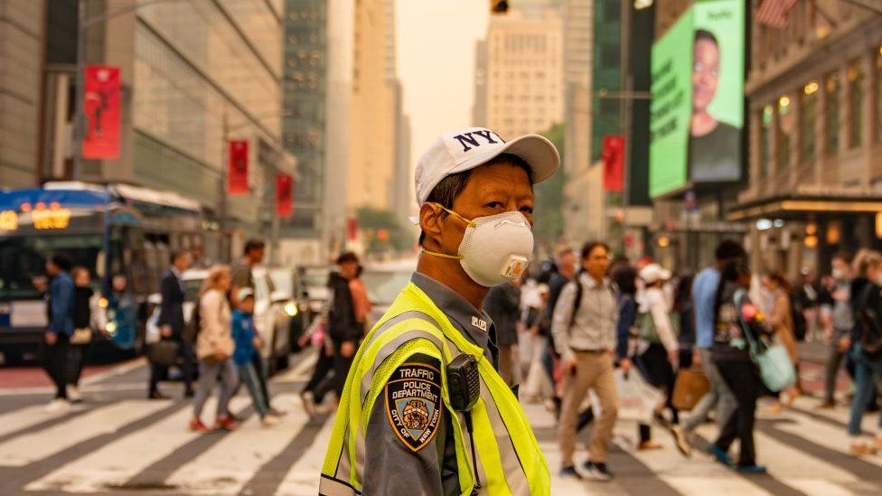
Published41 minutes agoShareclose panelShare pageCopy linkAbout sharingThis video can not be playedTo play this video you need to enable JavaScript in your browser.By Michelle Roberts & Gareth EvansBBC NewsMillions of people in North America have been warned about the health risks of poor air quality, as toxic wildfire smoke from Canada drifts across the continent.This will be a worrying problem for many and one that may persist for several days.Here’s a closer look at what you can do to stay safe.How do I check air quality near me? Air quality updates have become a routine part of many weather forecasts, and the US government has a site where you can check the air quality in your local area. Canadians can check local conditions here.Another useful tool is IQAir’s updated rankings for air quality in major metro areas around the world. AQI indicates how clean or polluted the air is around you. The index runs from 0-500 and the higher the number, the worse it is for your health. Anything from 151-200 is designated as “unhealthy”. How do I protect myself from wildfire smoke? It’s important to pay attention to local air quality reports like those above and listen to expert advice.When possible, limit your exposure. You might be advised to stay indoors, for example, and keep windows and doors closed. Matthew Adams, a professor at the University of Toronto and expert on air contaminants, suggests people living in cities far away from the fires but under current air advisories should limit outdoor exercise to avoid breathing in the smoke.”Don’t get so concerned about it,” he says, but: “Stay inside and reduce your exposure.”In areas closer to the fires, Prof Adams recommends wearing an N95 mask outside to block inhalation of most of the smoke particles. The US Centres for Disease Control gives the same advice and cautions “do not rely on simple dust masks for protection” since they will not protect your lungs from fine particles that are released into the air from wildfires.Get medical help if you need to. What are the symptoms of wildfire exposure? Wildfire smoke can irritate the eyes, nose, throat and lungs. It can cause:cough headachescratchy throatrunny nosestinging eyesirritated sinuseswheezing and shortness of breathInhaling it can also make your heart have to work harder, raising your pulse, and can sometimes cause chest pain, Prof Adams says.William Barrett from the American Lung Association says there is a range of negative health effects from breathing in smoke, dust and fine particles. “They can get deep into our lungs and actually bypass most of the body’s defences and they can create a whole range of negative health consequences for the respiratory system,” he says. “But also these particles can cross into the bloodstream and actually have an impact on cardiovascular health.”Older people, pregnant women and young children, as well as those with underlying health conditions, such as heart disease or asthma, are more likely to get sick. Even someone who is healthy can struggle if they are exposed to high levels, though. Should I wear an N95 mask or use an air purifier? Follow the advice for your local area. You can reduce your smoke exposure by wearing an N95 respirator mask – but you have to make sure you wear it correctly. It should fit tightly to your face to work well as a filter. They are not designed to fit children though, officials caution. They can also make it more difficult for people with certain lung conditions to breathe, says William Barrett, who advises people to check first before using them. You may consider using a portable air purifier or cleaner in your home or workplace, according to the CDC. These help remove airborne particles, called aerosols, from the air indoors. Where is the worst air quality in the world?Major cities in parts of Asia, Africa and Latin America often see dangerous air quality levels similar to what parts of North America are experiencing at the moment.South Asia is particularly polluted, with a World Health Organization report finding the region suffers an estimated two million premature deaths per year due to air pollution.The worst is Delhi, the capital city of India. It places first on IQAir’s rankings with an “unhealthy” Air Quality Index (AQI) score of 191.Several other Asian cities make IQAir’s top 10: Hanoi, Vietnam; Dhaka, Bangladesh; Jakarta, Indonesia; and, Shanghai, China.Several more Chinese cities feature further down the list.More on this storyMillions advised to mask up due to wildfire smokePublished19 minutes agoIs climate change fuelling Canada’s wildfires?Published1 hour agoJodie Comer stops stage show due to New York airPublished3 hours agoCanada could see worst wildfire season on recordPublished2 days ago

Read more →

Unexpected link between chromosomal instability and epigenetic alterations

A graduate student’s curiosity has uncovered a previously unknown link between two important hallmarks of cancer: chromosomal instability and epigenetic alterations.
The resulting study, which was published June 7 inNature, not only opens a fertile new area of basic science biology research, but has implications for clinical care as well.
Chromosomal instability has to do with changes to the number of chromosomes each cancer cell carries. Epigenetic alterations change which genes get turned on or off in a cell, but without modifying the cell’s DNA code.
In his first year as a doctoral student in pharmacology at Weill Cornell Medicine, Albert Agustinus did a rotation in the lab of Samuel Bakhoum, MD, PhD, whose research group at Memorial Sloan Kettering Cancer Center (MSK) studies how alterations in the number and structure of chromosomes drive cancer. Albert is also co-mentored by epigenetics expert Yael David, PhD, whose lab at MSK’s Sloan Kettering Institute takes a chemical-biology approach to studying the epigenetic regulation of transcription.
“He came to me and said, ‘I’m interested in understanding the link between chromosomal instability and epigenetic modifications,'” Dr. Bakhoum recalls. “And my response to him was, ‘Well, there isn’t a known link, but you’re welcome to find it!'”
And find one he did, expanding that initial inquiry into a 32-author, multi-institution collaboration published in one of science’s top journals. The study was jointly overseen by Bakhoum and David.

Recently, Agustinus recounted his first big “aha” moment in the project, for which he also received a   drug discovery fellowship from the PhRMA Foundation.
He was sitting next to a lab mate and peering through the microscope. The cells he was looking at had abnormal little mini-nuclei scattered throughout the cell — a common consequence of chromosomal instability. And they had been set up with fluorescent markers that would show the presence of epigenetic modifications.
“The micronuclei were glowing much brighter than the primary nucleus,” Agustinus says. “My lab mate said to me, ‘I’ve never seen you smile that wide before.'”
Chromosomes Gone Wild
Chromosomes are tightly packaged strands of DNA that carry our genetic information. Normally, each of our cells has 46 chromosomes — half from one parent and half from the other. When a cell divides to make a new copy of itself, all those chromosomes are supposed to end up in the new cell, but in cancer the process can go dreadfully awry.

“The big question that my lab is trying to answer is how chromosomal instability drives cancer evolution, progression, metastasis, and drug resistance,” Dr. Bakhoum says. “It’s a feature of cancer, especially advanced cancers, where the normal process of cell division goes haywire. Instead of 46 chromosomes, you can have a cell with 69 chromosomes right next to a cell with 80 chromosomes.”
The prevailing wisdom in the field has been that cancer cells increase their chance of survival by shuffling up their genetic material when they divide. This process increases the odds that some of the random changes will allow a cancer daughter cell to withstand the assaults of the immune system and medical interventions.
“This new research, however, suggests that’s only part of the story,” Bakhoum says.
That’s because you can have two cancer cells, each with the same number of extra copies of a given chromosome, but each have different genes that are turned off or on. This is due to additional epigenetic changes.
“Our work further suggests that you don’t actually need mutations in the genes that encode epigenetic-modifying enzymes for epigenetic abnormalities to happen. All you need is to have the ongoing chromosomal instability,” Dr. Bakhoum says. “It’s an unexpected finding, but really important. And it also explains why we often find chromosomal instability and epigenetic abnormalities in advanced, drug-resistant cancers, even when there is no evidence of the types of mutations that we would expect to create epigenetic havoc.”
There and Back Again — Or, What Micronuclei Have to Do With Cancer
Small, extra nuclei in cells — known as micronuclei — like the ones Agustinus saw through the microscope are usually rare and quickly get eliminated by the cell’s natural repair mechanisms. When you get a bunch of them, it’s a signal that something has gone horribly wrong, as happens in cancer.
Like a cell’s primary nucleus, these micronuclei contain packages of genetic material. And when these micronuclei burst — which they frequently do — it causes even more problems, the research team found.
Dr. Bakhoum uses the metaphor of a traveler who picks up a foreign accent and brings it back home. The research demonstrated that the sequestration of chromosomes into micronuclei disrupts the organization of chromatin — a complex of genetic components that get packaged into chromosomes during cell division.
This leads to ongoing epigenetic dysregulation, which continues long after a micronucleus is reintegrated into a cell’s primary nucleus.
And the repeated formation and reincorporation of micronuclei over many cycles of cell division leads to the buildup of epigenetic changes. These, in turn, lead to greater and greater differences between individual cancer cells.
The more variation between individual cancer cells within the same tumor, the more likely it is that some of the cells will be resistant to whatever treatment is being thrown at them — allowing them to survive and continue their runaway growth.
Analyzing Epigenetic Changes
To understand and quantify the epigenetic changes happening inside the cells, the researchers use a series of sophisticated experiments to isolate the micronuclei and examine changes occurring in them compared to the cells’ primary nuclei. This allowed them to see patterns of histone modification — changes to the spools around which DNA winds, which, in turn, change access to the underlying genes.
“This allowed us to ask some important questions, like do we actually get transcription of genes that are important in specific pathways?” Dr. David says. “And the answer is ‘yes.'”
They also compared intact versus ruptured micronuclei — revealing an even greater level of changes in the ones that had burst open.
“We also found there were a lot more accessible promoter regions in the micronuclei than in the primary nuclei,” she adds — promoter regions being DNA sequences near the beginning of a gene that help to initiate transcription, a critical step in gene expression.
In one key experiment, the researchers forced a chromosome to go out into a micronucleus and then allowed it to get reintegrated into the primary nucleus. They compared this adventuresome chromosome to one that stayed put.
“Our model chromosome, which happened to be chromosome Y, showed substantial changes in its epigenetic landscape and accessibility of its DNA,” Dr. David says. “This has major implications because of the significant impact the journey of a chromosome into a micronucleus have on the epigenetic changes of the primary nucleus, which we know play a role in tumor progression and evolution.”
The work, she adds, opens whole new avenues of study.
“Now that we’ve demonstrated that chromosomal instability and epigenetic changes are closely linked, we can go deeper and ask questions about precisely how and why,” Dr. David says.
Findings by another research team from Harvard University and the Dana-Farber Cancer Institute, and published in Nature at the same time found additional evidence that supports the MSK team’s discoveries.
Clinical Implications
More than just shedding light on the changes happening inside cancer cells, the research holds promise for treating patients, as well, the researchers note.
Epigenetic changes are a reversible form of gene regulation — and several classes of drugs have already been developed to work on them.
So, to begin with, chromosomal instability and the presence of micronuclei might be used as a biomarkers to help identify which patients might be more likely to be helped by epigenetic modifying drugs, Dr. Bakhoum says.
Additionally, the findings may pave the way for new therapeutic approaches.
“There’s a question of whether we should be treating chromosomally unstable cells with these epigenetic modifying therapies,” he says. “This research demonstrates epigenetic changes can occur without those mutations being present.”
Moreover, the study also suggests that ongoing research into drugs to target chromosomal instability directly might benefit from being combined with efforts to suppress epigenetic alternations, Dr. Bakhoum adds.
Longer term, another potential avenue would be to explore ways of targeting the micronuclei to preventing them from rupturing, which the research showed was a big driver of epigenetic changes, Dr. David notes.
“I think this is a great example of a fundamental, basic science research discovery that, over the next five years, will open multiple interesting avenues for exploration and potential translation to the clinical setting,” she says.
Agustinus, whose curiosity kicked off the entire project and who led the research effort, sums it up this way, “Chromosomal instability and epigenetic alterations help cancer achieve a population diversity that gives them a better chance to survive and develop. But armed with a new understanding of the relationship between these two phenomena, we should be better able to target them therapeutically.”

Read more →

Canada wildfires: Millions advised to mask up due to intense smoke

Published6 hours agoShareclose panelShare pageCopy linkAbout sharingThis video can not be playedTo play this video you need to enable JavaScript in your browser.By Nadine Yousif in Toronto & Sam Cabral in WashingtonBBC NewsMillions of people in North America have been advised to wear N95 masks outdoors due to poor air quality levels sparked by intense wildfires in Canada.New York will begin distributing free masks on Thursday. Canada has said that people should wear a mask if they are unable to remain indoors. Officials warn that the dangerously smoky conditions are expected to persist into the weekend.Much of the smoke is coming from Quebec, where 150 fires are burning. More than 15,000 residents are expected to be forced to evacuate in the province, officials said on Wednesday. It is already Quebec’s worst fire season on record. New York Governor Kathy Hochul announced on Wednesday that New York would distribute one million masks to state residents on Thursday.”This is a temporary situation. This is not Covid,” she said at a news conference. The governor added that New York City buses and trains have high-quality air filtration systems that make them safe forms of travel.Image source, Getty ImagesEnvironment Canada has said that conditions are worsening in Toronto on Thursday, as more smoke pours in.In a special weather bulletin on Wednesday, the agency recommended that anyone outdoors wear a mask. “These fine particles generally pose the greatest risk to health. However, respirators do not reduce exposure to the gases in wildfire smoke,” the Environment Canada statement said.Meanwhile, the US Environmental Protection Agency (EPA) classified the air quality in much of the north-east as “unhealthy” especially for people with respiratory concerns. In total, millions of people around North America are thought to be under a form of air quality warning.Four ways climate change is affecting the weatherIn New York, an orange haze blanketed the city’s skyline and shrouded landmarks including the Statue of Liberty.”We recommend all New Yorkers limit outdoor activity to the greatest extent possible,” Mayor Eric Adams warned on Wednesday.This video can not be playedTo play this video you need to enable JavaScript in your browser.Zoos have brought animals indoors, and in New York, carriage horse rides have been suspended.On Wednesday, schools in the Washington DC area also cancelled outdoor activities as air quality levels were labelled “code red”, while Detroit was listed as the fifth worst major metropolitan area in the world on IQAir’s air pollution rankings.Public health officials have cautioned people not to exercise outside and to minimise their exposure to the smoke as much as possible, as the air poses immediate and long-term health risks. Canadian officials say the country is shaping up for its worst wildfire season on record.Image source, Getty ImagesExperts have pointed to a warmer and drier spring than normal as the reason behind the trend. These conditions are projected to continue throughout the summer.Fires across Canada have already burned more than 3.8m hectares (9.4m acres) of land – an area 12 times the 10-year average for this time of year. More than 600 US firefighters have been sent to Canada to assist local officials, the White House announced on Wednesday.Also on Wednesday, US President Joe Biden and Canadian Prime Minister Justin Trudeau held a phone call to discuss the current situation. Climate change increases the risk of the hot, dry weather that is likely to fuel wildfires. The world has already warmed by about 1.2C since the industrial era began, and temperatures will keep rising unless governments around the world make steep cuts to emissions.How does wildfire smoke affect your health? Experts say exposure to wildfire smoke can cause a litany of health issues.Matthew Adams, a professor at the University of Toronto and the director of its Centre of Urban Environments, said immediate effects of inhaling wildfire smoke include shortness of breath, an elevated pulse, chest pain, or inflammation in the eyes, nose and throat. “On these elevated air pollution days, we’ll see an increased number of visits to hospital,” Prof Adams told the BBC. “And the people that are visiting the hospital typically have a pre-existing respiratory disease.”But wildfire smoke has also been linked to serious, long-term health issues like cancer or lung disease, Prof Adams said, specifically for people who live in areas that experience frequent forest fires.This is caused by small particles in the smoke haze, he said, which can enter the bloodstream and other parts of the human body, causing possible DNA mutations and other health issues.Some studies have also shown that prolonged wildfire smoke exposure can affect pregnant women and their unborn children, Prof Adams added. For people living in cities far away from the fires but under current air advisories, Prof Adams advised people limit outdoor exercise to avoid breathing in the wildfire smoke. “Don’t get so concerned about it,” he said. “Stay inside and reduce your exposure.”But in areas closer to the fires, Prof Adams recommended wearing an N95 mask outside to block inhalation of most of the smoke particles. More on this storyCanada could see worst wildfire season on recordPublished2 days agoCanada wildfire smoke reaches USPublished12 MayApocalyptic wildfire video captures driver’s near missPublished29 May

Read more →