Elizabeth Holmes Says She Can’t Afford $250 Payments to Theranos Victims

Elizabeth Holmes, the founder of a blood-testing company, was ordered to pay $250 a month to defrauded investors after her prison term. Her lawyers said she had “limited financial resources.”Lawyers for the disgraced entrepreneur Elizabeth Holmes said this week that she would be unable to afford to pay $250 each month to victims of her failed-blood testing start-up, Theranos, after leaving prison.Ms. Holmes, 39, began an 11-year, three-month prison sentence in Texas in May after she was found guilty last year of four counts of wire fraud and conspiracy for defrauding investors about her company’s technology and business dealings.Last month, a federal judge in California ordered Ms. Holmes and her former business partner, Ramesh Balwani, to pay $452 million in restitution to investors who were defrauded, including the media mogul Rupert Murdoch.Federal prosecutors asked the U.S. District Court for the Northern District of California last week to correct “clerical errors” in court records. One of the suggested corrections would require Ms. Holmes, as part of her restitution, to pay either $250 or at least 10 percent of her earnings, whichever is greater, each month after she is released from prison.Ms. Holmes’s lawyers objected to this proposed change in a filing on Monday and said the court had substantial evidence showing that she had “limited financial resources.”Her lawyers also disputed the government’s argument that the change would align with the payment schedule for Mr. Balwani, who was tried separately and is serving a nearly 13-year sentence on fraud charges. He has to pay at least $1,000 per month after he is released from prison, according to court records.Ms. Holmes and Mr. Balwani have appealed their cases.Ms. Holmes’s lawyers argued that it was appropriate that the two had been treated differently in sentencing. “There is no indication in the record that the absence of a change to the schedule after she is released was a clerical error,” the filing said.Lawyers representing Ms. Holmes and the U.S. government did not immediately respond to a request for comment on Thursday.Other entities listed as victims for the purpose of restitution include RDV Corporation, an investment firm representing Michigan’s wealthy DeVos family, which invested $100 million in Theranos, and several investment vehicles tied to Don Lucas, a Silicon Valley venture capitalist who died in 2019.Ms. Holmes raised $945 million for Theranos, a company she founded in 2003 after she dropped out of Stanford University. She promised that the company would revolutionize health care with tests that could detect a variety of ailments with just a few drops of blood. But the claims unraveled after a 2015 investigation by The Wall Street Journal revealed that its blood-testing technology did not work. Theranos dissolved in 2018.

Read more →
Better understanding of how genes make us prone to allergies

New research is bolstering scientific understanding behind why some people are more prone to allergies than others. Researchers in the Perelman School of Medicine at the University of Pennsylvania identified how genetic differences that alter a specific protein called ETS1 can affect our body’s response to allergies. They found that small changes in ETS1 in an animal model can lead to an increased likelihood for allergic reactions that cause inflammation. The findings were published recently in Immunity.
The United States Centers for Disease Control and Prevention reports that allergies rank as the sixth most prevalent cause of chronic illness in the U.S., resulting in an annual expenditure exceeding $18 billion. While previous research has established a strong genetic basis for allergies and identified specific genetic sequence variations which predispose for these chronic diseases, how our DNA can affect our chances of developing an allergy remains unclear. But understanding this could lead to improved research and potential new treatments.
By using modern genomics and imaging techniques, a collaborative team of researchers co-led by Penn’s Golnaz Vahedi, PhD, an associate professor of Genetics, and Jorge Henao-Mejia, MD, PhD, an associate professor of Pathology and Laboratory Medicine, found that the ETS1 protein plays a role in controlling a type of immune cell called CD4+ T helper cells, which are important in allergic reactions and help orchestrate the immune response by activating and coordinating other immune cells.
DNA interactions within the genomic segment encompassing the ETS1 gene control how much of the ETS1 protein is made.
“We discovered that these interactions, work like a dimmer switch,” said Vahedi. “When there are changes in the DNA in this area, it can mess up the dimmer switch, causing problems with controlling the ETS1 protein. This can lead to imbalances in our immune cells and cause allergic inflammations.”
While there has been progress in understanding genetic traits that follow predictable patterns, like those passed down from parents, it’s been more challenging to understand conditions that involve many different genes and are common in populations. These complex conditions cannot be explained by simply “turning off” one gene. Instead, they may be caused by small changes in the DNA that affect how genes work together. However, researchers still do not know much about how these changes in DNA relate to how our genes are organized or how they affect how genes are expressed in most complex diseases.
“This work demonstrates how small differences in our DNA can disturb the balance between our immune cells, resulting in significant observable characteristics in patients. This phenomenon may occur in other common diseases such as autoimmune disorders,” said Henao-Mejia.
Other co-authors of this study include Aditi Chandra, Sora Yoon, and Michael Michieletto. This research was funded by the National Institutes of Health (R01AI168240, UC4 DK112217, U01 DK112217, R01 HL145754, U01 DK127768, U01 DA052715, R01 HL136572), the Burroughs Welcome Fund, the Chan Zuckerberg Initiative Award, W. W. Smith Charitable Trust, the Sloan Foundation, and the PEW Charitable Trust.

Read more →

A 'spy' in the belly

To ensure that wounds remain tightly sealed in the abdomen after surgery, researchers at Empa and ETH Zurich have developed a patch with a sensor function. The polymer patch warns before the occurrence of dangerous leaks on sutures in the gastrointestinal tract take hold, while closes the areas on its own. A new material now enables a fast, easy and non-invasive leak diagnosis. The team recently published their findings in the journal Advanced Science.
After surgery in the abdominal cavity, they are dreaded: leaks at the sutures where the contents of the digestive tract can sip into the abdomen. “Even today, such leaks are a life-threatening complication,” explains Inge Herrmann, researcher at Empa and professor of Nanoparticulate Systems at ETH Zurich. The idea of sealing sutured tissue in the abdominal cavity with a plaster has already arrived in operating rooms. The problem is that clinical success is not always granted and varies depending on the adhesion of the tissue. This is because the patches made of protein-containing material dissolve too quickly when they come into contact with digestive juices. Motivated by this problem, Inge Herrmann and Andrea Schlegel, a surgeon at the University Hospital Zurich, pursued the idea of developing a new and innovative solution.
Teaching the patch to “see”
Addressing the challenge, Alex Anthis mentored by Herrmann from Empa’s Particles-Biology Interactions Laboratory in St. Gallen and the Nanoparticle Systems Engineering Laboratory at ETH Zurich, first developed a hydrogel-polymer patch that prevents the highly acidic digestive juices and germ-laden food residues from escaping from the digestive tract and triggering peritonitis or even life-threatening blood poisoning (sepsis).
But the Empa researchers wanted to go one step further: “Surgeons have told us that they keep a close eye on the surgical field during even the most complicated procedures — but as soon as the abdominal cavity is closed, they are “blind” and may not notice leaks until it is too late,” says Anthis. In order for the hydrogel patch to “learn to see,” the team has worked with hospitals in Switzerland and international research partners to develop a solution: The patch is equipped with non-electronic sensors, which already gives an “alarm” before digestive juices can leak into the abdominal cavity. The researchers reported on this novel technology in the journal Nature Communications.
Gas bubbles in ultrasound
The novel material achieves “vision” through a sensitive reaction to changes in the pH value and the occurrence of certain proteins in the vicinity of the wound. Depending on the location of the leak, the reaction takes place within minutes or a few hours. Until now, health care professionals have had to rely on physical reactions of those affected or laboratory tests that occur much later — both indications may come too late at providing a clear indication of a leaking seam.

The sensor patch, on the other hand, makes it possible to detect digestive fluid that escapes in the event of a leak due to its composite structure. For example, acidic gastric juice reacts with the sensor material so that fine gas bubbles appear in the matrix of the patch. The bubbles can then be visualized using ultrasound. “The patches can be equipped with tailor-made sensors for different parts of the digestive tract,” says Anthis. Moreover, the patch can even release medication if necessary, such as antibacterial agents.
Sensor with an eye-catching shape
In their latest development, Empa and ETH Zurich researcher Benjamin Suter, together with Anthis and Herrmann, has equipped the patch with additional capabilities: The sensor response now also entails a visible change during an examination of the patient using computed tomography (CT). If the operated site is leaking, contrast deviations on ultrasound and CT images indicate a leak. This detection is also made easier by the new material composition of the integrated sensor, which, thanks to an insoluble tantalum oxide compound, can be shaped into a suitable form that is conspicuous in the imaging process. On contact with digestive fluid, it changes its shape for example from circular to ring-shaped. “In future, a sensor whose shape clearly stands out from anatomical structures in CT and ultrasound images, could reduce ambiguity of impending leak diagnostics,” says team leader Inge Herrmann.
Biocompatible super glue
In addition, the material is equipped with the necessary properties for wound closure: a stable bond to the tissue, the formation of networks and stability against digestive fluids. In this way, the cost-effective, biocompatible super glue, which consists largely of water, could not only reduce the risk of complications after abdominal surgery, but also shorten hospital stays and save healthcare costs. “The intestinal patch project is already attracting a great deal of interest from the medical profession,” reports Herrmann. Now it is important to advance the application of the clinically relevant innovation in practice.
Awarded young researcher
The research team is currently founding a start-up, Veltist. As a spin-off of ETH Zurich and Empa, the future Medtech company aims to develop and bring to market materials that are intended to contribute to optimal wound closure and improved healing in surgery, thus helping to avoid the dreaded complications of sepsis or peritonitis. In addition to the MaP 2022 Award from ETH Zurich for the best dissertation in the field of Materials and Processes, Alexandre Anthis also received one of the coveted ETH Pioneer Fellowships as well as the Empa Research Award 2021.

Read more →

New discovery can help detect brain tumors

Folate-based radiopharmaceuticals can be used in positron emission tomography (PET) imaging to detect folate receptors in brain tumours. The discovery of folate receptors and their exploitation potential with respect to brain tumours is a new and significant finding in the field.
The discovery is related to gliomas, which are a group of serious brain tumours. Researchers discovered that brain tumours contain increased amount of folate receptor expression relative to adjacent brain tissue. This phenomenon has been observed in both experimental models and human tumour samples.
“Prior to this discovery, the presence of folate receptors and their increased presence in gliomas had not been recognised, and thus they have not yet been used for imaging nor treatment purposes,” summarises Doctoral Researcher Maxwell Miner from the Turku PET Centre at the University of Turku in Finland.
According to research group leader and InFLAMES PI Professor Anne Roivainen this presents an especially exciting target for potential future treatments.
“Our results show an average of 100-fold increase in folate-based radiopharmaceutical accumulation in glioma tissue versus that of adjacent healthy brain tissue,” says Professor Roivainen.
Urgent need for new chemotherapy treatments
Glioma brain tumours originate from the non-neuronal glial cells in the brain, which outnumber neurons in quantity. Gliomas comprise numerous subgroups, with even a high degree of morphological and receptor variability within a single cancerous lesion.
This exceptional cellular heterogeneity can make treatment difficult. There is an urgent need for new chemotherapy treatments particularly for the most malignant brain cancers as they often grow in an infiltrative web-like manner on their periphery making distinguishing the boundaries between glioma and non-glioma difficult. The researchers at the Turku PET Centre hope that this recent discovery will lead to further investigation into folate-targeted brain tumour detection and treatment.
The results were obtained in a multidisciplinary joint project involving researchers from the Turku PET Centre at the University of Turku, Turku University Hospital, InFLAMES Research Flagship, and collaborators from Purdue University, USA. The glioma samples were obtained from the Auria Biobank.

Read more →

A 'pinch' of mineral salts helps the noncaloric sweeteners go down

Perfect noncaloric replacements for sugar and high fructose corn syrup just don’t exist yet. For example, some alternatives have a lingering sweet aftertaste and lack a sugar-like mouthfeel, leaving consumers unsatisfied. Now, researchers in ACS’ Journal of Agricultural and Food Chemistry propose adding blends of nutritionally important mineral salts to make noncaloric sweeteners seem more like the real thing. Taste-testers indicated that these blends gave zero- and low-calorie drinks a better flavor.
Sugar substitutes are often used in sodas, baked goods and frozen desserts, to appeal to people who want lower-calorie or low-sugar treats. But many natural or synthetic noncaloric sweeteners, such as stevia and aspartame, have a delayed sweetness, which lasts long after a food or drink is consumed. These substances also don’t usually have the same mouthfeel as real sugar. Previously, Grant DuBois and colleagues observed that sodium chloride and potassium chloride could accelerate the onset of sweetness and eliminate its persistence for one stevia compound, rebaudioside A. They hypothesized that the salts compress the mucus hydrogel covering taste buds to allow rebaudioside A molecules to get through and then leave more quickly. But high concentrations were needed to achieve the desired effects, which led to off-tastes. So, the researchers wanted to test other mineral salts on commercially available noncaloric sweeteners to see if the products that they are used in could be improved.
In initial tests with a trained sensory panel, the researchers observed that calcium chloride, magnesium chloride and potassium chloride each separately reduced the perceived intensity of rebaudioside A after two minutes. However, again, high amounts of the mineral salt were needed to lower the intensity by more than 30%, which caused unpleasant saltiness or bitterness sensations. Next, mixing the three taste-modifying salts had synergistic effects, allowing the team to use lower amounts of each for the same effect. A blend of the potassium, magnesium and calcium salts reduced the lingering sweetness up to 79% and markedly increased the sugar-like mouthfeel of 10 noncaloric alternatives.
Some panelists still reported a slight saltiness in a few sugar substitute formulations with the all-chloride mineral salt blends. So, the team tested reduced-chloride versions in two commercial zero-calorie colas, resolving the faint salty off-taste issue and greatly improving the taste of the beverages. Additionally, they added salt blends to a reduced-calorie orange juice and a commercial citrus-flavored soft drink made with high fructose corn syrup, which made both beverages taste more like they contained sugar. The researchers say that they have a promising solution for replicating the taste of real sugar in low- and zero-calorie beverages.

Read more →

Ultra small molecule as a new target for Alzheimer's disease?

A new study from the Netherlands Institute for Neuroscience and the VIB-KU Leuven Center for Brain and Disease Research shows that a very small molecule called microRNA-132 can have a significant impact on different brain cells and may play a role in Alzheimer’s disease.
RNA is a molecule that, like DNA, consists of a series of linked building blocks. RNA was long thought to only serve as a messenger and copy of DNA, enabling the translation of DNA into proteins. However, there are also pieces of RNA that do not encode for proteins. MicroRNAs are an example of these non-coding RNA molecules. Despite being small in size, they can have a major function: they can bind to RNA and thereby influence the expression of genes and proteins. In many different diseases, including Alzheimer’s, microRNAs are often dysregulated.
Alzheimer’s patients often exhibit disrupted and altered microRNA profiles, particularly a significant decrease in microRNA-132. But does this molecule truly play a role in the disease, or is this decrease merely coincidental? Previous studies in mouse models have shown that increasing levels of microRNA-132 resulted in the generation of new brain cells and improved memory in the mice. While many researchers believe that the protein amyloid is the primary cause of Alzheimer’s disease, another protein called tau and inflammation also appear to play important roles. MicroRNA-132 has shown a positive effect on both amyloid and tau pathologies in mice. However, the exact mechanisms are still unknown.
Effect of microRNA-132 in different cell types
Researchers Hannah Walgrave, Amber Penning, Sarah Snoeck, Giorgia Tosoni, and their team, led by Evgenia Salta (in collaboration with the group of Bart De strooper at KU Leuven-VIB, Belgium) investigated the effects of microRNA-132 in different cell types. They manipulated the levels of microRNA-132 in a mouse model by both increasing and decreasing them. Subsequently, they used a special technique called single-cell RNA sequencing to examine the genes that changed in each cell type in the brain.
Amber Penning: “A microRNA can have numerous targets, which makes them interesting for diseases with multiple pathological aspects. However, this also makes them challenging to study because how do you find those targets? We know that microRNA-132 performs various functions in neurons, but surprisingly, we found that this microRNA also plays a role in microglia, the immune cells of the brain. This is interesting in the case of Alzheimer’s because we believe that neuroinflammation plays a significant role.”
Changes in cell state
“When we increase microRNA-132 in these microglia, we observe a shift from a disease-associated state to a more balanced homeostatic state. We see this result in both mouse brain and human cell lines. However, whether this change is positive or negative requires further investigation through follow-up experiments. There are different theories suggesting that this disease-associated state may initially aid in cell clearance during the early stages of the disease but becomes excessive later, leading to the death of healthy cells. We still need to determine how beneficial it is for the cells to become more homeostatic. Therefore, we need to be cautious in drawing conclusions.”
“The most significant aspect of this study is demonstrating that microRNA-132 also plays a role in microglia and can influence neuroinflammation. The next step is to examine whether increasing microRNA-132 in neurons and microglia in an Alzheimer’s mouse model has any actual effect. The same applies to the human cell lines we used. In this research, we only used a healthy control cell line, but we will conduct further tests in Alzheimer’s cell lines to see if there are any effects.”
Ultimate goal
“The ultimate goal would be to increase microRNA-132 in Alzheimer’s patients as a therapeutic strategy. Currently, we are using viruses (containing the microRNA) in Alzheimer’s mice that can be injected intravenously, directly into the veins. This makes it easier to eventually translate this strategy to the clinic, as we are utilizing a virus that, in theory, can also be injected into an arm. In addition to Alzheimer’s, there are other neurodegenerative diseases that exhibit a decrease in the same microRNA. Therefore, these results may also be relevant to other disease conditions.”

Read more →

Fewer meals may prevent Type 2 diabetes, obesity

When intermittent fasting became all the rage among Hollywood celebrities, skeptics balked at the idea of skipping meals. But new research from the University of Georgia suggests the celebs might not have been that far off.
The review found that a specific type of restricted eating may reduce the chances of developing Type 2 diabetes and improve your overall health. Known as time-restricted eating, this type of fasting means having regular but fewer meals, cutting out late-night snacks and not eating for 12 to 14 hours (often overnight).
After a comprehensive review of published, peer-reviewed studies, the researchers found a connection between number of meals and obesity and Type 2 diabetes.
“What we’ve been taught for many decades is that we should eat three meals a day plus snacking in between,” said Krzysztof Czaja, an associate professor of biomedical sciences in UGA’s College of Veterinary Medicine. “Unfortunately, this appears to be one of the causes of obesity.”
The three meals and snacks style of eating prevents insulin levels from going down during the day, and, with the amount of calories and sugars Americans consume on average, that can overload the body’s insulin receptors. That leads to insulin resistance and often Type 2 diabetes.
“That’s why it’s so hard to lose body fat,” Czaja said. “We are not giving our bodies a chance to use it. Having fewer meals a day will allow these fat deposits to be used as an energy source rather than the sugar we keep consuming.”
Modern eating approach disrupts body’s biological clock

The researchers found that time-restricted eating allows the body to relax and lower insulin and glucose levels, which in turn can improve insulin resistance, brain health and glycemic control. It can also reduce calorie intake by around 550 calories per day without the stress of calorie counting.
Previous studies have shown disruptions to sleep and meal schedules can change both the type and amount of bacteria and other microorganisms in the digestive tract. But fasting may positively alter the gut microbiome, potentially staving off inflammation and a variety of metabolic disorders.
Additionally, the review suggests time-restricted eating can help regulate hormones responsible for appetite regulation and energy levels.
Regular meal schedules, eating breakfast and decreasing meals and snacks can help guard against obesity and Type 2 diabetes, according to the publication. And all breakfasts aren’t created equal. Aim for healthy fats and protein, like eggs, and avoid the sugar-filled breakfast cereals and pastries.
Although time-restricted eating appeared to improve health, the researchers found that other types of restricted eating, such as fasting for days on end, provided few benefits.

Regular but fewer meals can stave off obesity and metabolic disorders
More than four in 10 Americans are clinically obese, meaning their weight is higher than what is considered a healthy range for their height. Almost 10% are severely obese, according to the Centers for Disease Control and Prevention.
Obesity may lead to a variety of health conditions, including Type 2 diabetes, heart disease and even some cancers.
“Obesity is an epidemic right now, especially in the United States,” Czaja said. “It is a preventable disease. When we started looking at the research, we found that ancient humans didn’t eat every day. That means our body evolved not needing food every day.”
The modern approach of three meals plus snacks became popular decades ago, and it’s a hard pattern to break.
“But our gut-brain signaling is not designed for this type of eating,” Czaja said.
The researchers caution that eating is not a one size fits all situation. Smaller, less active people need fewer calories on average than taller athletes, for example. So for some, one meal of nutrient-rich food might be another while others may need more.
But one thing was very clear from the literature they reviewed: Fewer meals of high-quality food is a good guideline for individuals at risk of developing Type 2 diabetes and obesity.
“Also definitely avoid late-night eating,” Czaja said. “Our midnight snacks spike insulin, so instead of us going into a resting state when we sleep, our GI is working on digestion. That’s why we wake up in the morning tired — because we don’t get enough resting sleep.”
Published in Nutrients, the study was co-authored by Carlee Harris, an undergraduate biology major in UGA’s Franklin College of Arts and Sciences.

Read more →

How a Toilet Plunger Improved CPR

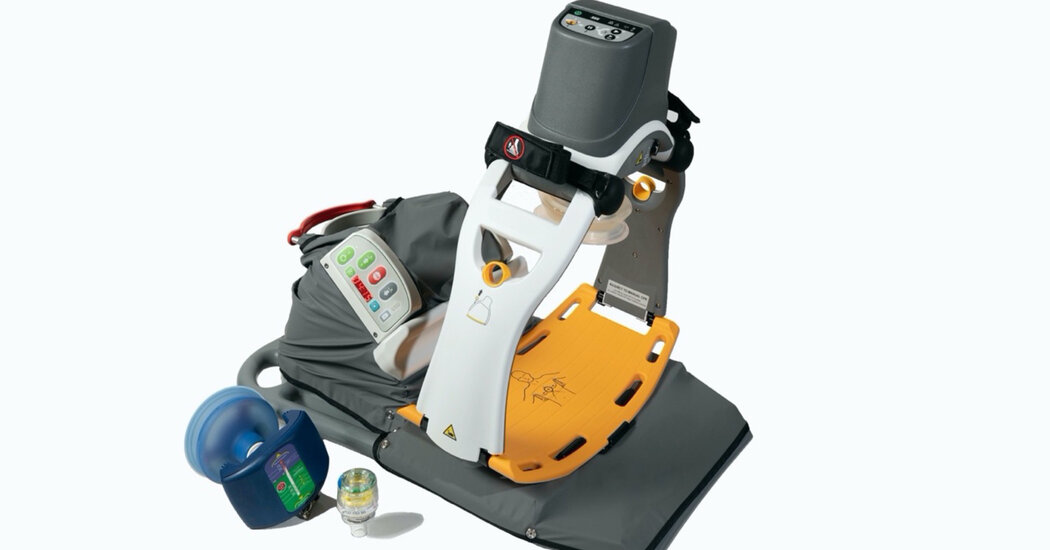
The conventional method for chest compressions doesn’t have a great success rate. Doctors are pumping it up with a high-tech plunger.In 1988, a 65-year-old man’s heart stopped at home. His wife and son didn’t know CPR, so in desperation they grabbed a toilet plunger to get his heart going until an ambulance showed up.Later, after the man recovered at San Francisco General Hospital, his son gave the doctors there some advice: Put toilet plungers next to all of the beds in the coronary unit.The hospital didn’t do that, but the idea got the doctors thinking about better ways to do CPR, or cardiopulmonary resuscitation, the conventional method for chest compressions after cardiac arrest. More than three decades later, at a meeting of emergency medical services directors this week in Hollywood, Fla., researchers presented data showing that using a plunger-like setup leads to remarkably better outcomes for reviving patients.Traditional CPR doesn’t have a great track record: On average, just 7 percent of people who receive it before getting to the hospital are ultimately discharged with full brain function, according to a national registry of cardiac arrests treated by emergency medical workers in communities across the country.“It is dismal,” said Dr. Keith Lurie, a cardiologist at the University of Minnesota Medical School who treated the plunger patient in 1988.The new procedure, known as neuroprotective CPR, has three components. First, a silicone plunger forces the chest up and down, not only pushing blood out to the body, but drawing it back in to refill the heart. A plastic valve fits over a face mask or breathing tube to control pressure in the lungs.The third piece is a body-positioning device sold by AdvancedCPR Solutions, a firm in Edina, Minn., that was founded by Dr. Lurie. A hinged support slowly elevates a supine patient into a partial sitting position. This allows oxygen-starved blood in the brain to drain more effectively and to be replenished more quickly with oxygenated blood.The three pieces of equipment, which fit into a backpack, cost about $20,000 and can be used for several years. The devices have been separately approved by the Food and Drug Administration.About four years ago, researchers began studying the combination of all three devices used in tandem. At this week’s meeting, Dr. Paul Pepe, a longtime CPR researcher and the director of Dallas County’s emergency medical services, reported results from 380 patients who could not be revived by defibrillation, making their odds of survival particularly bleak. Among those who received the new CPR method within 11 minutes of cardiac arrest, 6.1 percent survived with brain function intact, compared with just 0.6 percent who received traditional CPR.He also reported significantly better odds for a subgroup of patients who had no heartbeat but had random electric activity in their heart muscles. The typical odds of survival for people in those circumstances are about 3 percent. But the patients in Dr. Pepe’s study who received neuroprotective CPR had a 10 percent chance of leaving the hospital neurologically intact.Last year, a study carried out in four states found similar results. Patients who received neuroprotective CPR within 11 minutes of a 911 call were about three times as likely to survive with good brain function as those who received conventional CPR.“This is the right thing to do,” Dr. Pepe said.A couple of years ago, Jason Benjamin went into cardiac arrest after a workout at a gym in St. Augustine, Fla. A friend took him to a nearby fire department, where trained workers deployed the neuroprotective CPR gear. It took 24 minutes and multiple defibrillations to revive him.After he recovered, Mr. Benjamin, a former emergency medical technician himself, was amazed to learn about the new approach that had saved his life. He read the studies and interviewed Dr. Lurie. The three-part procedure had several complicated names at the time. It was Mr. Benjamin who came up with the term neuroprotective CPR “because that’s what it’s doing,” Mr. Benjamin recalled, adding that “the focus was on protecting my brain.”Dr. Karen Hirsch, a neurologist at Stanford University and a member of the CPR standards committee for the American Heart Association, said that the new approach was interesting and made physiological sense, but that the committee needed to see more research on patients before it could formally recommend it as a treatment option.“We’re limited to the available data,” she said, adding that the committee would like to see a clinical trial in which people undergoing cardiac arrests are randomly assigned to conventional CPR or neuroprotective CPR. No such trials are happening in the United States.Dr. Joe Holley, the medical director for the emergency medical service that serves Memphis and several surrounding communities, isn’t waiting for a larger trial. Two of his teams, he said, were getting neurologically intact survival rates of about 7 percent with conventional CPR. With neuroprotective CPR, the rates rose to around 23 percent.His crews are coming back from emergency calls much happier these days, too, and patients are even showing up at fire stations to thank them for their help.“That was a rare occurrence,” Dr. Holley said. “Now it’s almost a regular thing.”

Read more →

First synthetic human embryo raises ethical issues

Published13 minutes agoShareclose panelShare pageCopy linkAbout sharingImage source, Getty ImagesBy James GallagherHealth and science correspondentScientists have created the first synthetic human embryos – using no eggs or sperm – provoking deep ethical questions, according to reports.The synthetic embryos – only days or weeks old – could help researchers study the earliest stages of human development and explain pregnancy loss. Nobody is currently suggesting growing them into a baby. But the rapid progress has outpaced discussions on how they should be dealt with ethically and legally. Prof James Briscoe, from the Francis Crick Institute, said the field needed to “proceed cautiously, carefully and transparently” to avoid a “chilling effect” on the public.The development of human synthetic embryos was announced at the annual meeting of the International Society for Stem Cell Research. Synthetic embryos are also known as “embryo models”, as they resemble embryos, for the purposes of research, rather than being identical to them. Image source, University of CambridgeThe work comes from the laboratories of Prof Magdalena Zernicka-Goetz, from the University of Cambridge and the California Institute of Technology.The full details have yet to be published and made available for scientific scrutiny, leading many researchers to feel unable to comment on the significance of the reports.But the principle is the synthetic embryos are made from a stem cell rather than a fusion of egg and sperm.Beating heartStem cells have the capacity to become any cell-type in the body and if coaxed in just the right way can be persuaded to form embryos. This is the first time that has been achieved using human material. Although, they are not truly “synthetic”, as the starting material was cells cultured from a traditional embryo in the laboratory. “It’s beautiful and created entirely from embryonic stem cells,” Prof Zernicka-Goetz told the Guardian newspaper.She has already developed synthetic mouse embryos with evidence of a developing brain and beating heart. Meanwhile, scientists in China have implanted synthetic monkey embryos into female monkeys – although, all the pregnancies failed. Image source, Amadei and HandfordThe synthetic embryos do not behave in exactly the same way as normal embryos. And it is unclear how their use in research should be governed.Prof Briscoe said: “On the one hand, models of human embryos made of stem cells might offer an ethical and more readily available alternative to the use of IVF-derived [in-vitro fertilisation] human embryos. “On the other hand, the closer stem-cell-derived models of human embryos mirror human embryos, the more important it is to have clear regulations and guidelines for how they are used.” Most countries use the 14-day rule in human-embryo research. This allows an embryo created by fertilising a human egg to be grown for 14 days.However, these “embryo models” are not legally “embryos” and are not governed by the same laws. Dr Ildem Akerman, from the University of Birmingham, said: “These findings suggest that we would soon develop the technology to grow these cells beyond the 14-day limit, with potentially more insights to gain into human development. “Nevertheless, the ability to do something does not justify doing it.” ‘Understand infertility’Legal and ethical experts in the UK are drawing up a voluntary set of guidelines for how to proceed. Researchers hope these synthetic embryos will further understanding of the earliest stages of human’s lives.Prof Roger Sturmey, from the University of Manchester, said: “We know remarkably little about this step in human development but it is a time where many pregnancies are lost.”So models that can enable us to study this period are urgently needed to help to understand infertility and early pregnancy loss.” Follow James on Twitter.

Read more →

It’s Not Just You: Many People Confront Health Insurance Obstacles on Care and Bills

The NewsA majority of Americans with health insurance said they had encountered obstacles to coverage, including denied medical care, higher bills and a dearth of doctors in their plans, according to a new survey from KFF, a nonprofit health research group. As a result, some people delayed or skipped treatment.Those who were most likely to need medical care — people who described themselves as in fair or poor health — reported more trouble; three-fourths of those receiving mental health treatment experienced problems.“The consequences of care delayed and missed altogether because of the sheer complexity of the system are significant, especially for people who are sick,” said Drew Altman, the chief executive of KFF, formerly known as the Kaiser Family Foundation.The survey also underscored the persistent problem of affordability as people struggled to pay their share of health care costs. About 40 percent of those surveyed said they had delayed or gone without care in the last year because of the expense. People in fair or poor health were more than twice as likely to report problems with paying medical bills than those in better health, and Black adults were more likely than white adults to indicate they had trouble.Why It Matters: Delayed care can endanger health.Nearly half of those who encountered a problem with their insurance said they could not satisfactorily resolve it. Some could not obtain the care they had sought, while others said they paid more than expected. Among the nearly 60 percent who reported difficulty with their insurance coverage, 15 percent said their health had declined.“This survey shows it’s not enough to just get a card in your pocket — the insurance has to work or it’s not exactly coverage,” said Karen Pollitz, the co-director for KFF’s patient and consumer protections program.People have a hard time understanding their coverage and benefits, with 30 percent or more reporting difficulty figuring out what they will be required to pay for care or what exactly their insurance will cover.“Insurances are way more complicated than they should be,” said Amanda Parente, a 19-year-old college student in Nashville who is covered under her mother’s employer plan. She was surprised to find that her out-of-pocket costs spiked recently when she sought treatment for strep throat. While she realized her co-payments would be higher, “I guess we didn’t know how drastic it was going to be,” she said.Background: Insurance coverage is confusing to everyone.Navigating the intricacies of coverage and benefits were similar regardless of what kind of insurance people had. At least half of those surveyed with private coverage, through an employer, those with an Obamacare plan, or a government program like Medicare or Medicaid, said they experienced difficulties.Survey ResultsNearly 60 percent of Americans with health insurance had a problem with their coverage over the past year.

Read more →