Advanced universal control system may revolutionize lower limb exoskeleton control and optimize user experience

A team of researchers has developed a new method for controlling lower limb exoskeletons using deep reinforcement learning. The method, described in a study published in the Journal of NeuroEngineering and Rehabilitation on March 19, 2023, enables more robust and natural walking control for users of lower limb exoskeletons. “Robust walking control of a lower limb rehabilitation exoskeleton coupled with a musculoskeletal model via deep reinforcement learning” is available open access.
While advances in wearable robotics have helped restore mobility for people with lower limb impairments, current control methods for exoskeletons are limited in their ability to provide natural and intuitive movements for users. This can compromise balance and contribute to user fatigue and discomfort. Few studies have focused on the development of robust controllers that can optimize the user’s experience in terms of safety and independence.
Existing exoskeletons for lower limb rehabilitation employ a variety of technologies to help the user maintain balance, including special crutches and sensors, according to co-author Ghaith Androwis, PhD, senior research scientist in the Center for Mobility and Rehabilitation Engineering Research at Kessler Foundation and director of the Center’s Rehabilitation Robotics and Research Laboratory. Exoskeletons that operate without such helpers allow more independent walking, but at the cost of added weight and slow walking speed.
“Advanced control systems are essential to developing a lower limb exoskeleton that enables autonomous, independent walking under a range of conditions,” said Dr. Androwis. The novel method developed by the research team uses deep reinforcement learning to improve exoskeleton control. Reinforcement learning is a type of artificial intelligence that enables machines to learn from their own experiences through trial and error.
“Using a musculoskeletal model coupled with an exoskeleton, we simulated the movements of the lower limb and trained the exoskeleton control system to achieve natural walking patterns using reinforcement learning,” explained corresponding author Xianlian Zhou, PhD, associate professor and director of the BioDynamics Lab in the Department of Biomedical Engineering at New Jersey Institute of Technology (NJIT). “We are testing the system in real-world conditions with a lower limb exoskeleton being developed by our team and the results show the potential for improved walking stability and reduced user fatigue.”
The team determined that their proposed model generated a universal robust walking controller capable of handling various levels of human-exoskeleton interactions without the need for tuning parameters. The new system has the potential to benefit a wide range of users, including those with spinal cord injuries, multiple sclerosis, stroke, and other neurological conditions. The researchers plan to continue testing the system with users and further refine the control algorithms to improve walking performance.
“We are excited about the potential of this new system to improve the quality of life for people with lower limb impairments,” said Dr. Androwis. “By enabling more natural and intuitive walking patterns, we hope to help users of exoskeletons to move with greater ease and confidence.”
Read more →

Engineers develop a soft, printable, metal-free electrode

Do an image search for “electronic implants,” and you’ll draw up a wide assortment of devices, from traditional pacemakers and cochlear implants to more futuristic brain and retinal microchips aimed at augmenting vision, treating depression, and restoring mobility.
Some implants are hard and bulky, while others are flexible and thin. But no matter their form and function, nearly all implants incorporate electrodes — small conductive elements that attach directly to target tissues to electrically stimulate muscles and nerves.
Implantable electrodes are predominantly made from rigid metals that are electrically conductive by nature. But over time, metals can aggravate tissues, causing scarring and inflammation that in turn can degrade an implant’s performance.
Now, MIT engineers have developed a metal-free, jelly-like material that is as soft and tough as biological tissue and can conduct electricity similarly to conventional metals. The material can be made into a printable ink, which the researchers patterned into flexible, rubbery electrodes. The new material, which is a type of high-performance conducting polymer hydrogel, may one day replace metals as functional, gel-based electrodes, with the look and feel of biological tissue.
“This material operates the same as metal electrodes but is made from gels that are similar to our bodies, and with similar water content,” says Hyunwoo Yuk SM ’16 PhD ’21, co-founder of SanaHeal, a medical device startup. “It’s like an artificial tissue or nerve.”
“We believe that for the first time, we have a tough, robust, Jell-O-like electrode that can potentially replace metal to stimulate nerves and interface with the heart, brain, and other organs in the body,” adds Xuanhe Zhao, professor of mechanical engineering and of civil and environmental engineering at MIT.

Zhao, Yuk, and others at MIT and elsewhere report their results in Nature Materials. The study’s co-authors include first author and former MIT postdoc Tao Zhou, who is now an assistant professor at Penn State University, and colleagues at Jiangxi Science and Technology Normal University and Shanghai Jiao Tong University.
A true challenge
The vast majority of polymers are insulating by nature, meaning that electricity does not pass easily through them. But there exists a small and special class of polymers that can in fact pass electrons through their bulk. Some conductive polymers were first shown to exhibit high electrical conductivity in the 1970s — work that was later awarded a Nobel Prize in Chemistry.
Recently, researchers including those in Zhao’s lab have tried using conductive polymers to fabricate soft, metal-free electrodes for use in bioelectronic implants and other medical devices. These efforts have aimed to make soft yet tough, electrically conductive films and patches, primarily by mixing particles of conductive polymers, with hydrogel — a type of soft and spongy water-rich polymer.
Researchers hoped the combination of conductive polymer and hydrogel would yield a flexible, biocompatible, and electrically conductive gel. But the materials made to date were either too weak and brittle, or they exhibited poor electrical performance.

“In gel materials, the electrical and mechanical properties always fight each other,” Yuk says. “If you improve a gel’s electrical properties, you have to sacrifice mechanical properties, and vice versa. But in reality, we need both: A material should be conductive, and also stretchy and robust. That was the true challenge and the reason why people could not make conductive polymers into reliable devices entirely made out of gel.”
Electric spaghetti
In their new study, Yuk and his colleagues found they needed a new recipe to mix conductive polymers with hydrogels in a way that enhanced both the electrical and mechanical properties of the respective ingredients.
“People previously relied on homogenous, random mixing of the two materials,” Yuk says.
Such mixtures produced gels made of randomly dispersed polymer particles. The group realized that to preserve the electrical and mechanical strengths of the conductive polymer and the hydrogel respectively, both ingredients should be mixed in a way that they slightly repel — a state known as phase separation. In this slightly separated state, each ingredient could then link its respective polymers to form long, microscopic strands, while also mixing as a whole.
“Imagine we are making electrical and mechanical spaghetti,” Zhao offers. “The electrical spaghetti is the conductive polymer, which can now transmit electricity across the material because it is continuous. And the mechanical spaghetti is the hydrogel, which can transmit mechanical forces and be tough and stretchy because it is also continuous.”
The researchers then tweaked the recipe to cook the spaghettified gel into an ink, which they fed through a 3D printer, and printed onto films of pure hydrogel, in patterns similar to conventional metal electrodes.
“Because this gel is 3D-printable, we can customize geometries and shapes, which makes it easy to fabricate electrical interfaces for all kinds of organs,” says first-author Zhou.
The researchers then implanted the printed, Jell-O-like electrodes onto the heart, sciatic nerve, and spinal cord of rats. The team tested the electrodes’ electrical and mechanical performance in the animals for up to two months and found the devices remained stable throughout, with little inflammation or scarring to the surrounding tissues. The electrodes also were able to relay electrical pulses from the heart to an external monitor, as well as deliver small pulses to the sciatic nerve and spinal cord, which in turn stimulated motor activity in the associated muscles and limbs.
Going forward, Yuk envisions that an immediate application for the new material may be for people recovering from heart surgery.
“These patients need a few weeks of electrical support to avoid heart attack as a side effect of surgery,” Yuk says. “So, doctors stitch a metallic electrode on the surface of the heart and stimulate it over weeks. We may replace those metal electrodes with our gel to minimize complications and side effects that people currently just accept.”
The team is working to extend the material’s lifetime and performance. Then, the gel could be used as a soft electrical interface between organs and longer-term implants, including pacemakers and deep-brain stimulators.
“The goal of our group is to replace glass, ceramic, and metal inside the body, with something like Jell-O so it’s more benign but better performance, and can last a long time,” Zhao says. “That’s our hope.”
This research is supported, in part, by the National Institutes of Health.

Read more →

Scientists discover spiral-shaped signals that organize brain activity

University of Sydney and Fudan University scientists have discovered human brain signals travelling across the outer layer of neural tissue that naturally arrange themselves to resemble swirling spirals.
The research, published today in Nature Human Behaviour, indicates these ubiquitous spirals, which are brain signals observed on the cortex during both resting and cognitive states, help organise brain activity and cognitive processing.
Senior author Associate Professor Pulin Gong, from the School of Physics in the Faculty of Science, said the discovery could have the potential to advance powerful computing machines inspired by the intricate workings of the human brain.
The discovery opens up new avenues for understanding how the brain works and provides valuable insights into the fundamental functions of the human brain. Itcould help medical researchers understand the effects of brain diseases, such as dementia, by examining the role they play.
“Our study suggests that gaining insights into how the spirals are related to cognitive processing could significantly enhance our understanding of the dynamics and functions of the brain,” said Associate Professor Gong, who is a member of the Complex Systems research group in Physics.
“These spiral patterns exhibit intricate and complex dynamics, moving across the brain’s surface while rotating around central points known as phase singularities
“Much like vortices act in turbulence, the spirals engage in intricate interactions, playing a crucial role in organising the brain’s complex activities.

“The intricate interactions among multiple co-existing spirals could allow neural computations to be conducted in a distributed and parallel manner, leading to remarkable computational efficiency.”
PhD student Yiben Xu, the lead author of the research from the School of Physics, said the location of the spirals on the cortex could allow them to connect activity in different sections, or networks, of the brain — acting as a bridge of communication. Many of the spirals are large enough to cover multiple networks.
The cortex of the brain, also known as the cerebral cortex, is the outermost layer of the brain that is responsible for many complex cognitive functions, including perception, memory, attention, language and consciousness.
“One key characteristic of these brain spirals is that they often emerge at the boundaries that separate different functional networks in the brain,” Mr Xu said.
“Through their rotational motion, they effectively coordinate the flow of activity between these networks.

“In our research we observed that these interacting brain spirals allow for flexible reconfiguration of brain activity during various tasks involving natural language processing and working memory, which they achieve by changing their rotational directions.”
The scientists gathered their findings from functional magnetic resonance imaging (fMRI) brain scans of 100 young adults, which they analysed byadapting methods used to understand complex wave patterns in turbulence.
Neuroscience has traditionally focused on interactions between neurons to understand brain function. There is a growing area of science looking at larger processes within the brain to help us understand its mysteries.
“By unravelling the mysteries of brain activity and uncovering the mechanisms governing its coordination, we are moving closer to unlocking the full potential of understanding cognition and brain function,” Associate Professor Gong said.

Read more →

Tethering of shattered chromosomal fragments paves way for new cancer therapies

Healthy cells work hard to maintain the integrity of our DNA, but occasionally, a chromosome can get separated from the others and break apart during cell division. The tiny fragments of DNA then get reassembled in random order in the new cell, sometimes producing cancerous gene mutations.
This chromosomal shattering and rearranging is called “chromothripsis” and occurs in the majority of human cancers, especially cancers of the bones, brain and fatty tissue. Chromothripsis was first described just over a decade ago, but scientists did not understand how the floating pieces of DNA were able to be put back together.
In a study published on June 14, 2023 in Nature, researchers at University of California San Diego have answered this question, discovering that the shattered DNA fragments are actually tethered together. This allows them to travel as one during cell division and be re-encapsulated by one of the new daughter cells, where they are reassembled in a different order.
“It’s similar to a smashed car windshield, where the safety glass is designed to keep all of the broken pieces in place,” said senior study author Don W. Cleveland, PhD, Distinguished Professor and chair of the Department of Cellular and Molecular Medicine at UC San Diego School of Medicine. “What we’ve done here is find the safety glass and identify several of its core components, which we can now explore as therapeutic targets.”
When chromosomes break and rearrange themselves, this can initiate or exacerbate cancer in several ways. For example, if a tumor suppressor gene is broken in the process, the cell will become more vulnerable to tumor formation. In other cases, genes that aren’t usually close to each other on the chromosome can suddenly be stitched together to produce a new oncogenic fusion protein. During chromothripsis, many such changes occur simultaneously, rather than gradually, thus accelerating cancer development or its resistance to therapy.
Now that the researchers had identified an early step in this process — the tethering of shattered DNA fragments — they wondered if they could stop it. By destroying the tether, they might prevent the rearranged chromosomes from forming, thereby reducing the number of cells potentially carrying cancerous mutations.
To do this, postdoctoral fellow and first author of the study Prasad Trivedi, PhD, engineered a modified version of one of the tether proteins so that he could induce its destruction on demand. When he did so, the tether disintegrated, the DNA fragments did not cluster and the resulting cells showed reduced survival.
The authors suggest that the proteins in this tether complex, particularly cellular inhibitor of PP2A (CIP2A), may now be an attractive therapeutic target for chromosomally unstable tumors.
“The process of chromosomal care and repair contributes to cancer in many ways, so the more we understand how it works, the better we can fine-tune it to treat cancer,” said Cleveland.
Co-authors of the study include: Christopher D. Steele, Franco K. C. Au and Ludmil B. Alexandrov, all at UC San Diego.

Read more →

Astrocyte processing of serotonin regulates olfactory perception

To enjoy the scent of morning coffee and freshly baked cookies or to perceive the warning smell of something burning, the brain needs two types of cells, neurons and astrocytes, to work closely with each other. Research has shown a great deal of the changes that occur in neurons during olfactory, or smell, perception, but what are the astrocyte responses and how they contribute to the sensory experience remains unclear.
Researchers at Baylor College of Medicine and collaborating institutions report in the journal Science the responses of astrocytes to olfactory stimulation, revealing a new mechanism that is required to maintain astrocyte-neuron communication and process olfactory sensation.
“Previous studies have shown that under natural conditions in a living animal, olfactory stimulation of the brain activates neurons first, which changes the genes these neurons express to be able to mediate the olfactory sensation,” said first author Dr. Debosmita Sardar, a postdoctoral associate in Dr. Benjamin Deneen’s lab at Baylor. “In this study, we investigated what occurred to astrocytes following neural activity during olfactory stimulation and uncovered changes that had not been described before.”
Olfactory stimulation triggered an increase of serotonin transporter Slc22a3 on the astrocytes, which mediated serotonin transport into the cells. “We followed serotonin inside the astrocytes and were surprised to discover that it traveled to the cell nucleus, where it bound to histones, proteins attached to the DNA that help regulate astrocyte gene expression,” Sardar said. “Serotonin bound to DNA acted as a switch, which controlled gene expression.”
Interestingly, serotonin regulates the expression of astrocyte genes involved in the production of the neurotransmitter GABA, which then feeds back to neurons regulating the neural circuit fundamental to sensory perception.
“We showed that losing transporter Slc22a3 in astrocytes reduced serotonin levels in the cells and led to alterations in serotonin-bound DNA,” Sardar said. “In turn, this reduced the expression of genes involved in the synthesis of GABA and decreased astrocytic GABA release, which disturbed the neural circuits of olfactory sensation.”
Serotonin is well known for its contribution to normal brain function as well as being involved in addiction and depression. “Here we discovered a new function of serotonin in astrocytes. Serotonin triggers changes in astrocyte gene expression patterns, turning astrocytes into a hub of olfactory sensation processing,” Sardar said.
“This project has uncovered novel aspects of astrocyte function,” said Deneen, professor and Dr. Russell J. and Marian K. Blattner Chair in the Department of Neurosurgery and director of the Center for Cancer Neuroscience at Baylor. He also is the corresponding author of the work. “We are learning that astrocytes are very plastic, just as neurons are, meaning that astrocytes can change their characteristics and functions in response to environmental stimuli. They listen to neurons and respond, and their two-way communication is at the core of sensory processing and ultimately, animal behavior.”
Other contributors to this work include Yi-Ting Cheng, Junsung Woo, Dong-Joo Choi, Zhung-Fu Lee, Wookbong Kwon, Hsiao-Chi Chen, Brittney Lozzi, Alexis Cervantes, Kavitha Rajendran, Teng-Wei Huang, Antrix Jain, Benjamin Arenkiel and Ian Maze. The authors are affiliated with one or more of the following institutions: Baylor College of Medicine, Jan and Dan Duncan Neurological Research Institute at Texas Children’s Hospital and Icahn School of Medicine at Mount Sinai.
This work was supported by grants from the NIH (NINDS R01-NS071153, R01-AG071687, NIMH R01-MH116900, NIDCD 1K99-DC019668). Further support was provided by Dan L Duncan Comprehensive Cancer Center NIH award (P30 CA125123), CPRIT Core Facility Award (RP210227), NIH High End Instrument award (S10OD026804) and National Institutes of Health Cancer Center Grant (P30 CA125123).

Read more →

Close up on aging reveals how different cell types in the body age at different pace

As the body ages, organ function progressively declines and the risk for a wide range of diseases, including cardiovascular disease, cancer and neurodegenerative diseases, increases. Understanding how the body ages is an intense area of research as it will potentially illuminate ways to promote healthy aging.
Researchers at Baylor College of Medicine, Chan Zuckerberg Biohub San Francisco, Genentech, Inc. and collaborating institutions are breaking a path in that direction. They report in the journal Science, the first Aging Fly Cell Atlas (AFCA), a detailed characterization of the aging process in 163 distinct cell types in the laboratory fruit fly. Their in-depth analysis revealed that different cell types in the body age differently, each cell type following a process involving cell type-specific patterns. AFCA provides a valuable resource for researchers in the fruit fly and aging communities as a reference to study aging and age-related diseases and to evaluate the success of anti-aging strategies.
“Research has shown that, for instance, some cells like neurons in the brain live longer than cells in the gut lining, which are replaced by new ones often,” said co-corresponding author Dr. Hongjie Li, assistant professor of molecular and human genetics and the Huffington Center on Aging at Baylor. He is also a member of Baylor’s Dan L Duncan Comprehensive Cancer Center. “Our team is interested in better understanding how different cell types age differently, and to that end, we analyzed in detail several biological characteristics of individual cell types as fruit flies aged naturally in the lab. The fruit fly is a well-known model to study human conditions. About 75% of genes associated with human diseases have functionally similar counterparts in the fly.”
Dr. Stephen Quake, the Lee Otterson Professor of Bioengineering and Applied Physics at Stanford University and co-corresponding author of the Science paper, said the new atlas provides a powerful, open-access resource for scientists to better understand the biology of aging. “Through a highly productive collaboration, our team has created an exceptionally detailed map of how gene expression changes with aging across a wide range of cell types in the fly,” said Quake, also head of science at the Chan Zuckerberg Initiative (CZI). “Since a majority of these genes have similar roles in people, this dataset offers a unique vantage point to begin to decipher why many serious human diseases emerge in later life.”
As the flies aged, the researchers took samples when the animals were 30, 50 and 70 days old (the latter is equivalent to an 80-year-old person). At each time point, the team conducted single-nucleus RNA sequencing to analyze gene expression changes in individual cells in different organs and compared the results to those of young flies (5 days old). The team examined four different aging features: cell composition changes, number of differentially expressed genes, change in the number of expressed genes and decline of cell identity. They found that as flies age, these features change as a group according to cell type-specific patterns.
“We found that aging impacts cellular composition across the whole fly,” Li said. Fat body cells were among the cell types that increased in number the most, while muscle cells decreased the most. Neurons, however, did not show major changes in the number of cells during the fruit fly’s life. “In addition, the analysis of the genes expressed by different cell types in time revealed that fat cells show the largest difference between the number of genes expressed in young versus old fruit flies,” Li said.
The researchers also found that about 80% of all the cell types analyzed decreased the number of genes expressed, and 20% increased this number. “We plan to study the mechanism of this observation in the future,” said co-first author Dr. Tzu-Chiao Lu, postdoctoral associate in the Huffington Center on Aging.
The team also investigated whether cellular gene expression programs that define cell identity change as the animals age. “For example, compared to the flight muscle identity marker Nig1 in young flies, the marker in older flies is dramatically decreased, while other markers began to appear as the flies grew older,” Li said.
“We have learned that each of the four aging features we studied measures a different aspect of the cell and that not a single feature applies to all cell types,” Li said. “Combining all aging features led us to discover unique cell type-specific aging patterns and comparing them revealed useful and interesting findings. For instance, neurons in the brain age slowly, while muscle, fat and liver cells age much faster. Also, cell-type specific aging patterns may vary according to gender.”
“A critical observation of this study is that cell type-specific aging patterns in cells can be used to gauge biological age, that is the relative aging status of an organism, independent of its chronological age,” said co-corresponding author Dr. Heinrich Jasper, principal fellow at Genentech Inc. “This will provide further insight into factors, such as diets, drugs and diseases, that may change the aging trajectory and hence make an organism ‘younger’ or ‘older’ than its chronological age.”
“We hope that researchers will explore the possibilities AFAC offers to a variety of scientific fields, including genetics, cell biology and physiology,” Li said. The team has developed a user-friendly data portal and provide access via CZI’s CELLxGENE platform. All resources can be accessed at https://hongjielilab.org/afca.

Read more →

New study gives clues on why exercise helps with inflammation

Researchers have long known that moderate exercise has a beneficial impact on the body’s response to inflammation, but what’s been less understood is why. New research coming out of York University done on a mouse model suggests that the answers may lie at the production level of macrophages — white blood cells responsible for killing off infections, healing injury and otherwise acting as first responders in the body.
“Much like if you train your muscles through exercise, we showed that exercise of moderate intensity ended up training the precursors of those macrophages in the bone marrow,” says Faculty of Health Associate Professor and York Research Chair Ali Abdul-Sater with the School of Kinesiology and Health Science. “The way that exercise is doing this is by changing the way those cells breathe, essentially, how they use oxygen to generate energy and then changing the way they access their DNA.”
Associate Professor and York Research Chair Ali Abdul Sater
While many studies look at temporary boosts to the immune system immediately after exercise, this study, published in the journal AJP-Cell, found these changes occurred even a week later, suggesting that the changes were long term.
We often hear about inflammation in the body in the context of its negative effects, but inflammation is the body’s response to infection and other stressors, and some level of inflammation is necessary and desirable.
“Inflammation is amazing, it’s a very important part of our normal immune response,” says Abdul-Sater. “What we’re concerned about is excessive inflammation. Heart disease, diabetes, many cancers and autoimmune diseases, all essentially begin because there was an inappropriate inflammatory response.”
He says it is around the six-to-eight-week mark into the exercise regimen where changes really became apparent, compared with sedentary mice. “There’s a lot of rewiring that’s taking place in the circuitry of how the cells breathe, how the cells metabolize glucose, how the cells then accessDNA. So all that just takes time.”
Abdul-Sater says that because the inflammatory response is a very ancient one, this aspect of the immune system is generally very similar across mammals, and he expects the research would translate well to humans. In the next phase, Abdul-Sater and collaborators from the university will collect immune cells from human volunteers who will do exercises of various intensities to see which workout routines are most beneficial to balance the inflammatory response. They will also look at inflammation in mice in more complex infectious diseases similar to COVID-19 and autoimmune disease, where overactive inflammatory responses lead to poor outcomes.
“People that got seriously ill from COVID-19, went into what is called a cytokine storm essentially, they released this massive number of cytokines, those mediators that are produced by inflammatory cells, which then cause that accumulation of fluid in lungs.”
While the findings that exercise is beneficial will not come as a surprise, Abdul-Sater says he hopes that by finding the underlying mechanisms of the beneficial impact, this knowledge can be put to good use.
“The thing with humans is there’s no intervention that will work on everyone. We know that, but what this study suggests is that moderate and persistent exercise not only improves metabolic health, but also will improve immune health in the long run.”

Read more →

Pregnancy hormone repairs myelin damage in MS mouse model

Treating a mouse model of multiple sclerosis with the pregnancy hormone estriol reversed the breakdown of myelin in the brain’s cortex, a key region affected in multiple sclerosis, according to a new UCLA Health study.
In multiple sclerosis, inflammation spurs the immune system to strip away the protective myelin coating around nerve fibers in the brain’s cortex, hampering electrical signals sent and received by the brain. Atrophy of the cortex in MS patients is associated with permanent worsening of disability, such as cognitive decline, visual impairment, weakness and sensory loss.
No currently available treatments for MS can repair damage to myelin. Instead, these treatments target inflammation to reduce symptom flare-ups and new nerve tissue scarring. Previous UCLA-led research found that estriol, a type of estrogen hormone produced in pregnancy, reduced brain atrophy and improved cognitive function in MS patients.
In the new study, researchers treated a mouse model of MS with estriol and found that it prevented brain atrophy and induced remyelination in the cortex, indicating that the treatment can repair damage caused by MS, rather than just slow the destruction of myelin.
This is the first study to identify a treatment that could repair myelin in the cortex, undoing some of the damage caused by MS.
Allan MacKenzie-Graham, an associate professor of neurology, is the study’s corresponding author. Other authors include Cassandra Meyer, Andrew Smith, Aitana A. Padilla-Requerey, Vista Farkhondeh, Noriko Itoh, Yuichiro Itoh, Josephine Gao, Patrick Herbig, Quynhanh Nguyen, Katelyn Ngo, Mandavi Oberoi, Prabha Siddarth and Rhonda R. Voskuhl, all of UCLA.

Read more →

FDA Panel Recommends a Covid Shot Aimed at Only One Virus Variant

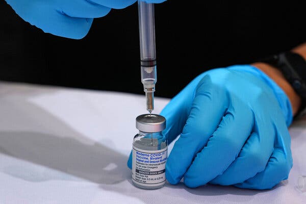
An F.D.A. advisory panel told manufacturers to aim the latest coronavirus vaccine for the fall at the dominant SARS-CoV-2 variant.The NewsVaccine makers should target the XBB variant of the coronavirus in a shot to be available in the fall, moving away from the existing formula that protected against the Omicron variant and an early form of the virus, an advisory panel to the Food and Drug Administration agreed on Thursday.The 21-member panel unanimously recommended that manufacturers should aim at the most dominant variant of the coronavirus this summer. If the F.D.A. agrees, the advice would start the manufacturing of millions of shots.Agency officials had earlier said they hoped to move toward an annual vaccine against the coronavirus. But the discussion on Thursday did not involve any timetables as to how often adults should receive new shots, or which populations should be offered the latest vaccine, though the initial advice last fall was for those age 12 and older to get the shot.Jim Wilson/The New York TimesWhy It Matters: Some people remain at risk.Pfizer, Moderna and Novavax have made it clear that they need time to make tens of millions of doses of the shots that would be available in the fall.“I think that’s what today’s discussion is about — how to best to come up with what goes into people’s arms to offer the best protection during a period when we think we’ll have waning immunity,” said Dr. Peter Marks, the F.D.A.’s vaccine chief. He added that the winter may also bring “further evolution of the virus.”Since the beginning of the pandemic, 6.2 million hospitalizations and 1.1 million deaths have been attributed to the virus in the United States, according to data presented by Natalie Thornburg, a vaccine expert at the Centers for Disease Control and Prevention.She said the picture had improved this year, but those who remain vulnerable include the unvaccinated, people who are immunocompromised and those who have diabetes or chronic kidney, lung, cardiovascular or neurologic diseases. People 65 and older are also at risk, and that rises with age.Background: Changes are afoot in who gets the shots and when.The bivalent shots offered last fall included protection against the Omicron variant and an early Covid variant. About 20 percent of adults, or about 53 million, in the United States got the booster shot, with the rates higher among older adults.Moving ahead with a shot targeted at only an XBB variant means that newborns and people with compromised immune systems may not have immunity against the earliest coronavirus variants. That should not be a problem, according to a briefing given by a World Health Organization official, who said those variants were no longer in circulation.What’s Next: A vaccine may be offered alongside flu and R.S.V. jabs.The F.D.A. is expected to make a more official recommendation to vaccine makers soon. The manufacturers will be expected to study the new formulas and submit data to the agency. If approvals are granted, the C.D.C. will advise health providers on which age groups should get the jab.An F.D.A. spokesman said it expected that an updated vaccine would be available by late September, assuming the data support safe and effective vaccines.It remains unclear whether or when the vaccine makers or the F.D.A. will examine the potential effects of administering multiple vaccines in the fall, including those for the flu and respiratory syncytial virus, or R.S.V., which are expected to be available for pregnant people and older adults. Agency advisers have also endorsed the use of an R.S.V. antibody shot to protect infants.

Read more →

FDA Panel Recommends a Covid Vaccine Aimed at Only the XBB Variant

An F.D.A. advisory panel told manufacturers to aim the latest coronavirus vaccine for the fall at the dominant SARS-CoV-2 variant.The NewsVaccine makers should target the XBB variant of the coronavirus in a shot to be available in the fall, moving away from the existing formula that protected against the Omicron variant and an early form of the virus, an advisory panel to the Food and Drug Administration agreed on Thursday.The 21-member panel unanimously recommended that manufacturers should aim at the most dominant variant of the coronavirus this summer. If the F.D.A. agrees, the advice would start the manufacturing of millions of shots.Agency officials had earlier said they hoped to move toward an annual vaccine against the coronavirus. But the discussion on Thursday did not involve any timetables as to how often adults should receive new shots, or which populations should be offered the latest vaccine, though the initial advice last fall was for those age 12 and older to get the shot.Jim Wilson/The New York TimesWhy It Matters: Some people remain at risk.Pfizer, Moderna and Novavax have made it clear that they need time to make tens of millions of doses of the shots that would be available in the fall.“I think that’s what today’s discussion is about — how to best to come up with what goes into people’s arms to offer the best protection during a period when we think we’ll have waning immunity,” said Dr. Peter Marks, the F.D.A.’s vaccine chief. He added that the winter may also bring “further evolution of the virus.”Since the beginning of the pandemic, 6.2 million hospitalizations and 1.1 million deaths have been attributed to the virus in the United States, according to data presented by Natalie Thornburg, a vaccine expert at the Centers for Disease Control and Prevention.She said the picture had improved this year, but those who remain vulnerable include the unvaccinated, people who are immunocompromised and those who have diabetes or chronic kidney, lung, cardiovascular or neurologic diseases. People 65 and older are also at risk, and that rises with age.Background: Changes are afoot in who gets the shots and when.The bivalent shots offered last fall included protection against the Omicron variant and an early Covid variant. About 20 percent of adults, or about 53 million, in the United States got the booster shot, with the rates higher among older adults.Moving ahead with a shot targeted at only an XBB variant means that newborns and people with compromised immune systems may not have immunity against the earliest coronavirus variants. That should not be a problem, according to a briefing given by a World Health Organization official, who said those variants were no longer in circulation.What’s Next: A vaccine may be offered alongside flu and R.S.V. jabs.The F.D.A. is expected to make a more official recommendation to vaccine makers soon. The manufacturers will be expected to study the new formulas and submit data to the agency. If approvals are granted, the C.D.C. will advise health providers on which age groups should get the jab.An F.D.A. spokesman said it expected that an updated vaccine would be available by late September, assuming the data support safe and effective vaccines.It remains unclear whether or when the vaccine makers or the F.D.A. will examine the potential effects of administering multiple vaccines in the fall, including those for the flu and respiratory syncytial virus, or R.S.V., which are expected to be available for pregnant people and older adults. Agency advisers have also endorsed the use of an R.S.V. antibody shot to protect infants.

Read more →