Sharp rise in teenage girls with eating disorders during Covid

Published5 hours agoShareclose panelShare pageCopy linkAbout sharingImage source, AnnabelleBy Philippa Roxby and Annabel RackhamHealth reporterGP records show a sharp rise in teenage girls in the UK developing eating disorders and self-harming during the Covid pandemic, a study has found.The increases were greatest among girls living in the wealthiest areas, which could be due to better GP access. Young women have told the BBC that the lack of control over their lives during lockdown was a behavioural trigger.The government says it is investing in eating disorders services to help more children and young people.Charities maintain everyone needs access to early support for mental health issues, no matter where they live.’Lack of control’Annabelle, 19, from Surrey, recalls how difficult she found lockdown.”We had very little control over our lives – our GCSEs were cancelled, we had no say in what our grades were going to be. “We couldn’t see people, we couldn’t control where we went.”The only thing we could control was what you ate and how you looked – so that’s what I chose to focus on.”Annabelle received help overcoming bulimia and is feeling better, but her family is still paying for therapy privately.She says people don’t realise how common eating disorders are: “I don’t know a single girl or female friend who hasn’t had some sort of struggle with eating.””It’s incredibly hard, but there isn’t enough help for everyone on the NHS.Image source, Sophie RowlandSophie Rowland, 18, from South Shields, has been posting about her recovery from anorexia on TikTok.She loved food before the pandemic, but being stuck in the house during lockdown made her obsessive about exercise and watching workouts online.”I just realised I couldn’t stop tracking calories. It had taken over my life.””Everything was just food, food, food – and it was food that became the enemy.”She told her mum one day, and says she was “very lucky” with the help she received from nurses, friends and family. Positive feedback from her videos have also aided her recovery and now she wants to help others.’Staggering rise’Eating disorders and self-harming have been rising among children and young people for a number of years but “increased substantially” between 2020 and 2022, the study found.Over that period, around 2,700 diagnoses of eating disorders were anticipated among 13-16-year-olds, but 3,862 were actually observed – 42% more than the expected figure.In the same age group, 6,631 cases of self-harm were expected but 9,174 were recorded by GPs – 38% more than predicted.Among 17-19-year-olds eating disorders also rose above expectations.The analysis, by the University of Manchester, Keele University and University of Exeter, looked at nine million records belonging to patients aged 10-24 years, from nearly 2,000 GP practices across the UK.Dr Shruti Garg, from the University of Manchester – a child and adolescent psychiatrist and the study author – called it a “staggering rise” which highlighted an urgent need to improve early access to support.An eating disorder – most commonly anorexia or bulimia – is a mental health condition where control over food is used as a means of coping with distress and other difficult situations. During the pandemic, prolonged access to social media, more focus on body image and less face-to-face contact may have led to feelings of low self-esteem and psychological distress, particularly among adolescent girls, the study says.Social media may also have exposed young people to content which increased the risk of developing an eating disorder.In the media “there was a lot of emphasis on food availability and restriction, and also on the message that being overweight was a risk for Covid,” Dr Garg notes.The research also suggests young people might self-harm as a coping strategy in times of uncertainty.’Huge strain’Even before the pandemic, there had been a gradual decline in mental health generally among teenagers and young people – and a recent study found five children in every classroom had a probable mental disorder in 2022.Since March 2020, GP records show a big rise in eating disorders among the richest in the UK population – with 52% of flagged eating disorders occurring in the least deprived areas and 22% in the most deprived.Tom Quinn, director of external affairs at charity Beat, says there is still “a postcode lottery” for care and everyone needs to get “the help they need as quickly as possible”.”These figures are shocking but sadly not surprising,” he adds.”We also know that the NHS is treating more children and young people than ever before, with healthcare professionals under huge amounts of strain.” The study found no sign of increasing rates of eating disorders among boys or young men. Researchers say males have a higher suicide risk than females, suggesting mental health issues manifest in different ways.The research, which was supported by mental health research charity The McPin Foundation, is published in the Lancet Child and Adolescent Health. A spokesperson for the Department of Health and Social Care said they recognised “the devastating impact eating disorders can have on an individual and [their] family’s life”. The government says it will invest an additional £2.3 billion a year in NHS mental health services by March 2024, alongside £54m a year to increase capacity at children and young people’s community eating disorder services.If you’ve been affected by the issues raised in this article, help and support is available via BBC Action Line.More on this storySelf-harm hospital admissions up 22% for childrenPublished23 MarchQuarter of 17-19-year-olds may have mental disorderPublished29 November 2022Huge leap in children in mental health crisisPublished4 February 2022Related Internet LinksWhat are eating disorders? – NHSThe UK’s Eating Disorder Charity – BeatYoungMinds – Mental Health Charity For Children And Young People – YoungMindsThe McPin FoundationThe Lancet Child & Adolescent Health Home PageThe BBC is not responsible for the content of external sites.

Read more →
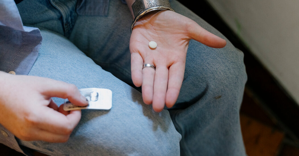
New York Passes Bill to Shield Abortion Providers Sending Pills to States With Bans

The measure is one of several recently enacted by states aiming to expand access to medication abortion for patients who live where it is outlawed.The New York State Legislature gave final approval on Tuesday to legislation that provides legal protection for New York doctors to prescribe and send abortion pills to patients in states that have outlawed abortion.The measure, along with similar new laws in several other states controlled by Democrats, could significantly expand medication abortion access by allowing more patients in states that restrict abortion to end pregnancies at home, without traveling to states where abortion is legal.The New York bill now goes to the desk of Gov. Kathy Hochul, who has indicated that she supports the idea of such a shield law. The bill stipulates that New York courts and officials will not cooperate if a state with an abortion ban tries to prosecute, sue or otherwise penalize a New York health care provider who offers abortion via telemedicine to a patient in that state, as long as the provider complies with New York law. It passed the State Assembly by a vote of 99 to 45 on Tuesday evening after clearing the Senate by a vote of 39 to 22 last month.Since the Supreme Court ended the nationwide right to an abortion last year, legislation known as telemedicine abortion shield laws have been enacted in Massachusetts, Colorado, Vermont and Washington. But New York’s legislation is expected to have a notable impact. Several providers in New York say they plan to send abortion pills to patients in all restrictive states, and a few providers are speaking publicly, which those in other states with shield laws have so far not done.“I’m going to mail pills as soon as the governor signs the bill,” said Dr. Linda Prine, a New York physician and co-founder of the Miscarriage and Abortion Hotline, which answers patients’ questions about medication abortion. “This is the first time we’re able to do something to fight back,” she added.Dr. Prine said she and at least three other doctors would mail pills to patients in all states with restrictions or bans. A telemedicine service in New York, Juniper Midwifery, also said it hoped to use the shield law to mail pills to patients in states with abortion bans.“It’s definitely something that we would want to explore and make it happen,” Jillian Barovick, a midwife and co-founder of Juniper, said.Assembly member Karines Reyes, a registered nurse who introduced the bill to the assembly.Cindy Schultz for The New York TimesAbortion is now mostly outlawed in 14 states. But most bans penalize people who assist with an abortion, not those who take abortion pills.More than half of abortions in the United States are now carried out with medication, using two drugs, mifepristone followed by misoprostol. Data indicates the process is safe and effective.Abortion opponents are trying to force mifepristone off the market by challenging the Food and Drug Administration’s approval of the drug 23 years ago in a lawsuit expected to be decided by the Supreme Court. The suit also challenges the F.D.A.’s decision in 2021 allowing patients to receive prescribed pills by mail.Since the overturning of Roe v. Wade, tens of thousands of patients in states with bans or severe restrictions have taken abortion pills. Many have traveled to states where abortion is legal to obtain pills at clinics or at addresses in those states where pills were sent by mail. But many patients cannot afford the cost and time of traveling.“The telemedicine option and protecting providers who are providing telemedicine abortion services is going to help tremendously the people here in Mississippi and other restricted states,” said Michelle Colón, the executive director of SHERo Mississippi, an organization focused on supporting reproductive rights for people of color. “This will expand access, which we so desperately need.”Other patients receive pills in the mail from overseas, either prescribed by doctors abroad, through a European telemedicine service, or ordered directly from online pharmacies in India or other countries. By the time the pills arrive, often two to three weeks later, patients may be past the 12-week threshold of pregnancy that the World Health Organization endorses for using medication abortion. Mailing pills from within the United States, as doctors operating under shield laws are doing, cuts delivery time to a few days.The telemedicine shield laws are not a guarantee of full legal protection for providers. Under the legislation, if a state with a ban issued an arrest warrant for a physician like Dr. Prine, New York would refuse to extradite her to that state. But if she traveled there, or to another state that agreed to extradite her, she could face criminal charges.A law that would shield telehealth abortions disrupts the usual model for telemedicine law and policy, which assumes a doctor is providing care in the same place where the patient is located.Johnny Milano for The New York Times“Texas could prosecute them for murder,” said Jonathan Mitchell, a former Texas solicitor general and the architect of a 2021 Texas law that banned abortion after six weeks into pregnancy and deputized private citizens to enforce it by suing for cash judgments of $10,000 per abortion.“Under Texas law, killing a fetus through an illegal abortion is no different from killing a baby, except that the mother cannot be prosecuted (or sued) for death of a fetus,” Mr. Mitchell wrote in an email.Because of the substantial risks, only about 10 health care providers in states with shield laws are known to have begun sending pills to patients in states that restrict abortion. These providers have proceeded cautiously.“Most of the people who have been using the shield laws so far have been just dipping their toes in the water, going to states that maybe aren’t quite as litigious, but I think we just need to start doing it to all of them,” said a doctor in New York’s Hudson Valley who is planning to send pills under the new shield legislation and spoke on the condition of anonymity because of the risk involved.Other abortion providers have indicated interest in using shield laws if they could send prescriptions to a pharmacy that would mail the pills rather than stocking and shipping the medications themselves. That option could become available in the coming months if California enacts a telehealth abortion shield bill that passed the state Senate last month. An online pharmacy based in California, Honeybee Health, hopes to use that law to ship to all 50 states, said Jessica Nouhavandi, co-founder and president of Honeybee.“We are anxiously awaiting the passing of the California shield law,” she said.As providers test their states’ telehealth shield laws, many legal questions could arise, including the possibility of civil suits and challenges to a provider’s medical license for unauthorized practice of medicine.“It’s not a matter of if a provider will be challenged in court,” said Rachel Rebouché, the dean of Temple University Law School, who has written in support of shield laws. “It’s a matter of when.”State Senator Shelley Mayer, the bill’s senate sponsor, in the senate chamber on Friday.Cindy Schultz for The New York TimesLawyers on each side of the issue say that state shield laws undermine basic premises of interstate cooperation. Rather than recognizing one state’s arrest warrant or court order, another state effectively throws a wrench into the enforcement of that state’s laws.And a law that shields telehealth abortions disrupts the usual model for telemedicine law and policy, which “presumes that you’re providing care in the place where the patient is,” Professor Rebouché said.To treat a patient in Texas, for example, a New York physician would normally first get a Texas medical license. If the doctor practiced without one, New York would generally help Texas initiate disciplinary proceedings through its medical board. Without the shield law, an out-of-state ruling could have consequences for the doctor’s license in New York.In addition, in some cases, the U.S. Constitution requires states to enforce the civil judgments of another state. An abortion provider in New York could be sued in Texas, for example, by a family member of an abortion patient, claiming damages for wrongful death of the embryo or fetus. If the plaintiff won, the Full Faith and Credit Clause in the Constitution could obligate New York to comply in collecting damages.“It’s very clear that in other states, citizens can still sue doctors who engage in the unlawful practice of medicine,” said Denise Harle, senior counsel for the Alliance Defending Freedom, a conservative Christian legal organization that represents the anti-abortion plaintiffs seeking to remove mifepristone from the market in their lawsuit against the F.D.A. “They can sue to protect against bad acts by people who are committing crimes.”Malpractice insurance can also be challenging to arrange. New York’s shield bill, however, says that an insurer cannot drop or penalize a provider who complies with New York law in providing abortions.To date, there have been no known examples of prosecutions, lawsuits or other actions against providers working under telemedicine abortion shield laws. Dr. Prine and others said that they were prepared to be the guinea pigs but that they were also taking steps to protect themselves.Most providers using shield laws say they are avoiding travel to states with abortion bans. Dr. Prine said she would even make sure not to have a flight connection in an airport in a restrictive state.

Read more →

What role does alternative splicing play in neurodegenerative disease?

Alternative splicing, a clever way a cell generates many different variations of messenger RNAs — single-stranded RNAs involved in protein synthesis — and proteins from the same stretch of DNA, plays an important role in molecular biology. Progressing rapidly, the field of alternative splicing is a complex topic and the scientific literature on it is already extensive.
David Nikom, a student in the UC Riverside Neuroscience Graduate Program, and his advisor, Sika Zheng, an associate professor of biomedical sciences in the UCR School of Medicine and director of the Center for RNA Biology and Medicine, have written a review in Nature Reviews Neuroscience to discuss emerging research and evidence of the roles of alternative splicing defects in major neurodegenerative diseases. They also summarize the latest advances in RNA-based therapeutic strategies to target these disorders.
According to them, the topic of alternative splicing in neurodegenerative disease is particularly relevant in view of the increasing frequency of neurodegenerative disease worldwide and the urgent need for novel approaches for their treatment and management. They argue that since aberrant splicing dysregulation occurs commonly in neurodegenerative disease, the promise of using RNA therapies is important to understand and well-suited for a review.
Titled “Alternative Splicing in Neurodegenerative Disease and the Promise of RNA Therapies,” their review aims at providing comprehensive, all-inclusive knowledge for a scientific audience interested in the topic. It synthesizes knowledge and discoveries from decades of research made by many labs worldwide on Alzheimer’s disease, Parkinson’s disease, Huntington’s disease, ALS, frontal temporal dementia, etc. The work is supported by grants to Zheng from the National Institutes of Health.
In the following Q&A, Zheng and Nikom unpack key aspects of the review.
Q: What is alternative splicing dysregulation?

Once the DNA of a gene is transcribed into a pre-messenger RNA (RNA before it is spliced), only a small fraction of the pre-messenger RNA makes into the final messenger RNA, or mRNA, transcript that encodes protein. Alternative splicing is a process by which a cell can select which of those protein-coding parts to include in the resulting RNA or protein. Alternative splicing dysregulation is when this process goes wrong in some way. The cell chooses to include the wrong protein-coding parts or exclude some correct parts. This can cause all sorts of problems with the resulting protein: it could be shorter than it is supposed to be which would disrupt its normal function in the cell, or it could result in the protein not being produced at all.
Q: What role does alternative splicing play in molecular biology?
Alternative splicing greatly expands the diversity of the proteins that can be made from a single gene. This is important because multicellular organisms make so many different types of cells that compose the diverse tissue types of their body. But each cell only has the same genetic code. To produce the dazzling complexity of multicellular life, cells depend on alternative splicing to give them the flexibility to make large families of similar proteins with different tissue-specific and developmental stage-specific functions. For example, certain alternative splicing networks are only activated during embryonic development and get shut down when the organism matures.
Q: Briefly, how does it contribute to the molecular pathology of a wide range of neurodegenerative diseases?
Certain organs rely on alternative splicing to generate cellular diversity more than others. We don’t know why for sure, but the brain has more alternative splicing going on than any other organ in the body. Scientists speculate this might be due to the brain’s unique complexity, rapid evolution, or the extraordinary diversity of cell types it contains. What we do know is that there are a lot of brain-specific alternative splicing events that consistently go wrong in neurological diseases. These include neurodevelopmental disorders, like autism spectrum disorder, or neurodegenerative diseases, like Alzheimer’s Disease or ALS. The best understood example we have so far has to do with dysregulated alternative splicing in ALS. Scientists found these erroneous splicing events lead to production of aberrant proteins or reduction of normal proteins, which ultimately affect neuronal health and function. Some other neurodegenerative diseases with dysregulated alternative splicing include frontotemporal dementia, Parkinson’s disease, Familial dysautonomia, Huntington’s disease, spinal muscular atrophy, and Duchenne muscular dystrophy.

Q: Does alternative splicing play a role in other diseases?
Alternative splicing has been linked to about 15% of human genetic diseases and cancers. Mutations in the components that regulate alternative splicing are causative for many diseases, both common and rare. Myotonic dystrophy, myelodysplastic syndromes (bone marrow cancers), retinal degenerative disorders like retinitis pigmentosa, and progeria (rare premature aging syndrome) are prominent examples of diseases caused by splicing defects.
Q: You conclude the review with the latest advances in RNA-based therapeutic strategies developed to target the underlying splicing mechanisms. What are some of these advances?
A good example of targeting underlying splicing mechanisms to treat diseases is with a disease called spinal muscular atrophy, a major genetic disease of children and infants. Humans carry two near identical copies of the Survival Motor Neuron gene: SMN1 and SMN2 which are essential for the survival of all animal cells. Patients with Spinal Muscular Atrophy have loss of SMN1; SMN2 is the only source of the SMN protein in patients. The critical difference between SMN1 and SMN2 is splicing of exon 7, a small fragment of protein-coding sequence within the SMN gene. Unlike SMN1 exon 7, SMN2 exon 7 is usually not included in most tissues. The exon 7-skipped transcript generated by SMN2 produces a partially functional and unstable protein. The first therapeutic approval for SMA targets the SMN2 pre-mRNA and binds to a region that is accessed by the splicing machinery to remove exon 7. This ultimately leads to blocking of the removal of exon 7 and promotes the formation of functional SMN protein. By promoting splicing of exon 7, this drug (Spinraza) increased SMN expression in the cell from the SMN2 gene, compensating for the loss of SMN1, and preventing the loss of cells in the central nervous system.
This story is a textbook example of a splicing mechanism that can be targeted to treat an otherwise fatal disease in children. The hope is to understand many more splicing mechanisms and find new ways to target them to treat other diseases.
Some of the latest advances: Splice-switching oligonucleotides (like Spinraza) for tauopathies — neurodegenerative disorders with abnormal tau protein deposition — that can correct the balance of disease-causing isoforms (tau-RNA variants) in the brain Splice-splicing oligonucleotides targeting amyloid proteins that can reduce brain plaques in Alzheimer’s mice Spliceosome-mediated RNA trans-splicing (SMaRT) — gene reprogramming system designed to correct aberrantly spliced mRNAs by replacing the entire coding sequence upstream or downstream of a splice site RNA-targeted CRISPR approaches that can reverse splicing defects without altering the patient’s genome like traditional gene therapies.

Read more →

Hidden mechanism connects cancer and diabetes

Back in the 1920s, researchers discovered that cancer patients had sweet-smelling urine. First, the doctors were puzzled, but they soon realised that it was a result of elevated blood sugar levels.
“This was one of the first things we learned about cancer patients,” says Associate Professor Lykke Sylow.
The sweet-smelling urine suggested that cancer affects the body’s blood sugar level. But how? A new study is ready to answer that question. Where previous studies have examined the connection between cancer and insulin, Lykke Sylow and colleagues’ new study is the first to compile the best research on the topic, and the answer seems to be clear:
“In cancer patients, the cells do not respond well to the hormone insulin. It therefore takes more insulin to create the same effect in cancer patients. If you suffer from insulin resistance, your body has to produce more insulin than usual to be able to regulate the blood sugar,” says Lykke Sylow, who is one of the main authors of the new study.
And the body’s ability to respond to insulin is impaired in both cancer patients and people with type 2 diabetes.
Symptoms of type 2 diabetes such as fatigue and increased thirst and urination develop gradually and can therefore be hard to spot. And in cancer patients, insulin resistance can be even harder to identify as they already experience some of these symptoms, e.g. fatigue.

Insulin can cause cancer cells to multiply
Aside from the negative consequences of insulin resistance, the condition can also cause cancer cells to multiply.
“We know from cell studies, animal studies and some human studies that insulin is a growth hormone, and that it has the same effect on cancer cells. That is, a high level of insulin can make cancer cells grow faster,” says the second main author of the study, Joan Màrmol, and adds:
“Of course, this can be a huge problem for cancer patients.”
Furthermore, insulin resistance can influence the build-up of proteins in the muscles. That is, if the body fails to respond to insulin, it will lose muscle mass and strength, and that is a huge problem for a lot of cancer patients.

All in all, cancer and insulin resistance is a really bad combination.
Lykke Sylow hopes oncologists will begin to check patients’ blood sugar level — even when it appears to be normal, because insulin resistance can be hard to spot as the body will simply compensate by producing more insulin.
“And if they do find that the patient suffers from insulin resistance, they need to start treating it. We are able to treat insulin resistance because we have in-depth knowledge of the condition — we are just used to associating it with type 2 diabetes.”
Aspects of the connection require more research, though.
“The next step is trying to determine who develops insulin resistance. Which cancer patients are at risk here? Do they have a particular type of cancer or specific risk factors? Or is it perhaps connected with the treatment?” Lykke Sylow says and adds:
“And once we have identified those at high risk of developing the condition, I hope to see more long-term studies of insulin resistance treatment and whether it has a positive effect on the patients.”
You can read the full study, “Insulin resistance in patients with cancer: a systematic review and meta-analysis,” in Acta Oncologica.

Read more →

Scientists discover new embryonic cell type that self-destructs to protect the developing embryo

Scientists studying gene activity data of the early human embryo have discovered an overlooked type of cell which self-destructs within days of forming, as part of a quality control process to protect the developing foetus. The findings give insights on what happens at the very first stages of life after fertilisation which could in the future help improve IVF or regenerative medicine treatments.
A new study published on 20 June 2023 in PLoS Biology by an international team of scientists including researchers at the University of Bath, finds that our earliest development in the womb may be rather different to what we have always assumed.
While human adults are made up of trillions of cells, we all started out as just one cell, the fertilized egg. This divides to become 2 cells which in turn divide to become four, which become 8 and so on. At some point the cells then start to specialise in their function. Like trains sent to different end stations, some will be shunted off to become the placenta while others will become the embryo.
Self-destructing embryonic cell
The team of scientists analysed previously published data on gene activity of each individual cell from 5-day old embryos and discovered around a quarter of the cells didn’t fit the profile of any of the known cell types (pre-embryo, pre-placenta etc).
Investigating further, they discovered that these cells contained so-called “Young transposable elements” or “jumping genes.” These are rogue elements of DNA that can copy themselves and insert themselves back into our DNA, often causing damage in the process.

Staining of embryos by project collaborators in Spain confirmed the existence of the cells with proteins derived from the jumping genes.
Looking a little further forward in time, the team found their descendants both have DNA damage and undergo a process of programmed cell death.
Quality control mechanism
This process, the researchers suggest, looks like a form of quality control: selection between cells in favour of the good ones.
Dr Zsuzsanna Izsva?k, co-senior author from the Max Delbrück Center and an expert on mobile DNA, said: “Humans, like all organisms, fight a never-ending game of cat and mouse with these harmful jumping genes.

“While we try and suppress these jumping genes by any means possible, very early in development they are active in some cells, probably because we cannot get our genetic defences in place fast enough.”
Co-lead author Professor Laurence Hurst, from the Milner Centre for Evolution at the University of Bath, said: “If a cell is damaged by the jumping genes — or any other sort of error such as having too few or too many chromosomes — then the embryo is better off removing these cells and not allowing them to become part of the developing baby.
“We are used to the idea of natural selection favouring one organism over another. What we are seeing within embryos also looks like survival of the fittest but this time between almost identical cells. It looks like we’ve uncovered a novel part of our arsenal against these harmful genetic components.”
Using old genetic enemies to fight new ones
Conversely, the single-cell data showed that the key cells that will become the embryo (the inner cell mass or ICM) don’t contain jumping genes but instead express a virus-like gene called human endogenous virus H. This helps suppress the young jumping genes in the inner cell mass, fitting with an emerging pattern that we use our old genetic enemies to fight our new ones.
The authors suggest that if the quality control process is too sensitive, the embryo as a whole may die. This might explain why some mutations in our system to detect damage in early embryos are also associated with infertility.

Read more →

Ex-medical officer close to tears over Covid deaths

Published20 June 2023Shareclose panelShare pageCopy linkAbout sharingThis video can not be playedTo play this video you need to enable JavaScript in your browser.By Kate WhannelPolitical reporter, BBC NewsEngland’s former chief medical officer Dame Sally Davies was close to tears at the Covid Inquiry as she apologised to families bereaved by the pandemic.”It wasn’t just the deaths, it was the way they died… it was harrowing and it remains horrible,” she said. She also said the UK did not have enough resilience to cope with the pandemic, with fewer doctors, nurses or hospital beds than similar countries.The inquiry is currently examining the UK’s preparedness ahead of Covid.In her evidence, Dame Sally also expressed concern about the impact of the lockdowns on children and students. “We have damaged a generation, and it is awful… watching these people struggle,” she said.The former chief medical officer told the inquiry the UK did not have plans in place to cope with a Covid pandemic, but she added “it didn’t have resilience either”.Compared with similar countries, the UK was at the bottom of the table for numbers of doctors, nurses, beds, IT units and ventilators per 100,000, she said.During questions about preparation exercises for pandemics, Dame Sally broke off to say: “Maybe this is the moment to say how sorry I am to the relatives who lost their families.””I heard a lot about it from my daughter who was on the front line as a doctor in Scotland,” she added. Dame Sally Davies became chief medical officer in 2010 and left in 2019 to be replaced by Sir Chris Whitty. He is due to give evidence on Thursday along with Sir Patrick Vallance, who was the government’s chief scientific adviser during the pandemic. Ex-PM Cameron admits mistake over pandemic planningWhat is the UK Covid inquiry and how long will it take?Covid inquiry criticises government evidence The NHS crisis – decades in the makingAt the same hearing, George Osborne said his spending cuts meant the UK was better able to cope with the pandemic. The former chancellor argued that without austerity Britain would have been “more exposed” and rejected claims his approach left the health and social care “depleted” ahead of the Covid pandemic. Last week Sir Michael Marmot, a professor of epidemiology at University College London told the inquiry that the UK had entered the pandemic with “depleted” public services. Asked by inquiry lawyer Kate Blackwell KC if he agreed with the statement, Mr Osborne said: “Most certainly not, I completely reject that.”He accepted more money could have been spent on the NHS, but said as chancellor he had to balance demands for resources from other public services.”You can’t just say we like public spending to be higher without explaining where you get money from,” he told the inquiry.He said the public had elected the Conservatives to government in 2010 and 2015 knowing the party was planning to cut public spending. During the period, cuts were introduced in welfare spending, school building programs, local government, police, courts and prisons. There was also an overall squeeze on health spending. Mr Osborne – who was chancellor from 2010 to 2016 – said: “If we had not done that Britain would have been more exposed, not just to future things like the coronavirus pandemic, but indeed to the fiscal crisis which very rapidly followed in countries across Europe.”If we had not had a clear plan to put the public finances on a sustainable path then Britain might have experienced a fiscal crisis, we would not have had the fiscal space to deal with the coronavirus pandemic when it hit.” The British Medical Association said Mr Osborne’s “denial” of a connection between austerity and the impact of the pandemic on the most vulnerable was “staggering”. On Monday, the Trades Union Congress (TUC) produced a report,

Read more →

Covid Inquiry: Former chief medical officer close to tears over pandemic deaths

Published3 minutes agoShareclose panelShare pageCopy linkAbout sharingThis video can not be playedTo play this video you need to enable JavaScript in your browser.By Kate WhannelPolitical reporter, BBC NewsEngland’s former chief medical officer Dame Sally Davies was close to tears at the Covid Inquiry as she apologised to families bereaved by the pandemic.”It wasn’t just the deaths, it was the way they died… it was harrowing and it remains horrible,” she said. She also said the UK did not have enough resilience to cope with the pandemic, with fewer doctors, nurses or hospital beds than similar countries.The inquiry is currently examining the UK’s preparedness ahead of Covid.In her evidence, Dame Sally also expressed concern about the impact of the lockdowns on children and students. “We have damaged a generation, and it is awful… watching these people struggle,” she said.The former chief medical officer told the inquiry the UK did not have plans in place to cope with a Covid pandemic, but she added “it didn’t have resilience either”.Compared with similar countries, the UK was at the bottom of the table for numbers of doctors, nurses, beds, IT units and ventilators per 100,000, she said.During questions about preparation exercises for pandemics, Dame Sally broke off to say: “Maybe this is the moment to say how sorry I am to the relatives who lost their families.””I heard a lot about it from my daughter who was on the front line as a doctor in Scotland,” she added. Dame Sally Davies was chief medical officer from 2010 and 2019 and is currently a master at Trinity College Cambridge.Ex-PM Cameron admits mistake over pandemic planningWhat is the UK Covid inquiry and how long will it take?Covid inquiry criticises government evidence The NHS crisis – decades in the makingAt the same hearing, George Osborne said his spending cuts meant the UK was better able to cope with the pandemic. The former chancellor argued that without austerity Britain would have been “more exposed” and rejected claims his approach left the health and social care “depleted” ahead of the Covid pandemic. Last week Sir Michael Marmot, a professor of epidemiology at University College London told the inquiry that the UK had entered the pandemic with “depleted” public services. Asked by inquiry lawyer Kate Blackwell KC if he agreed with the statement, Mr Osborne said: “Most certainly not, I completely reject that.”He accepted more money could have been spent on the NHS, but said as chancellor he had to balance demands for resources from other public services.”You can’t just say we like public spending to be higher without explaining where you get money from,” he told the inquiry.He said the public had elected the Conservatives to government in 2010 and 2015 knowing the party was planning to cut public spending. During the period, cuts were introduced in welfare spending, school building programs, local government, police, courts and prisons. There was also an overall squeeze on health spending. Mr Osborne – who was chancellor from 2010 to 2016 – said: “If we had not done that Britain would have been more exposed, not just to future things like the coronavirus pandemic, but indeed to the fiscal crisis which very rapidly followed in countries across Europe.”If we had not had a clear plan to put the public finances on a sustainable path then Britain might have experienced a fiscal crisis, we would not have had the fiscal space to deal with the coronavirus pandemic when it hit.” The British Medical Association said Mr Osborne’s “denial” of a connection between austerity and the impact of the pandemic on the most vulnerable was “staggering”. On Monday, the Trades Union Congress (TUC) produced a report which said austerity had led to unsafe staffing in public services leaving the UK “hugely unprepared” for Covid. During the one hour 20 minute question session, Mr Osborne was also asked about the Treasury’s planning for potential national lockdown. He said the department had plans for an outbreak of influenza but added “given what subsequently happened that was very small scale”.”There was no planning done by Treasury – or any western Treasury – for asking the entire population to stay at home for months and months on end.”If someone had said to you the UK government should be preparing for a lockdown that might last for months, then I have no doubt the Treasury would have developed schemes it did subsequently develop around the furlough and the Covid loans. “Planning could have been done for a furlough scheme in advance – I’m not clear that would have made a better furlough scheme than the one we as a country actually saw.”Earlier in the day, Sir Oliver Letwin, a senior minister in David Cameron’s government, told the inquiry a rapid turnover of civil service staff hindered the government’s ability to plan for pandemics. He also warned that the UK was “wildly under-resilient” and said there should be a minister “solely devoted” to the subject.Labour said the admissions were “too little, too late”, adding the Conservatives “cannot be trusted to protect the public from the emergencies of tomorrow”.More on this storyEx-PM Cameron admits mistake over pandemic planningPublished21 hours agoWhat is the UK Covid inquiry and how long will it take?Published5 days agoCovid inquiry criticises government evidencePublished6 June

Read more →

Restoring the blood-brain barrier?

There’s a bouncer in everyone: The blood-brain barrier, a layer of cells between blood vessels and the rest of the brain, kicks out toxins, pathogens and other undesirables that can sabotage the brain’s precious gray matter.
When the bouncer is off its guard and a rowdy element gains entry, a variety of conditions can crop up. Barrier-invading cancer cells can develop into tumors, and multiple sclerosis can occur when too many white blood cells slip pass the barrier, leading to an autoimmune attack on the protective layer of brain nerves, hindering their communication with the rest of the body.
“A leaky blood-brain barrier is a common pathway for a lot of brain diseases, so to be able to seal off the barrier has been a long sought-after goal in medicine,” said Calvin Kuo, MD, PhD, the Maureen Lyles D’Ambrogio Professor and a professor of hematology.
Methods of repairing the blood-brain barrier remain understudied, according to Kuo. But a recent paper he and colleagues led describes a treatment that could be instrumental in restoring the barrier’s normal function. Kuo is the senior author of the paper, published in Nature Communications on June 2.
“We have evaluated a new therapeutic class of molecules that can be used to treat a leaky blood-brain barrier; previously, there were no treatments directed at the blood-brain barrier specifically,” Kuo said.
The researchers started their quest by looking at WNT signaling, a communication pathway used by cells to promote tissue regeneration and wound healing. WNT signaling helps maintain the blood-brain barrier by promoting cell-to-cell communication that lines brain blood vessels.

“There’s a lot of historical data that indicated that the WNT signaling pathway would be important for maintaining the blood-brain barrier,” Kuo said. “The opportunity arose to test a novel WNT signaling pathway that would turn on signaling in the blood-brain barrier by binding very selectively to a receptor called frizzled.”
Scientists have been focusing on frizzled, a protein receptor that initiates the WNT pathway, for blood-brain barrier therapies since mouse mutations in the frizzled gene cause blood-brain barrier abnormalities.
How it’s made
Many different molecules bind to frizzled protein receptors, so to narrow their search for a potential therapeutic molecule, the researchers selected only those that specifically target cells that line the brain’s blood vessels.
Chris Garcia, PhD, a professor of molecular and cellular physiology as well as the Younger Family Professor, developed prototype therapeutic WNT pathway molecules in the lab, including a molecule that activates the frizzled receptor FZD4. Building off of the work of Garcia and Kuo, collaborators at a research company created L6-F4-2, a FZD4 binding molecule that activates WNT signaling 100 times more efficiently than other FZD4 binders.

When the team, including Jie Ding, a research scientist and the lead author of the paper, activated WNT signaling at a higher rate, they saw an increase in blood-brain barrier strength.
Keeping the bouncer on duty
The researchers wanted to study what happens when the natural molecular key for frizzled is missing, and whether it can be replaced successfully with L6-F4-2. So they turned to Norrie disease, a genetic abnormality that results in a leaky blood-retinal barrier.
The blood-retinal barrier performs the same function for the eye as the blood-brain barrier does for the brain. In Norrie disease, the development of blood vessels of the retina — the layer of light-sensitive cells in the back of the eye — is hindered, resulting in leaky blood vessel connections, improper development and blindness.
Norrie disease results from mutations in the NDP gene, which provides instructions for making a protein called Norrin, which is the key that fits the lock of the FZD4 receptor and turns it on. In the study’s mice, the gene is inactivated, and the key is missing causing a leaky barrier and blindness. The scientists replaced the missing Norrin protein with L6-F4-2, which they call a surrogate.
When L6-F4-2 replaced the missing Norrin protein, the blood-retinal layer was restored in the mice. Researchers knew this because they imaged the blood vessels and found them to be denser, and less leaky, than before treatment. Scientists also showed that, for the blood-brain barrier surrounding the mice cerebellum — a region responsible for muscle coordination — L6-F4-2 replaced Norrin and activated WNT signaling.
Next, the researchers wanted to study a more common human condition — ischemic stroke (in which blood vessels and the blood-brain barrier are damaged, and fluid, blood and inflammatory proteins involved in cellular communication can leak into the brain. They found that L6-F4-2 reduced the severity of stroke and improved survival of mice compared with mice that had untreated strokes. Importantly, L6-F4-2 reversed the leakiness of brain blood vessels after stroke. Mice treated with L6-F4-2 had increased stroke survival, compared to those that were not treated.
The finding shows that, in mice, the blood-brain barrier could be restored by drugs that activate FZD receptors and the WNT signaling pathway.
Because a variety of disorders have their origin in blood-brain barrier dysfunction, Kuo is excited about the treatment potential for a variety of other neurological diseases, such as Alzheimer’s, multiple sclerosis and brain tumors.
“We hope this will be a first step toward developing a new generation of drugs that can repair the blood-brain barrier, using a very different strategy and molecular target than current medications,” Kuo said.

Read more →

The speed of life: A zoo of cells to study developmental time

In humans, pregnancy lasts around nine months. In mice, only 20 days, and in rhinoceroses, as long as 17 months. Although many mammalian species go through the same stages during embryo development, the speed of development differs substantially across animals. Another example of an event that differs in time across species is the formation of the vertebrate body axis, the spine. The formation of the body segments that will give rise to the vertebrae and ribs, called somites, is controlled by a mechanism called segmentation clock. The segmentation clock is a group of genes that oscillates. Each oscillation controls the formation of a pair of somites. The frequency of the oscillations differs across species, taking two to three times longer in humans compared to mice.
The segmentation clock is a convenient system to study differences in species, and the Ebisuya group has been studying it for a long time, recently revealing that thedifferences in biochemical reaction speeds are responsible for the differences in the mouse and human clocks. However, in order to establish whether this is a general principle of development, researchers needed to broaden the species that have been studied, which up to now has been relatively limited to human and mouse.
Now, researchers from the Ebisuya Group have recapitulated in the lab the segmentation clock of four novel mammalian species, in addition to mouse and human: marmoset, rabbit, cattle and rhinoceros. This work has been done in collaboration with research groups based in Europe, Japan and the United States.
What is a stem cell zoo?
A stem cell zoo is like a library of stem cells from several species to study and compare different developmental events. The collaboration group collected embryonic stem cells and induced pluripotent stem cells from marmoset, rabbit, cattle and rhinoceros, which added to the already existing library of human and mouse. This diverse sampling of species is unprecedented for developmental studies, and aims to constitute a platform for comparison of developmental processes.
“We wanted to create a platform of cells from several mammalian species to study why their developmental time is different. We wanted to have as wide a range as possible, so we chose species with body weights spanning from 50 grams to 2 tonnes, gestation lengths from 20 days to 17 months, and three different evolutionary histories or phylogenies: Primates (human and marmoset), Glires (mouse and rabbit) and Ungulates (cattle and rhino).” said Jorge Lázaro, pre-doctoral student at Ebisuya Group and first author of the paper.

The group focused on studying the differences in the segmentation clock of the four new species. They applied experimental protocols to differentiate the embryonic and induced pluripotent stem cells into pre-somitic mesoderm like cells, the cells that will give rise to the spine, ribs and skeleton muscles.
“Our stem cell zoo serves as an ideal platform to investigate the cause of interspecies differences in the segmentation clock period, as well as to determine whether there is any general relationship between segmentation tempo and the characteristics of the organism.” said Miki Ebisuya, Group Leader at EMBL Barcelona and at the Cluster of Excellence Physics of Life, TU Dresden.
Correlating the segmentation clock
The gestation length, as well as many other bodily parameters are known to scale with the animal body weight. Larger species tend to have a longer gestation period. The group thus hypothesized that the differences in the segmentation clock could be related to body weight. However, surprisingly they found no correlation between the average body weight of each of the species and its segmentation clock period. Similarly, the gestation length did not correlate with the segmentation clock period.
Instead, the group found that the segmentation clock period was highly correlated with the duration of embryogenesis. Embryogenesis is the time between fertilisation until the end of organogenesis, when all organs are formed in an embryo. This could mean that the segmentation clock can serve as a good system to understand how general embryonic developmental time is established across species.

Furthermore, the group found that the three different evolutionary histories — Primates, Glires and Ungulates -, corresponded with slow, fast and intermediate segmentation clock periods respectively, pointing to a relation between developmental tempo and evolutionary groups.
In previous studies, the Ebisuya group already found thatbiochemical reaction speeds scale with the segmentation clock period. However, those studies focused on mice and human. The group has now extended the species under study and has confirmed that the four new mammals also show differences in the biochemical reactions speeds, correlating very well with the segmentation clock period. That indicates that changes in the biochemical rates might be a general mechanism to control developmental tempo.
Moreover, they found that genes related to biochemical processes show an expression pattern that correlates with the segmentation clock period, providing a concrete clue for a potential molecular mechanism underlying the differences in developmental speeds across species.
“Our aim is to keep adding species in our stem cell zoo,” said Ebisuya. “If we want to confirm whether the findings of our research could constitute a universal principle of mammalian development, we need to expand the zoo and include a wider range of species and phylogenies.”
In the current study published in Cell Stem Cell, the group focused on the segmentation clock, but the stem cell zoo approach opens the possibility to study other biological times such as the heart rate or the lifespan. The more researchers know about how biological time works, the more they might be able to control it. For example, in the field of organoids, if one could accelerate the time required to develop organoids, it could speed up regenerative medicine studies.
“Another aspect that I really like about the stem cell zoo is the possibility to learn from different species outside of human and mouse,” said Lázaro. “Many animals have particular features that make them interesting to study, but due to practical or ethical reasons we don’t have access to them in the lab. Features like for example the size of a rhino, or the long neck of giraffes. Who knows, perhaps in our next project we can use stem cells to try to understand how do giraffes develop their long neck — and longer somites!”

Read more →

Focus on function helps identify the changes that made us human

Humans split away from our closest animal relatives, chimpanzees, and formed our own branch on the evolutionary tree about seven million years ago. In the time since — brief, from an evolutionary perspective — our ancestors evolved the traits that make us human, including a much bigger brain than chimpanzees and bodies that are better suited to walking on two feet. These physical differences are underpinned by subtle changes at the level of our DNA. However, it can be hard to tell which of the many small genetic differences between us and chimps have been significant to our evolution.
New research from Whitehead Institute Member Jonathan Weissman; University of California, San Francisco Assistant Professor Alex Pollen; Weissman lab postdoc Richard She; Pollen lab graduate student Tyler Fair; and colleagues uses cutting edge tools developed in the Weissman lab to narrow in on the key differences in how humans and chimps rely on certain genes. Their findings, published in the journal Cell on June 20th, may provide unique clues into how humans and chimps have evolved, including how humans became able to grow comparatively large brains.
Studying function rather than genetic code
Only a handful of genes are fundamentally different between humans and chimps; the rest of the two species’ genes are typically nearly identical. Differences between the species often come down to when and how cells use those nearly identical genes. However, only some of the many differences in gene use between the two species underlie big changes in physical traits. The researchers developed an approach to narrow in on these impactful differences.
Their approach, using stem cells derived from human and chimp skin samples, relies on a tool called CRISPR interference (CRISPRi) that Weissman’s lab developed. CRISPRi uses a modified version of the CRISPR/Cas9 gene editing system to effectively turn off individual genes. The researchers used CRISPRi to turn off each gene one at a time in a group of human stem cells and a group of chimp stem cells. Then they looked to see whether or not the cells multiplied at their normal rate. If the cells stopped multiplying as quickly or stopped altogether, then the gene that had been turned off was considered essential: a gene that the cells need to be active-producing a protein product-in order to thrive. The researchers looked for instances in which a gene was essential in one species but not the other as a way of exploring if and how there were fundamental differences in the basic ways that human and chimp cells function.
By looking for differences in how cells function with particular genes disabled, rather than looking at differences in the DNA sequence or expression of genes, the approach ignores differences that do not appear to impact cells. If a difference in gene use between species has a large, measurable effect at the level of the cell, this likely reflects a meaningful difference between the species at a larger physical scale, and so the genes identified in this way are likely to be relevant to the distinguishing features that have emerged over human and chimp evolution.

“The problem with looking at expression changes or changes in DNA sequences is that there are many of them and their functional importance is unclear,” says Weissman, who is also a professor of biology at the Massachusetts Institute of Technology and an Investigator with the Howard Hughes Medical Institute. “This approach looks at changes in how genes interact to perform key biological processes, and what we see by doing that is that, even on the short timescale of human evolution, there has been fundamental rewiring of cells.”
After the CRISPRi experiments were completed, She compiled a list of the genes that appeared to be essential in one species but not the other. Then he looked for patterns. Many of the 75 genes identified by the experiments clustered together in the same pathways, meaning the clusters were involved in the same biological processes. This is what the researchers hoped to see. Individual small changes in gene use may not have much of an effect, but when those changes accumulate in the same biological pathway or process, collectively they can cause a substantive change in the species. When the researchers’ approach identified genes that cluster in the same processes, this suggested to them that their approach had worked and that the genes were likely involved in human and chimp evolution.
“Isolating the genetic changes that made us human has been compared to searching for needles in a haystack because there are millions of genetic differences, and most are likely to have negligible effects on traits,” Pollen says. “However, we know that there are lots of small effect mutations that in aggregate may account for many species differences. This new approach allows us to study these aggregate effects, enabling us to weigh the impact of the haystack on cellular functions.”
Researchers think bigger brains may rely on genes regulating how quickly cells divide
One cluster on the list stood out to the researchers: a group of genes essential to chimps, but not to humans, that help to control the cell cycle, which regulates when and how cells decide to divide. Cell cycle regulation has long been hypothesized to play a role in the evolution of humans’ large brains. The hypothesis goes like this: Neural progenitors are the cells that will become neurons and other brain cells. Before becoming mature brain cells, neural progenitors divide multiple times to make more of themselves. The more divisions that the neural progenitors undergo, the more cells the brain will ultimately contain — and so, the bigger it will be. Researchers think that something changed during human evolution to allow neural progenitors to spend less time in a non-dividing phase of the cell cycle and transition more quickly towards division. This simple difference would lead to additional divisions, each of which could essentially double the final number of brain cells.

Consistent with the popular hypothesis that human neural progenitors may undergo more divisions, resulting in a larger brain, the researchers found that several genes that help cells to transition more quickly through the cell cycle are essential in chimp neural progenitor cells but not in human cells. When chimp neural progenitor cells lose these genes, they linger in a non-dividing phase, but when human cells lose them, they keep cycling and dividing. These findings suggest that human neural progenitors may be better able to withstand stresses — such as the loss of cell cycle genes — that would limit the number of divisions the cells undergo, enabling humans to produce enough cells to build a larger brain.
“This hypothesis has been around for a long time, and I think our study is among the first to show that there is in fact a species difference in how the cell cycle is regulated in neural progenitors,” She says. “We had no idea going in which genes our approach would highlight, and it was really exciting when we saw that one of our strongest findings matched and expanded on this existing hypothesis.”
More subjects lead to more robust results
Research comparing chimps to humans often uses samples from only one or two individuals from each species, but this study used samples from six humans and six chimps. By making sure that the patterns they observed were consistent across multiple individuals of each species, the researchers could avoid mistaking the naturally occurring genetic variation between individuals as representative of the whole species. This allowed them to be confident that the differences they identified were truly differences between species.
The researchers also compared their findings for chimps and humans to orangutans, which split from the other species earlier in our shared evolutionary history. This allowed them to figure out where on the evolutionary tree a change in gene use most likely occurred. If a gene is essential in both chimps and orangutans, then it was likely essential in the shared ancestor of all three species; it’s more likely for a particular difference to have evolved once, in a common ancestor, than to have evolved independently multiple times. If the same gene is no longer essential in humans, then its role most likely shifted after humans split from chimps. Using this system, the researchers showed that the changes in cell cycle regulation occurred during human evolution, consistent with the proposal that they contributed to the expansion of the brain in humans.
The researchers hope that their work not only improves our understanding of human and chimp evolution, but also demonstrates the strength of the CRISPRi approach for studying human evolution and other areas of human biology. Researchers in the Weissman and Pollen labs are now using the approach to better understand human diseases — looking for the subtle differences in gene use that may underlie important traits such as whether someone is at risk of developing a disease, or how they will respond to a medication. The researchers anticipate that their approach will enable them to sort through many small genetic differences between people to narrow in on impactful ones underlying traits in health and disease, just as the approach enabled them to narrow in on the evolutionary changes that helped make us human.

Read more →