Researchers discuss the ethical challenges of studying DNA from a 18th–19th century African American community

A population genetics team recently identified the genetic relationship between over 40,000 23andMe users and a population of enslaved and free African Americans that lived in Catoctin Furnace, Maryland between 1776-1850. Over the course of this study, the researchers considered how best to inform descendants and other genetic relatives of their genetic connection to the site. The group has published their considerations and the ethical questions they have encountered on August 3rd in the American Journal of Human Genetics.
“This study required us to consider several ethical issues that had not been explicitly addressed in the existing literature on ethics in ancient DNA,” say the authors, led by Éadaoin Harney, a population geneticist at 23andMe. “This work has contributed to community and national conversations about the role that scientific approaches can play in restoring information about the lives of enslaved people that would otherwise be lost to history.”
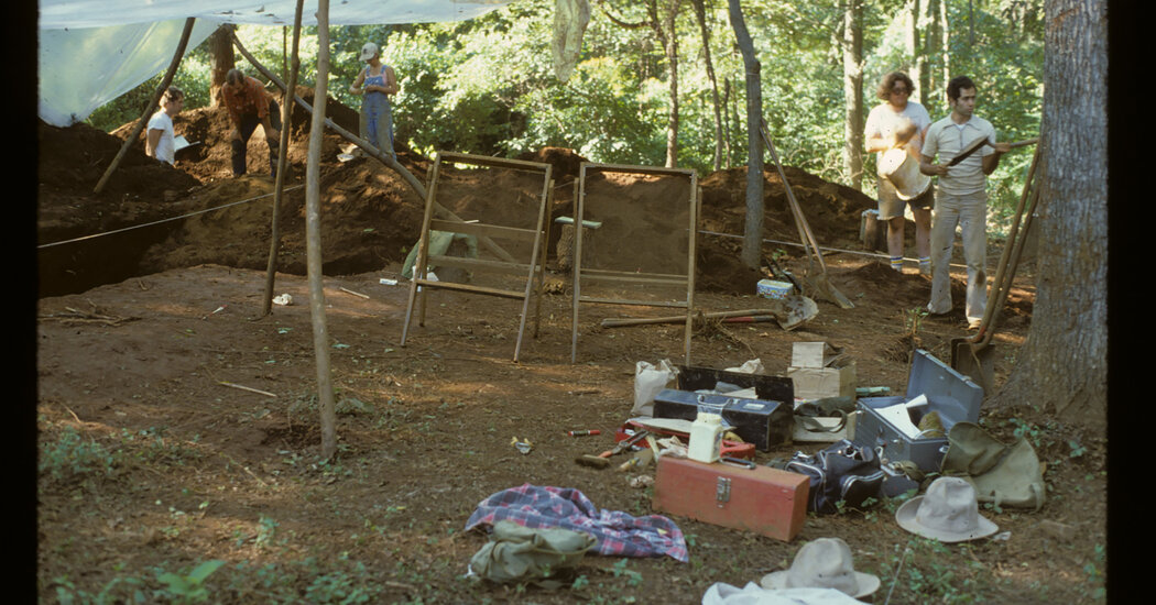
The team’s genetic analysis hoped to further the goals of community stakeholders who wish to “identify and foster a Catoctin descendent community” by identifying previously unknown connections to living descendants. They analyzed human remains from an undocumented African American cemetery under stewardship of the Smithsonian and in coordination with The Catoctin Furnace Historical Society and self-identified descendants of the Catoctin Furnace community.
Most stakeholders agreed that these genetic results should be shared with relevant 23andMe users, but with proper user consent. The researchers note that people might be distressed to find out they have an ancestral connection to slavery, so an opt-in program could be an optimal way of letting users decide for themselves whether they want to learn about their genetic links to Catoctin Furnace or not.
In addition, the team warns that geneticists need to be wary of “biologically gatekeeping” who is and is not a Catoctin Furnace stakeholder. There is already a self-identified community of stakeholders who are connected by cultural and kinship bonds, and any member’s genetic results should not minimize their group identity.
“Returning inaccurate results or returning results in a confusing way may actively harm community stakeholders, people or organizations who serve as stewards for human remains, those who serve in other roles that involve educating the public about specific historical individuals or sites, or broader public understanding,” say the authors. “Reports created by genetic ancestry companies should be created with enough educational content and detail so that customers with shared genetic connections can interpret their results without the need for guidance from outside organizations.”
Even though 23andMe has a genetic dataset more than an order of magnitude larger than any publicly available genetic dataset and thus was an optimal organization for identifying Catoctin descendants and other relatives, many genetic researchers argue that companies should not be able to benefit financially from ancient genetic datasets tied to living descendants. To address this concern, 23andMe and collaborators at Harvard and the Howard Hughes Medical Institute agreed that the Catoctin data could not be used for non-research purposes until after it was made publicly available.
23andMe also refrains from sharing individual-level genetic data in order to protect their customers’ privacy, but this can make it difficult for other researchers to reproduce published results. To address this dilemma, 23andMe has agreed to rerun genetic comparisons of this study upon request for academic and non-profit researchers for a limited amount of time.
“There is no perfect solution to this problem, but weighed against this is the fact that leveraging a genetic database as large as 23andMe’s in ancient DNA powers research that otherwise would be impossible. In fact, it is possible that 23andMe’s high research participation rate can be attributed in part to the strong privacy protections that are offered to research participants,” the authors write in the paper.

Read more →

A gut hormone for controlling appetite doubles as an immune regulator for the fungal microbiome

Peptide YY (PYY), a hormone produced by gut endocrine cells that was already known to control appetite, also plays an important role in maintaining the balance of fungi in the digestive system of mammals, according to new research from the University of Chicago.
In a study published this week in Science, researchers found that specialized immune cells in the small intestine called Paneth cells express a form of PYY that prevents the fungus Candida albicans from turning into its more virulent form. PYY was already known to be produced by endocrine cells in the gut as a hormone that signals satiety, or when an animal has had enough to eat. The new research shows that it also functions as an antimicrobial peptide that selectively allows commensal yeast forms of C. albicans to flourish while keeping its more dangerous forms in check.
“So little is known about what regulates these fungi in our in our microbiome. We know that they’re there, but we have no idea what keeps them in a state that provides health benefit to us,” said Eugene B. Chang, MD, Martin Boyer Professor of Medicine at UChicago and senior author of the study. “We now think that this peptide we discovered is actually important for maintaining fungal commensalism in the gut.”
Regulating the ‘mycobiome’
Chang and his team didn’t set to explore the fungal side of the gut microbiome, or “mycobiome” as he calls it. Joseph Pierre, PhD, a former postdoctoral scholar in Chang’s lab who is now an Assistant Professor of Nutritional Sciences at the University of Wisconsin-Madison, was studying the enteroendocrine cells in mice that produce PYY when he noticed that it was also present in Paneth cells. These are important immune system defenders in the gut of mammals, secreting several antimicrobial compounds to prevent dangerous bacteria from flourishing.
At first this didn’t make sense, because until then, PYY was only recognized as an appetite hormone. When they tested it against a variety of bacteria, it wasn’t very good at killing them either. But when they ran a computer search for other classes of peptides with a similar structure, they discovered one similar to PYY called magainin 2, which is found on the skin of the African clawed frog. This peptide protects the frogs from infection by both bacteria and fungi, so Chang’s team thought to test PYY’s antifungal properties too. As it turns out, it is not only an effective antifungal agent, but a very specific one as well.
C. albicans is a yeast that typically grows in small amounts in the mouth, on the skin, and in the intestines. The basic yeast form is commensal, or coexists peacefully in the body, but given the right conditions it transforms into what are called hyphae that branch out to form biofilms. When too much grows, it causes thrush, an infection in the mouth and throat, vaginal yeast infections, or more serious generalized infections in the body. When Chang’s team tested PYY against both forms of the fungus, it effectively prevented growth and killed the more dangerous hyphae while sparing the commensal Candida yeast.

Read more →

New, simple and accessible method creates potency-increasing structure in drugs

Chemical structures called cyclopropanes can increase the potency and fine-tune the properties of many drugs, but traditional methods to create this structure only work with certain molecules and require highly reactive — potentially explosive — ingredients. Now, a team of researchers from Penn State has identified and demonstrated a safe, efficient and practical way to create cyclopropanes on a wide variety of molecules using a previously undescribed chemical process. With additional development, the new method — described in a paper publishing Aug. 4 in the journal Science — could transform how this important process occurs during drug development and creation.
Cyclopropanes are a key feature in many drugs currently approved by the U.S. Food and Drug Administration, including those used to treat COVID-19, asthma, hepatitis C, and HIV/AIDs. These structures can increase a drug’s potency, alter its ability to dissolve in the body, minimize its interactions with unintended targets, and otherwise fine-tune performance. Cyclopropanes are a ring of three connected carbon atoms, with one carbon attached to the rest of the drug molecule and the other two each attached to two hydrogen atoms.
“Cyclopropanes are an essential component of many drugs and adding them to drug candidates can be an important part of the drug discovery process,” said Ramesh Giri, professor of chemistry in the Eberly College of Science at Penn State and leader of the research team. “Previous efforts to improve the creation of cyclopropanes have focused on altering a mechanistic pathway devolved more than 60 years ago. We approached this from a different angle and identified a completely new pathway that is a simple, practical, and broadly applicable.”
The new method transforms a specific chemical structure on compounds called alkenes — used in the synthesis of many molecules — into cyclopropanes. The method takes advantage of “radical chemistry,” where intermediate steps of reactions leave some carbon atoms with unpaired electrons called free radicals that propel the reaction forward. This specific method uses visible light to initiate the reaction and uses common chemical ingredients, including oxygen.
Traditional methods to create cyclopropanes require highly reactive and difficult-to-acquire ingredients and must be conducted under controlled conditions, and the resulting compounds often have a very short shelf life. These unstable ingredients are critical to producing an intermediate compound in the process called a carbene — a highly reactive carbon atom with two unpaired electrons. The new method completely bypasses the carbene intermediate, producing the unpaired electrons one at a time as radicals.
“All of the ingredients used in this pathway are commercially available or easy to create in the lab and do not require any special safety precautions, and the end product can be stored for prolonged periods,” Giri said. “We can add all the ingredients together in one mixture while exposed to air with as little as 10% oxygen, and it proceeds in one step. The reaction is simple and safe enough that we are even planning to include it as part of an undergraduate chemistry lab.”
Another shortcoming of traditional methods is that they generally do not work with complex molecules. For this reason, cyclopropanes are typically installed early in the synthesis when the molecule is less complex, but following steps can cause the ring to open up, and later attempts to make derivatives of the molecule would require backtracking to those early steps. Using the new method, the researchers successfully transformed a variety of alkenes with a wide range of complexities into cyclopropanes, including pharmaceutically relevant compounds such as the steroid estrone, penicillin and vitamin B.

Read more →

Study finds a surprising new role for a major immune regulator

A signaling protein known as STING is a critical player in the human immune system, detecting signs of danger within cells and then activating a variety of defense mechanisms.
STING is primarily on the lookout for DNA, which can indicate either a foreign invader such as a virus or damage to the host tissue or cell. When STING detects that danger signal, it can turn on at least three different pathways — one leading to interferon production, one to non-canonical autophagy (involved in recycling cell components and clearing pathogens), and a third to formation of the inflammasome, a complex of proteins that activates inflammatory responses. The mechanism by which STING stimulates interferon production is well characterized, but it has not been understood how it activates the other two processes.
Now, a team of MIT and Harvard Medical School researchers has discovered how STING activates those two pathways. They found that STING has a surprising and previously unknown function: It can act as an ion channel that allows protons to leak out of an organelle known as the Golgi body. This makes it the first human immune sensor that can translate danger signals into ion flow.
“Arriving at this new idea that STING is a proton channel required connecting prior findings by other labs that either STING or proton flux could activate the inflammasome and non-canonical autophagy, which led us to hypothesize that STING initiates or mediates proton flux to trigger both downstream processes,” says Nir Hacohen, a member of the Broad Institute of MIT and Harvard, a professor of medicine at Massachusetts General Hospital and Harvard Medical School, and a senior author of the study.
“Because of its importance to host immunity, there is a great interest in developing drugs that can activate or suppress STING activity, and the discovery of STING’s ion channel activity will provide new ways to think about designing therapeutics to modulate STING,” says Darrell Irvine, the Underwood-Prescott Professor at MIT with appointments in the departments of Biological Engineering and of Materials Science and Engineering; a member of MIT’s Koch Institute for Integrative Cancer Research and the Ragon Institute of MGH, MIT, and Harvard; and a senior author of the study.
MIT biology PhD student Bingxu Liu and Rebecca Carlson PhD ’23, a recent graduate of the Medical Engineering and Medical Physics program through the Harvard-MIT Division of Health Sciences and Technology, are the lead authors of the paper, which appears today in Science. Paul Blainey, the Karl Van Tassel Associate Professor of Biological Engineering at MIT and a member of the Broad Institute and the Koch Institute, is also an author of the paper.
A surprising role
STING (short for stimulator of interferon genes) is considered one of the major factors that triggers the immune response in the context of infection, autoimmunity, and cancer. Drugs that activate STING have been developed and tested in clinical trials as cancer immunotherapy drugs that would help stimulate the immune system to destroy tumors.

Read more →

Enslaved Black People in Maryland Linked to 42,000 Living Relatives

The analysis marks the first time historical DNA has been used to trace the descendants and distant cousins of enslaved people, researchers said.A construction team working on a highway expansion in Maryland in 1979 discovered human remains on the grounds of an 18th-century ironworks. Eventually, archaeologists uncovered 35 graves in a cemetery where enslaved people had been buried.In the first effort of its kind, researchers now have linked DNA from 27 African Americans buried in the cemetery to nearly 42,000 living relatives. Almost 3,000 of them are so closely related that some people might be direct descendants.Henry Louis Gates Jr., a historian at Harvard University and an author of the study, published on Thursday in the journal Science, said that the project marked the first time that historical DNA had been used to connect enslaved African Americans to living people.“The history of Black people was intended to be a dark, unlit cave,” Dr. Gates said. With the new research, “you’re bringing light into the cave.”In an accompanying commentary, Fatimah Jackson, an anthropologist at Howard University, wrote that the research was also significant because the local community in Maryland worked alongside geneticists and archaeologists.“This is the way that this type of research should be performed,” Dr. Jackson wrote.The cemetery was located at a former ironworks called the Catoctin Furnace, which started operating in 1776. For its first five decades, enslaved African Americans carried out most of the work including chopping wood for charcoal and crafting items like kitchen pans and shell casings used in the Revolutionary War.Elizabeth Comer, an archaeologist and the president of the Catoctin Furnace Historical Society, said that some of the workers were most likely skilled in ironworking before being forced into slavery.“When you’re stealing these people from their village in Africa and bringing them to the United States, you were bringing people who had a background in iron technology,” she said.Elizabeth Comer, an archaeologist, at an unmarked gravestone in an old African American cemetery in Thurmont, Md., in 2020.Katherine Frey/The Washington Post, via Getty ImagesUpon their discovery, some of the remains were taken to the Smithsonian for curation. In 2015, the historical society and the African American Resources Cultural and Heritage Society in Frederick, Md., organized a closer look.Smithsonian researchers documented the toll that hard labor at the furnace took on the enslaved people. Some bones had high levels of metals like zinc, which workers inhaled in the furnace fumes. Teenagers suffered damage to their spines from hauling heavy loads.The identities of the buried African Americans were a mystery, so Ms. Comer looked through diaries of local ministers for clues. She assembled a list of 271 people, almost all of whom were known only by a first name. One family of freed African Americans, she discovered, supplied charcoal to the furnace operators.From that list, Ms. Comer has managed to trace one family of enslaved workers to living people and one family of freed African Americans to another set of descendants.At Harvard, researchers extracted DNA from samples of the cemetery bones. Genetic similarities among 15 of the buried people revealed that they belonged to five families. One family consisted of a mother laid alongside her two sons.Following Smithsonian guidelines, the researchers made the genetic sequences public in June 2022. They then developed a method to reliably compare historical DNA to the genes of living people.Éadaoin Harney, a former graduate student at Harvard, continued the genetic research after she joined the DNA-testing company 23andMe, focusing on the DNA of 9.3 million customers who had volunteered to participate in research efforts.Dr. Harney and her colleagues looked for long stretches of DNA that contained identical variants found in the DNA of the Catoctin Furnace individuals. These stretches reveal a shared ancestry: Closer relatives share longer stretches of genetic material, and more of them.The researchers found 41,799 people in the 23andMe database with at least one stretch of matching DNA. But a vast majority of those people were only distant cousins who shared common ancestors with the enslaved people.“That person might have lived several generations before the Catoctin individual, or hundreds or thousands of years,” Dr. Harney said.The researchers also found that the people buried at the Catoctin Furnace mostly carried ancestry from two groups: the Wolof, who live today in Senegal and Gambia in West Africa, and the Kongo, who now live 2,000 miles away in Angola and the Democratic Republic of Congo.The Catoctin Furnace in 1933.E.H. Pickering, Historic American Buildings Survey/Library of CongressAbout a quarter of the individuals in the cemetery had only African ancestry. DNA from the rest typically showed traces of ancestry from Britain — the legacy of white men who raped Black women, as the authors noted in their study.Most of the living people with links to the furnace reside in the United States. Almost 3,000 people had especially long stretches of matching DNA, which could mean they are direct descendants or can trace their ancestry to cousins of the Catoctin Furnace workers.A strong concentration of these close relatives is in Maryland, Dr. Gates noted. That continuity contrasts with the Great Migration, which brought millions of African Americans out of the South in the early 20th century.“The thing about Maryland is that it’s a border state,” Dr. Gates said. “What this means is that a lot of people didn’t leave, which is quite interesting.”In advance of the publication of their paper, the researchers shared the results with the two families that Ms. Comey identified through her own research, as well as with the African American Resources Cultural and Heritage Society.Andy Kill, a spokesman for 23andMe, said that the company was willing to share genetic results with relatives who participated in the new study. So far, the company hasn’t been asked.But 23andMe does not have plans to notify the thousands of other customers who have a connection to the enslaved people of the Catoctin Furnace. When customers consent for their DNA to be used for research, the data is stripped of their identities to protect their privacy.“We still have work to do on thinking about the best way to do that, but it’s something we would like to do at some point,” Mr. Kill said.Jada Benn Torres, a genetic anthropologist at Vanderbilt University who was not involved in the research, said rushing out the results would be a mistake.“To take this process slowly gives us time to think about what the different repercussions might be,” she said, “in terms of opening these boxes and looking in and finding answers that we didn’t even know we had questions about.”The Catoctin Furnace is only one of many African American burial grounds scattered across the country. Alondra Nelson, a social scientist at the Institute for Advanced Study in Princeton, N.J., said that similar studies could be carried out with the remains found in them, so long as scientists partner with the people caring for the cemeteries.“If these kinds of projects go forward, it is going to require researchers to have a real engagement with these well-established communities,” Dr. Nelson said.

Read more →

DNA tilts and stretches underlie differences in mutation rates across genomes

Each cell in the body stores its genetic information in DNA in a stable and protected form that is readily accessible for the cell to carry on its activities. Nevertheless, mutations — changes in genetic information — occur throughout the human genome and can have a powerful influence on human health and evolution.
“Our team is interested in a classical question about mutation — why do mutation rates in the genome vary so tremendously from one DNA location to another? We just do not have a clear understanding of why this occurs,” said Dr. Md. Abul Hassan Samee, assistant professor of integrative physiology at Baylor College of Medicine and corresponding author of the work.
Previous studies have shown that the DNA sequences flanking a mutated position — the sequence context — play a strong role in the mutation rate. “But this explanation still leaves unanswered questions,” Samee said. “For example, one type of mutation occurs frequently in a specific sequence context while a different type of mutation occurs infrequently in that same sequence context. So, we think that a different mechanism could explain how mutation rates vary in the genome. We know that each building block or base that makes up a DNA sequence has its own 3D chemical shape. We proposed, therefore, that there is a connection between DNA shape and mutations rates, and this paper shows that our idea was correct.”
The genetic code is “written” as a string of bases that is furthermore underwound or overwound and constrained into loops, all of which is known to influence every aspect of DNA activity. Surprisingly, most genome analyses treat DNA merely as a string of bases and ignore the fact that each base has a shape.
“We built a statistical model using only DNA structural information, otherwise ignoring the sequence data,” said first author Zian Liu, a graduate student in the Samee lab. “We used the model to pinpoint which DNA shape features, such as stretches, twists or tilts, underlie variations of mutation rates in the human genome. Surprisingly, we found that although the sequence context may look very different from one mutation to another, the structural properties are remarkably similar.”
“We found that stretch — the distance between paired building blocks in the two DNA strings forming the double helix — is one of the top structural properties that defines whether a location is mutable,” Liu said.
“Although we were expecting these results, we did not anticipate that for all types of mutations the same DNA structural feature, stretch, would be similarly important in affecting the mutation rate,” Samee said. “DNA tilt was the second structural feature that most influenced mutation rate of all types. We confirmed that DNA shape is important in functionally relevant regions of the human genome, such as protein-DNA binding sites that regulate gene expression, and that this structural mechanism is conserved across many species.”
The DNA-shape models of mutation rates developed by Liu and Samee showed similar or improved performance when compared to sequence-based models and accurately characterized mutation hotspots. This study supports considering DNA shape when studying mechanisms of mutation rate variations in the human genome.

Read more →

Potential new tool for early identification of dementia risk

Research at the Florida State University College of Medicine has identified a potential low-cost method for predicting if a person is at risk of developing dementia.
By analyzing data from nearly 13,000 subjects who participated in a long-term aging study, researchers found that an interviewer’s rating of a cognitively healthy person’s memory successfully predicted the likelihood of developing dementia over a 15-year period. Their findings will be published in the Journal of Alzheimer’s Disease.
“Our findings show that interviewers were able to detect deficits in the memory of participants that predicted higher risk of developing dementia over time,” said research author Angelina Sutin, professor of Behavioral Sciences and Social Medicine. “The interviewer ratings of memory were particularly important for participants who were among the top performers on objective memory tests.”
The results show that ratings of a person’s memory performance by an interviewer could be a valuable alternative or addition to other methods of detection, such as self reporting or cognitive testing.
Sutin’s team analyzed 15 years of data involving nearly 13,000 people without cognitive impairment at baseline who participated in the University of Michigan Health and Retirement Study, or HRS, and whose memory was rated by their interviewer. The longitudinal study surveys a representative sample of adults aged 50 and older about their health, financial situation and well-being every two years for as long as they choose to remain in the study.
Sutin’s sample included participants who were interviewed in 2006 and scored within the normal range of cognitive function during their first interview and had at least one follow-up assessment of cognition between 2008-2020.
Interviewers were trained research assistants working for the HRS who conducted the 2-3 hour interviews and rated the item, “How much difficulty did the respondent have remembering things that you asked (him/her) about?” from 1 (no difficulty) to 5 (could not do at all).

Read more →

Newly identified lipid in breast milk might reduce cerebral palsy in infants

While it’s known that the white matter loss will lead to neurological deficits, there is currently no treatment to help these infants avoid the outcome.
In experiments using neonatal mice, researchers at Duke Health have identified a fatty molecule in breast milk that triggers a process in which stem cells in the brain produce cells that create new white matter, reversing the injury.
The study appears Aug. 3 in the journal Cell Stem Cell. Eric Benner, M.D., Ph.D., is the study’s corresponding author and is a distinguished assistant professor in the Department of Pediatrics at Duke University School of Medicine. Benner said further study in a clinical trial is needed, but the finding is promising.
“Developing therapies for children — especially such medically fragile children — is very difficult to do because there are justifiably strict safety concerns,” Benner said. “The fact that this molecule is already found in something that is safe for premature babies — breast milk — is extremely encouraging.
“It’s been known that fats in breast milk benefit a child’s brain development, but there are many types of fats in breast milk,” Benner said. “This work has identified a lipid molecule in breast milk that promotes white matter development. Now, we can begin to develop a therapy that isolates and delivers this lipid in a way that is safe for the unique challenges of these infants.”
Benner is a neonatologist at Duke University and one of the co-founders of Tellus Therapeutics, a Duke spinout company developed with the help of the Duke University Office for Translation & Commercialization to bring this therapy from the bench into the neonatal intensive care unit.
The fatty molecule identified in the study will be administered intravenously to patients in an upcoming clinical trial. This is significant because many of the infants who are part of this vulnerable population also have gastrointestinal issues and cannot safely be given milk or medication by mouth.

Read more →

How the gut signals to the brain

The gut-brain connection is a complicated two-way signaling cascade that is responsible for keeping the digestive system working properly and can cause problems when it breaks down. A key part of that axis is the colon, which extracts water and nutrients from food and transports waste out of the body. This crucial organ is implicated in a range of gastrointestinal conditions, including constipation, diarrhea, pain, and inflammation.
Now, in a first, researchers at Harvard Medical School have defined five distinct subtypes of sensory neurons in the colon that carry signals to the brain.
In a new study, conducted in mice and published Aug. 3 in Cell, the researchers found that some neurons are dedicated to sensing gentle forces, such as substances moving through the colon, while others sense more intense ones, such as pain.
The researchers say that, if confirmed in humans, their findings could help scientists develop more effective therapies to treat conditions that arise when this colon-brain sensing system goes awry.
“Patients often complain about sensation and pain in the gastrointestinal system, yet we don’t know a lot about the sensory neurons that innervate the gut and allow us to respond to different stimuli,” said lead author Rachel Wolfson, a research fellow in neurobiology at HMS and a gastroenterology fellow at Massachusetts General Hospital.
From skin to colon
David Ginty and scientists in his lab have spent many years studying how sensory neurons in the skin communicate with the brain to form our sense of touch. They have developed precise genetic tools that label subtypes of sensory neurons and used these tools to uncover basic information about the structure, organization, and function of skin-sensing neurons.

Read more →

Dopamine controls movement, not just rewards

Dopamine: It’s not just for rewards anymore.
In a new Northwestern University-led study, researchers identified and recorded from three genetic subtypes of dopamine neurons in the midbrain region of a mouse model.
Although there is a long-standing, common assumption that most — if not all — dopamine neurons solely respond to rewards or reward-predicting cues, the researchers instead discovered that one genetic subtype fires when the body moves. And, even more surprisingly, these neurons curiously do not respond to rewards at all.
Not only does this finding shed new light on the mysterious nature of the brain, it also opens new research directions for further understanding and potentially even treating Parkinson’s disease, which is characterized by the loss of dopamine neurons yet affects the motor system.
The study will be published on Thursday (Aug. 3) in the journal Nature Neuroscience.
“When people think about dopamine, they likely think about reward signals,” said Northwestern’s Daniel Dombeck, who co-led the study. “But when the dopamine neurons die, people have trouble with movement. That’s what happens with Parkinson’s disease, and it’s been a confusing problem for the field. We found a subtype that are motor signaling without any reward response, and they sit right where dopamine neurons first die in Parkinson’s disease. That’s just another hint and clue that seems to suggest that there’s some genetic subtype that’s more susceptible to degradation over time as people age.”
“This genetic subtype is correlated with acceleration,” added Northwestern’s Rajeshwar Awatramani, who co-led the study with Dombeck. “Whenever the mouse accelerated, we saw activity, but in contrast we did not see activity in response to a rewarding stimulus. This goes against the dogma of what most people think these neurons should be doing. Not all dopamine neurons respond to rewards. That’s a big change for the field. And now we found a signature for that dopamine neuron that does not show reward response.”
Dombeck is a professor of neurobiology at Northwestern’s Weinberg College of Arts and Sciences. Awatramani is the John Eccles Professor of Neurology at Northwestern University Feinberg School of Medicine. The paper’s first authors are Maite Azcorra and Zachary Gaertner, both graduate students in Dombeck’s and Awatramani’s laboratories.

Read more →