A new ally in fighting brain diseases: Our very own skull

Alzheimer’s, stroke, multiple sclerosis and other neurological diseases cause severe damage due to neuroinflammation mediated by immune cells. Managing this inflammation poses a significant medical challenge because the brain is protected by the skull and additional surrounding membranes that make the brain less accessible for treatment approaches. Scientists had previously discovered pathways going from the bone marrow of the skull towards the brain, allowing immune cell movement. Now, new research revealed that cells in the skull’s bone marrow are unique in their composition and in their disease response. These findings offer new possibilities for the diagnosis and treatment of neurological diseases and revolutionize brain health monitoring in the future with non-invasive skull imaging. The results are now published in Cell.
Neurological diseases such as Alzheimer’s, stroke, and multiple sclerosis have a devastating impact on the lives of millions worldwide. A common feature is neuroinflammation, an internal “fire” in the brain that can cause severe damage by activation of immune cells and release of inflammatory molecules. However, due to the brain’s relative inaccessibility, as it is shielded by the skull and three additional layers of protection in the form of membranes, controlling and monitoring this inflammation has been a major challenge. A team of scientists around Prof. Ali Ertürk at Helmholtz Munich in collaboration with researchers from the Ludwig-Maximilians-Universität München (LMU) and the Technical University of Munich (TUM) sought to address this unmet need.
Not Just a Helmet: The Intricate Connection Between the Skull and Brain
Defying traditional understanding that the skull and the brain have no direct interchange, recent studies have unveiled direct connections between the skull’s bone marrow and the brain’s outermost surface of the protective membranes, the meningeal surface. These connections act as conduits, facilitating the movement of immune cells back and forth. The team of scientists found that these connections often traverse even through the outermost and toughest layer of membrane, the dura, opening up even closer to the brain surface than previously thought. To achieve these significant findings, the team utilized a specialized method called tissue clearing in combination with 3D imaging to visualize the conduits. During the tissue clearing process biological tissues are treated with a specific solution to render them transparent enabling the passage of light for the examination of both brain tissue and the skull under a microscope. As a result, 3D images of structures and cells were generated, leading to a comprehensive visual analysis.
The research team probed even deeper into the distinct role the skull-based immune cells play in brain physiology and diseases. They began by questioning if the skull harbors unique brain-specific cells and molecules that cannot be found in other bones. Extensive analysis of the RNA and protein content in the form of transcriptomics and proteomics analyses of both mouse and human bones affirmed this — the skull is indeed exceptional, hosting unique neutrophil immune cells, which are a type of white blood cell that play a critical role in the immune system’s defense. “These findings carry profound implications, suggesting a far more complex connection between the skull and the brain than previously believed” highlights the first author of the study Ilgin Kolabas, Ph.D.-student at the Ertürk lab at Helmholtz Munich.
Ali Ertürk, corresponding author, adds: “This opens up a myriad of possibilities for diagnosing and treating brain diseases and has the potential to revolutionize our understanding of neurological diseases. This breakthrough could lead to more effective monitoring of conditions such as Alzheimer’s and stroke, and potentially even aid in preventing the onset of these diseases by enabling early detection.”
Envisioning a New Future: From Research to Clinical Practice
Another impactful finding was that using PET imaging, the researchers discovered that signals from the skull mirrored those from the underlying brain, with changes in these signals corresponding to disease progression in patients with Alzheimer’s and stroke. Thereby showcasing a new potential to monitor brain inflammation simply by scanning the surface of the patient’s head.
Looking forward, the researchers envision that their findings could translate to clinical practice in the form of non-invasive skull imaging. Ali Ertürk explains the impact on disease monitoring: “This could potentially be done using portable and wearable devices, offering a more accessible and practical way to monitor brain health.” The team hopes that this approach will greatly improve the diagnosis, monitoring, and possibly even treatment of neurological disorders, bringing us a step closer to more effective management of these devastating conditions.

Read more →

Brain cells of males and females respond differently to chronic stress

Scientific excellence requires diversity — research conducted by men and women, by people from different backgrounds and with varied worldviews. The need for diversity extends to scientific experiments themselves, but even today the vast majority of studies in the life sciences are done on male mice only, which could harm the findings, as well as our ability to extrapolate from them to humans. A new study by researchers from the Weizmann Institute of Science addresses this challenge, revealing in unprecedented detail how the brains of male and female mice respond differently to stress. In the study, published in Cell Reports, researchers from Prof. Alon Chen’s joint laboratory at the Weizmann Institute and the Max Planck Institute of Psychiatry in Munich discovered that a subcategory of brain cells responds to stress in a totally different manner in males and females. The findings could lead to a better understanding of health conditions affected by chronic stress, such as anxiety, depression and even obesity and diabetes, and they could pave the way toward personalized therapies for these disorders.
Mental and physical disorders caused by chronic stress are constantly on the rise, putting a significant strain on society. They affect both men and women, but not necessarily in the same way. Although plenty of evidence suggests that men and women deal differently with stress, the causes of these differences are not yet fully understood, and in any event, personalized treatments for men and women are still beyond the reach of medicine. But researchers from Chen’s laboratory, which specializes in studying the response to stress, hypothesized that innovative research methods could help to change the picture. Previous studies in other labs had uncovered certain sex differences in the response to stress, but those findings were obtained using research methods that could mask significant differences in the responses of specific cells or even entirely erase the roles played by relatively rare cells. Chen’s laboratory, in contrast, uses advanced methods that allow scientists to analyze brain activity at an unprecedented resolution — on the level of the individual cell — and could therefore shed new light on the differences between the sexes.
“We turned the most sensitive research lens possible onto the area of the brain that acts as a central hub of the stress response in mammals, the paraventricular nucleus (PVN) of the hypothalamus,” says Dr. Elena Brivio, who led the study. “By sequencing the RNA molecules in that part of the brain on the level of the individual cell, we were able to map the stress response in male and female mice along three main axes: how each cell type in that part of the brain responds to stress, how each cell type previously exposed to chronic stress responds to a new stress experience and how these responses differ between males and females.”
The researchers mapped out gene expression in more than 35,000 individual cells, generating a huge amount of data that provides a picture of stress response that’s unprecedented in its scope and in highlighting the differences between how males and females perceive and process stress. As part of the study, and in keeping with the principles of open-access science, the researchers decided to make the entire detailed mapping publicly available on a dedicated interactive website, which went live at the same time the study was published, providing other researchers with convenient, user-friendly access to the data. “The website will, for example, allow researchers who are focusing on a specific gene to see how that gene’s expression changes in a certain cell type in response to stress, in males as well as females,” Brivio explains.
The comprehensive mapping has already allowed the researchers to identify a long list of differences in gene expression — between males and females, and between chronic and acute stress. The data showed, inter alia, that certain brain cells respond differently to stress in males and females: Some cells are more susceptible to stress in females and some to stress in males. The most significant difference was found in a type of brain cell called the oligodendrocyte — a subtype of glial cell that provides support to nerve cells and plays an important role in regulating brain activity. In males, exposure to stress conditions, especially chronic stress, changed not only the gene expression in these cells and their interactions with surrounding nerve cells but also their very structure. In females, however, no significant change was observed in these cells, and they were not susceptible to stress exposure. “Neurons attract most of the scientific attention, but they only make up approximately a third of all cells in the brain. The method we implemented allows us to see a much richer and fuller picture, including all the cell types and their interactions in the part of the brain under study,” says Dr. Juan Pablo Lopez, a former postdoctoral fellow in Chen’s group and now the head of a research group at the Department of Neuroscience of the Karolinska Institute in Sweden.
Basic diversity
Until the 1980s, clinical trials of new drugs were conducted on men alone. The accepted view was that including women was unnecessary, and that it would only complicate the research, bringing into play new variables such as menstruation and hormonal changes. For the same reasons, preclinical studies avoided using female animals until very recently. But it’s now known that the variability among male animals, on a molecular and behavioral level, is usually greater than among females, so there is no reason to suppose that females would complicate the experiments any more than males. Nonetheless, in basic research it’s still common to conduct experiments only on males. “Our findings show that, when it comes to stress-related health conditions, from depression to diabetes, it’s very important to take the sex variable into account, since it has a significant impact on how different brain cells respond to stress,” Chen explains. “Even if a study does not specifically focus on the differences between males and females, it’s essential to include female animals in the research, especially in neuroscience and behavioral science, just as it is important to implement the most sensitive research methods, in order to obtain as complete a picture of brain activity as possible,” Brivio adds.
Also participating in the study were Dr. Aron Kos, Stoyo Karamihalev, Andrea Ressle, Rainer Stoffel and Dr. Mathias V. Schmidt of the Max Planck Institute of Psychiatry in Munich; Dr. Alessandro Francesco Ulivi of the Leibniz Institute for Neurobiology in Magdeburg, Germany; Dana Hirsch of Weizmann’s Veterinary Resources Department; and Dr. Gil Stelzer of Weizmann’s Life Sciences Core Facilities Department.

Read more →

Treatments for poxviruses — including those causing mpox and smallpox — may already exist in licensed drugs

Scientists studying how poxviruses evade natural defences in human cells have identified a new approach to treatment that may be more durable than current treatments.
This follows their discovery of how poxviruses exploit a cellular protein to evade the host cell defences, and thereby replicate and spread effectively.
Existing drugs developed to be immunosuppressive, or treat other viral infections target this cellular protein. The team found that these drugs can also restrict the replication and spread of poxviruses.
This approach to treatment, in which the drug does not directly target the virus, means that it will be much more difficult for the virus to evolve drug-resistance.
And because this hijacking mechanism is the same across many poxviruses, the drugs will be effective in treating a range of diseases such as mpox and smallpox.
The research is published today in the journal Nature.
Despite the fact that smallpox has been eradicated as a disease since 1979, the virus that causes it, variola, is still held in two high security labs — one in the United States, and one in Russia. The threat of variola virus being used in bioterrorism has led to a drug, tecovirimat, being licensed to treat smallpox.

Read more →

Exercise apps a good prescription to boost healthcare workers' mental health

Simple home workouts using exercise apps can effectively reduce depressive symptoms in healthcare workers and could be a major tool to combat the global mental health crisis in the sector, says new University of British Columbia research.
The study, published today in JAMA Psychiatry, divided participants into either a waitlisted control group or an exercise group who were given free access to a suite of home exercise apps called DownDog, that included yoga, cardio and strength training. They were asked to aim for at least 80 minutes of moderate-intensity exercise per week over 12 weeks.
“The exercise group reported significantly lower levels of depressive symptoms compared to the control group as the study progressed,” said first author Dr. Vincent Gosselin-Boucher, a postdoctoral fellow at UBC’s school of kinesiology. “The majority of the participants in the study started very high on the scale for depressive symptoms compared to the general population so the changes were actually quite amazing to see.”
The positive effects were most pronounced among participants who completed an average of at least 80 minutes of exercise per week.
In addition to depressive symptoms, the researchers measured burnout symptoms and sick days over the two-week period prior to the beginning of their participation in the study, and every two weeks during the trial, while the exercise apps tracked participants’ workouts.
Exercise improved two measured facets of burnout, namely cynicism and emotional exhaustion. The exercise group also reported fewer sick days than the control group.
Using physical activity to boost mental health
While previous research has shown that exercise can improve depressive symptoms, researchers said behavioural approaches such as exercise were missing in the mental health initiatives offered by healthcare institutions around the world to address covid-19’s toll on their staff’s mental health.

Read more →

Engineered probiotic developed to treat multiple sclerosis

Brigham researchers are working on a new approach to target autoimmunity in the brain leverages designer bacteria to make treatment safer and more effective
Researchers from Brigham and Women’s Hospital, a founding member of the Mass General Brigham healthcare system, have designed a probiotic to suppress autoimmunity in the brain, which occurs when the immune system attacks the cells of the central nervous system. Autoimmunity in the brain is at the core of several diseases, including multiple sclerosis. In a new study, researchers demonstrated the treatment’s potential using preclinical models of these diseases, finding that the technique offered a more precise way target brain inflammation with reduced negative side effects compared to standard therapies. The results are published in Nature.
“Engineered probiotics could revolutionize the way we treat chronic diseases,” said lead author Francisco Quintana, PhD, of the Ann Romney Center for Neurologic Diseases at Brigham and Women’s Hospital. “When a drug is taken, its concentration in the bloodstream peaks after the initial dose, but then its levels go down. However, if we can use living microbes to produce medicine from within the body, they can keep producing the active compound as its needed, which is essential when we consider lifelong diseases that require constant treatment.”
Autoimmune diseases affect approximately 5-8% of the U.S. population. Despite their widespread prevalence, there are limited treatment options for most of these diseases. Autoimmune diseases that affect the brain, such as MS, are particularly challenging to treat due to their location — many pharmacological therapies can’t effectively access the brain due to the blood-brain barrier, a protective mechanism that separates the brain from the circulatory system.
To look for new ways to treat autoimmune diseases, the researchers studied dendritic cells, a type of immune cell that is abundant in the gastrointestinal tract and in the spaces around the brain. These cells help control the rest of the immune system, but scientists don’t yet know their role in autoimmune diseases. By analyzing dendritic cells in the central nervous system of mice, they were able to identify a biochemical pathway that dendritic cells use to stop other immune cells from attacking the body.
“The mechanism we found is like a brake for the immune system,” said Quintana. “In most of us, it’s activated, but in people with autoimmune diseases, there are problems with this brake system, which means the body has no way to protect itself from its own immune system.”
The researchers found that this biochemical brake can be activated with lactate, a molecule involved in many metabolic processes. The researchers were then able to genetically engineer probiotic bacteria to produce lactate.

Read more →

Unlocking the power of citizen science to monitor health and wellbeing targets

The World Health Organization’s Triple Billion Targets, which are largely based on the SDGs, aim to ensure that one billion more people benefit from universal health coverage; one billion more people are better protected from health emergencies, and that one billion more people are able to enjoy better health and wellbeing by 2025. To achieve these ambitious goals, informed decision making based on robust data collection and monitoring efforts is essential.
While data availability has improved in recent years, gaps in routine surveillance persist, impeding effective policy formulation and action. In addition, the COVID-19 pandemic has further emphasized the critical need for enhanced data and strengthened health information systems to enable timely decisions that can save lives. Traditional data sources, such as nationally representative surveys, are insufficient and securing funding for monitoring systems using conventional data sources presents significant challenges.
In this context, citizen science, which refers to public participation in scientific research and knowledge production, emerges as a promising avenue to address these data gaps efficiently and sustainably. By involving citizens in data collection and analysis, as well as in mobilizing action, this approach can contribute to better monitoring and ultimately achieving health and wellbeing-related SDGs and Triple Billion Targets.
The systematic review, which has just been published in the journal Frontiers in Public Health, demonstrates that citizen science has the potential to monitor 48 out of 58 health and wellbeing-related indicators covered. These contributions primarily occur at the local and community levels and can be scaled up to the national and global levels. According to the authors, citizen science has the power to increase the availability, quality, granularity, applicability, and timeliness of health-related data, effectively filling critical gaps in monitoring efforts.
“Establishing trusted partnerships with key stakeholders is crucial to integrating citizen science with official health and wellbeing statistics. Collaboration with National Statistical Offices, governments, academia, and custodian agencies, including the WHO, is essential to showcase the value of citizen science in health monitoring,” explains Dilek Fraisl, lead author of the study and a researcher in the Novel Data Ecosystems for Sustainability Research Group of the IIASA Advancing Systems Analysis Program. “By forging partnerships, citizen science data can seamlessly integrate with official statistics, thereby strengthening the overall monitoring processes.”
The findings of the review highlight that citizen science can directly contribute to or complement the monitoring of 83% of health and wellbeing-related indicators. Notably, the examples analyzed primarily come from low- and middle-income countries, aligning with the “leaving no one behind” principle of the SDG agenda and considering the needs of the most vulnerable populations.
“This is an important study. It provides a systematic review of citizen science data for health-related indicators, providing valuable insights to where the WHO can explore potential alternative data sources. Given the pioneering nature of this study, the WHO was very happy to collaborate with a credible institution like IIASA,” reflected Steve MacFeely, Director of Data and Analytics at WHO.
The authors encourage future research to build on their findings by identifying citizen science initiatives with the greatest potential based on the data gaps and needs of custodian agencies such as the WHO and National Statistical Offices. By focusing on data management processes and data sets of specific projects, they say, further studies can provide valuable insights into optimizing citizen science for monitoring health and wellbeing-related targets.
“Citizen science presents a promising approach to monitor health and wellbeing-related indicators of the SDGs and Triple Billion Targets. By engaging citizens in data collection and analysis, citizen science can help to bridge critical data gaps, particularly at the local and community levels. Leveraging the power of citizen science effectively will advance the achievement of health and wellbeing goals outlined in the SDG framework and WHO’s Triple Billion Targets, creating a healthier and more sustainable future for all,” concludes Linda See, a study coauthor and senior researcher in the Novel Data Ecosystems for Sustainability Research Group.

Read more →

Tau-PET : Future of Alzheimer's patients

Alzheimer’s disease, one of the most common neurodegenerative diseases, leads to progressive loss of memory and autonomy. It is characterised by the accumulation of neurotoxic proteins in the brain, namely amyloid plaques and tau tangles. Due to the silent development of pathology over decades, very early diagnosis is of utmost importance to be able to take action as early as possible in the disease process. A team from the University of Geneva (UNIGE) and the Geneva University Hospitals (HUG) has demonstrated that tau PET — a novel imaging technique for visualising the tau protein — can predict cognitive decline in patients much better than the imaging techniques normally used. These results, published in Alzheimer’s & Dementia: The Journal of the Alzheimer’s Association, argue in favour of the rapid introduction of tau PET into the clinical routine to provide patients with early and individualised solutions.
Today, one of the main diagnostic tools for Alzheimer’s disease is positron emission tomography (PET), an imaging technique in which tracers are injected to visualise specific pathological processes in the brain. “PET involves injecting patients with low-level radioactive tracers that disappear within a few hours. They are designed to bind to the human molecules that we want to detect, making them visible with the PET tomographs,” explains Valentina Garibotto, Associate Professor in the Department of Radiology and Medical Informatics at UNIGE Faculty of Medicine and Head Physician in the Division of Nuclear Medicine and Molecular Imaging at the HUG, who directed this research.
“Specific tracers for amyloid exist since two decades, and tracers to monitor glucose metabolism, which indicates the brain’s ability to use its energy resources correctly, have long existed. However, Alzheimer’s disease is complex and these two techniques are not enough to provide all the answers.”
Comparing imaging techniques
Flortaucipir is a radiotracer that binds to the tau protein. It was developed by a pharmaceutical company and approved by the Food and Drug Administration (FDA) in 2020. It allows the detection of tau accumulation as well as its distribution in the brain to precisely assess its role in the clinical manifestation of the disease. Scientists from the UNIGE and the HUG wanted to determine which imaging modality — amyloid PET, glucose metabolism PET, or tau PET — would best predict future cognitive decline due to Alzheimer’s disease. Around 90 participants were recruited at the HUG Memory Centre.
“Our results show that while the various PET measures were all associated with the presence of cognitive symptoms, confirming their role as strong indicators of Alzheimer’s disease, tau PET was the best to predict the rate of cognitive decline, even in individuals with minimal symptoms,” summarises Cecilia Boccalini, a PhD student in Professor Garibotto’s team and first author of this study.
Detecting individual variations
Amyloid plaques are not necessarily accompanied by cognitive or memory loss. However, the presence of tau goes hand in hand with clinical symptoms. Its absence or presence is the main determinant of whether a patient’s condition remains stable or deteriorates rapidly. It has been more difficult to develop imaging techniques to visualise tau, mainly because of its lower concentration and particularly complex structure.
“This breakthrough is crucial for better management of Alzheimer’s disease. Recently, drugs targeting amyloid have shown positive results. New drugs targeting the tau protein also look promising. By detecting the pathology as early as possible, before the brain is further damaged, and thanks to new treatments, we hope to be able to make a greater impact on patients’ future and quality of life,” Valentina Garibotto points out. “Similarly, we are beginning to map the distribution of tau in order to understand how its location in the different regions of the brain influences symptoms.” Indeed, the causes and different stages of the disease are proving to be much less uniform than previously thought, and individual susceptibility to the same phenomena needs to be better understood.
These results are a strong argument in favour of incorporating tau PET into the routine clinical evaluation to assess individual prognosis and select the most appropriate therapeutic strategy for each patient.

Read more →

'Humanized' liver in mice reveals roots of chronic diseases

Yale researchers have created a functional “humanized” liver in living mice that will help scientists find human-specific mechanisms for regulating cholesterol levels and potentially for treating chronic liver diseases afflicting tens of millions of people in the United States.
The findings are published Aug. 9 in the journal Cell.
Chronic liver diseases such as alcoholic and non-alcoholic liver disease, cancer, viral hepatitis, fibrosis, and cancer affect more than 1.5 billion people worldwide. In the U.S., an estimated 30 to 40% of the population has been diagnosed with non-alcoholic fatty liver disease alone. Yet liver disease has been difficult to study in animal models. The livers of mice, for instance, perform different functions than those of humans.
“Inside the liver multiple human cell types talk in their own language,” said senior author Richard Flavell, Sterling Professor of Immunobiology at Yale School of Medicine and investigator for the Howard Hughes Medical Institute. “Mouse and human cells talk in different languages, but we have enabled human liver cells to speak in their own language within living mice.”
For the study, a team of scientists led by Eleanna Kaffe, an associate research scientist in Flavell’s Lab, used progenitor stem cells and mature cells known as hepatocytes from a human liver to create a complete human liver in a mouse model. The humanized liver, researchers said, developed into similar size-adjusted shape and carried out similar cellular functions as a healthy human liver. The cellular functions in the humanized liver could also be manipulated to mimic human fibrosis and non-alcoholic fatty liver disease, the researchers report.
The researchers also found that essential liver metabolism is controlled by activity in endothelial cells, which line blood vessels that feed the liver. Those endothelial cells, they said, secrete a signaling molecule called Wnt which regulates cholesterol transport to hepatocytes for the synthesis of bile acid. The transport of cholesterol to hepatocytes is an important mechanism that reduces excess blood cholesterol levels in humans.
According to the researchers, the humanized liver model can be used immediately by drug companies seeking to assess safety of experimental drugs designed to treat chronic diseases.
“However, our long-term goal is to find ways to predict, prevent, and treat all liver diseases, which take such a huge toll on individuals,” the authors said.

Read more →

Alice K. Ladas, Author of Landmark Book on Female Sexuality, Dies at 102

Working with collaborators, she wrote “The G Spot,” which became a cultural sensation and sold more than a million copies.Alice Kahn Ladas, a psychologist and psychotherapist whose best-selling 1982 book, “The G Spot and Other Recent Discoveries About Human Sexuality,” created a tipping point for female sensual autonomy by introducing ways for women to experience greater sexual pleasure, died on July 29 at her home in Santa Fe, N.M. She was 102.Her daughter Robin Janis confirmed the death, adding that Dr. Ladas was still seeing patients at her home office the day before she died.Her book, written with the researchers Beverly Whipple and John Perry, examined the existence of the G-spot, a patch of erectile tissue that can be felt through the front wall of the vagina, behind the pubic bone. (The tissue is named for Ernst Gräfenberg, a German physician who was the first person to write about it in modern medical literature.) The book compared the G-spot to the male prostate: Each, when stimulated, can produce a sexual response similar to an orgasm.For their research, Dr. Whipple and Dr. Perry interviewed and tested some 400 women in Florida, all of whom all were able to locate their G-spots.“My role was to see the connection,” Dr. Ladas told The Santa Fe Reporter in 2010. “There was a vaginal orgasm, there was a clitoral orgasm, but they’re not exclusive.”The book, which has been translated into multiple languages and has sold more than one million copies, was revolutionary in helping women understand their sexual function, especially regarding female ejaculation.Still, the book proved controversial within the medical community, as women flocked to doctors wondering if they were experiencing ejaculation or urinary incontinence during intercourse. Some doctors questioned the depth of the authors’ research and whether the book was meant to be a medical tool or simply a “how to” handbook for women.“‘The G Spot’ reads like a scientific study, when it isn’t,” Dr. Martin Weisberg, then an assistant professor of obstetrics, gynecology and psychiatry at Jefferson Medical College, told The New York Times after the book was published.But Dr. Robert Francoeur, then a professor of human sexuality at Fairleigh Dickinson University, argued differently: “The professional jealousy is incredible in terms of sex educators, therapists and doctors. The nasty comments from professionals sound like they’re upset that they didn’t write the book.”In 2021, the National Institutes of Health published a review of 31 studies on the G spot and found that they “did systematically agree” on its existence.“Among the studies in which it was considered to exist, there was no agreement on its location, size, or nature,” the N.I.H. review said, concluding, “The existence of this structure remains unproved.”“The G Spot and Other Recent Discoveries About Human Sexuality,” published in 1982, sold over a million copies.Holt/Metropolitan BooksAlice Kahn was born in Manhattan on May 30, 1921, to Rosalie Heil Kahn, an early supporter of the ethical culture movement, an effort to develop humanist codes of behavior, and Myron Daniel Kahn, a cotton merchant. Her parents divorced when she was 2, and she spent winters with her mother in Manhattan and extended summer vacations with her father in Montgomery, Ala.She attended the Ethical Culture Fieldston School in Manhattan from kindergarten through high school and enrolled at Smith College in Massachusetts, graduating cum laude in 1943 with a Bachelor of Arts Degree in political science and as a member of the honor society Phi Beta Kappa. She received a master’s in social work from Smith in 1946.While at Smith, Dr. Ladas met Eleanor Roosevelt while participating in a student leadership program at Campobello, the presidential summer retreat in New Brunswick. Inspired by the first lady’s feminism and activism, Dr. Ladas marched for civil rights in the South and in Washington.Dr. Ladas became a follower of the controversial Austrian psychologist Wilhelm Reich, developer of psychosexual theories centered on the orgasm, and joined his staff in New York in the early 1950s. In 1956, she helped Reich’s student Alexander Lowen found the Institute for Bioenergetic Analysis, with its focus on the bodily underpinnings of mental health.Intrigued by infants and breastfeeding, Dr. Ladas soon went to France to study the Lamaze method of childbirth, whereby women are encouraged to move around and use controlled breathing and relaxation as tools to begin labor. Returning to the United States, she became, in 1959, one of the first to teach Lamaze classes there.She received her doctorate in education from Teachers College at Columbia University in 1970. Her dissertation on breastfeeding had initially been refused by faculty members until she persuaded the anthropologist Margaret Mead to sit on her dissertation committee. Dr. Ladas’s research was ultimately published in peer-reviewed journals in medicine and sociology.“That’s what I’m most proud of,” she told a Smith alumni magazine for a profile about her this year. “I believe it influenced — in the United States, at least — more women to breastfeed.”She married Harold Ladas, a psychology professor at Hunter College in New York, in 1963; he died in 1989. In addition to her daughter Robin, she is survived by another daughter, Pamela Ladas, and three grandchildren.In the 1970s, Dr. Ladas served on the boards of the Society for the Scientific Study of Sexuality, in Allentown, Pa., and the International Institute of Bioenergetic Analysis, based in Barcelona, Spain. A study she conducted with her husband about the effects of body psychotherapy on women’s sexuality led to her collaboration with Dr. Whipple and Dr. Perry.Dr. Ladas was a protégé of Adelle Davis, a nutritionist who taught her about organic foods and the importance of exercise. Dr. Ladas snorkeled and played tennis into her 90s and played piano even after she turned 100, her daughter said.Two nights before she died, she and a friend went to see the movie “Oppenheimer,” about the developer of the atomic bomb. It was “not history to her,” her daughter said, because “that was what she lived.”

Read more →

California Battles Fentanyl With a New Tactic: Treating Addiction in Prison

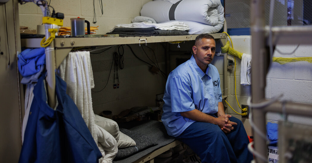
In response to soaring overdose deaths in state prisons, California started a sprawling addiction program. But keeping people on medication during and after a sentence can be difficult.In blistering 100-degree heat one recent afternoon at Valley State Prison in California’s Central Valley, inmates crowded around small windows in a prison yard to pick up their daily doses of buprenorphine, an opioid addiction medication.At one window, Quennie Uy, a nurse, scanned inmate identification cards, then retrieved strips of the medication, slipping them through a sliding panel below the window. One by one, inmates deposited the strips in their mouths, then flashed their palms — proof they had not pocketed the drug that was helping to stanch their cravings.The daily ritual is part of a sprawling health experiment in California that aims to unwind the often lasting damage of opioid use before, during and after incarceration. The state’s efforts also reflect the beginnings of a potential transformation in the nation’s approach to treating addiction in a part of American society that is often neglected.“For the first time, there is a trend toward expanding access to treatment in jails and prisons,” said Dr. Justin Berk, an addiction medicine physician at Brown University and the former medical director for Rhode Island’s Department of Corrections. “There’s this better understanding that if we’re going to treat the opioid overdose crisis, one of the high-target populations to treat is people in jails and prisons.”The federal government estimates that a majority of incarcerated Americans have substance use disorder, many of them with opioid addiction that can be complicated to manage in the age of potent synthetic opioids like fentanyl. Deaths in state prisons from drug or alcohol intoxication rose by more than 600 percent from 2001 to 2019, according to the Justice Department.Inmates waiting for prescriptions at Valley State Prison.Inmates must show their empty hands after taking opioid addiction medication.But addiction treatment is still only sporadically available in the nation’s prisons and jails. As of 2021, only about 630 of the roughly 5,000 correctional facilities in the United States provided medication for opioid use, according to the Jail and Prison Opioid Project, a group led in part by Dr. Berk that studies treatment among incarcerated people.The Biden administration is seeking to change that, aiming to increase the number of prisons and jails offering opioid addiction treatment and working to install treatment programs in all federal prisons by this summer. In Congress, lawmakers from both parties are seeking to expand coverage of treatment in the weeks leading up to an inmate’s release.Not treating opioid addiction in correctional facilities, said Dr. Ruth Potee, the medical director for the Franklin County Jail in Western Massachusetts, is “like running a psychiatric hospital without treating psychiatric disease.”Expanding TreatmentIn 2019, California prisons recorded their highest rate of overdose deaths and the highest overdose mortality rate for a state prison system nationwide. The same year, state lawmakers approved a far-reaching plan by Gov. Gavin Newsom, a Democrat, for substance use treatment in prisons.Today, the state is one of only a small number in the nation with a comprehensive treatment program across its prison system, an effort that has led to a significant reduction in overdose deaths. The program is costly, with a budget of $283 million for the current fiscal year. But in January, California became the first state to secure permission from the Biden administration to use Medicaid for health care in correctional facilities, which will allow officials to use federal funds to cover opioid treatment.People who are incarcerated have a constitutional right to health care. But the standards of care can vary between states, said Regina LaBelle, who served as the acting director of the Office of National Drug Control Policy under President Biden. Some inmates can get treatment only if they also received it before incarceration, while others move between jails and prisons without consistent treatment. Shorter jail stays can frequently lead to withdrawal symptoms.At Valley State Prison in Chowchilla, Calif., near vast fields of almond trees northwest of Fresno, inmates are screened for substance use upon entering the facility, allowing staff members to prescribe buprenorphine early in a prisoner’s sentence.Medication, inmates said, has allowed them to become more engaged students or employees in the prison. But there is still reluctance among some in need of treatment to use it, said Alberto Barreto, an inmate who counsels others on their substance use.Carlos Meza said he overdosed twice on fentanyl in a different prison.Trevillion Ward said he relapsed on drugs and was back in prison roughly three years after finishing his first sentence.Rachel Bujalski for The New York TimesPrison staff members and inmates need to “help them get to where they feel comfortable enough to at least listen to somebody else talk about their addiction,” he said as he leaned against the bathroom in a cell he shares with several inmates.Current and former inmates in California said in interviews that visitors could still sometimes smuggle opioids into state prisons. Some inmates said that scents like vinegar or those emanating from machinery could prompt memories of or cravings for drugs.The penal culture of incarceration can also lead to suspicions around drug use that discourage treatment, some inmates said. Carlos Meza, a Valley State Prison inmate doing push-ups in a prison yard on a recent morning, said he overdosed twice on fentanyl in a different prison, prompting staff members at the facility to suspect he was suicidal. He just wanted a high, he told them. They eventually started him on addiction treatment, Mr. Meza said.A class on apologizing at Valley State Prison. Inmates said that opioid addiction medication had allowed them to become more engaged students or employees in the prison.California is one of only a small number of states with a comprehensive treatment program across its prison system, an effort that has led to a significant reduction in overdose deaths.At Valley State Prison, addiction treatment is paired with group behavioral therapy. The same morning that Mr. Meza did his push-ups, a group of inmates lined the walls of a small classroom to practice the act of apologizing, including in one scenario in which an inmate stole part of someone’s daily allotment of phone time.Across the hallway, with textbooks sprawled across desks, another class discussed the science of substance use, an effort to understand the roots of addiction.Going without treatment can leave the incarcerated vulnerable to recidivism once free, some inmates said. “They go hand in hand — they’re intertwined,” said Trevillion Ward, an inmate who works in a prison cafeteria, referring to how drug use can increase the risk of incarceration. Mr. Ward said he relapsed on drugs and was back in prison roughly three years after finishing his first prison sentence.“I didn’t have any coping skills to go out and deal with life stressors,” he said. “And as a result, as soon as things got serious and hectic, I turned back to drugs.”Dangers Upon ReleasePeople in jails and prisons are especially vulnerable to fatal overdoses shortly after they are released, when tolerance for potent opioids like fentanyl can be weaker.When inmates leave Valley State Prison and other state prisons in California, they are offered naloxone, and those being treated for opioid addiction also receive a 30-day supply of buprenorphine. That continuity is needed for treatment to be effective, said Dr. Shira Shavit, a physician at the University of California, San Francisco, and the executive director of the Transitions Clinic Network, a set of clinics offering health care to people who have left jail or prison.The shift to the outside world can be harrowing, with parole-mandated activities squeezed among the responsibilities of working, moving into housing, securing benefits and attending medical appointments.Patients can get treatment for opioid addiction at a mobile clinic in San Jose, Calif.Sharon Fennix, who spent nearly 40 years in prison, operates a hotline for the Transitions Clinic Network, a set of clinics offering health care to people who have left jail or prison.Robert Banuelos, who left a California prison in June, said that when his 30-day post-release supply of buprenorphine ran out, an urgent care clinic near San Diego could not confirm his insurance status. With the help of Sharon Fennix, who operates a hotline for the Transitions Clinic Network after spending nearly 40 years in prison, he verified that he had Medicaid. More recently, Mr. Banuelos moved to Los Angeles and struggled to obtain a new buprenorphine prescription, worried that any break in treatment could lead to a relapse.“The loneliness is scary,” he said of his efforts to find friends and a job. Even with his daily strips of buprenorphine, he added: “I feel like I can’t move. I feel like my hands are tied up behind my back.”On a recent afternoon, Delilah Sunseri, a wedding bartender who spent time in prison and now lives in her car, reported to a mobile health clinic in San Jose where health workers were administering injectable buprenorphine to formerly incarcerated patients. Ms. Sunseri was there for her monthly dose of the medication.Ms. Sunseri said she chose to live in her car because she was worried about living near other drug users, whether at a friend’s house or in transitional housing.“There’s people out there that are like: ‘Oh, you did this to yourself. You know, you got yourself in this mess. You need to get yourself out of it,’” she said. “But it’s a disease.”Just before she arrived at the clinic, her daughter Blaise Sunseri received the same injection, determined not to relapse with fentanyl. The younger Ms. Sunseri had spent time in a series of California jails, she said. For both women, it took treatment after being released to stabilize their drug use. Delilah Sunseri said that addiction medication was not available during her time in prison, where she said inmates would overdose in the yard and die.Delilah Sunseri, left, and her daughter Blaise Sunseri are receiving opioid treatment together.At the mobile clinic in San Jose, Blaise Sunseri received injectable buprenorphine, which helps her stave off fentanyl cravings. Treatment upon release is like a “safety net,” said Nicholas Brady, a recent jail inmate who received a buprenorphine injection at the San Jose clinic.During his time in jail, he said, he saw inmates vulnerable to immediate relapse. Some people would plot their drug use for when they left jail, thinking they could avoid overdosing, he said. Inmates spent their time “thinking about it, fantasizing about it,” Mr. Brady said.Karen Souder, a former food truck owner, has been piecing her life together after a prison sentence with the help of buprenorphine, which she stayed on after her release with Dr. Shavit’s help. The medication “really makes me be able to go throughout my day,” said Ms. Souder, who now cleans roads for California’s Department of Transportation.Stable on buprenorphine, Ms. Souder said she found joy in the freedom to take a bath or put on makeup.The day she was released this year, she drove to lunch with a woman who helped run a gardening class she took in prison. At the Red Lobster where they dined, Ms. Souder spotted flowers and plants outside the restaurant, marveling at their beauty. The sky was blue. They took a photo in front of the plantings. There were no fences surrounding them, Ms. Souder said. “We sat there for a minute,” she added, “and just took a deep breath.”Karen Souder recalled marveling at the beauty of flowers on the day she was released from prison this year.

Read more →