Use of ADHD Drugs Surged During Pandemic, Study Finds

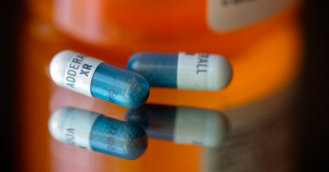
The NewsPrescriptions for drugs used to treat attention deficit hyperactivity disorder surged during the coronavirus pandemic, particularly among women and patients ages 20 to 39, according to new research compiled by scholars affiliated with the U.S. Food and Drug Administration.The increase came as prescription rates held relatively steady for other key classes of behavioral health medications used to treat conditions such as depression and anxiety, according to the study, which was published on Wednesday in JAMA Psychiatry.The reasons are not totally clear, the researchers found, and could include pandemic-related stress, recognition of undiagnosed cases, over-prescription and online marketing of medications.Lisa Cosgrove, a clinical psychologist at the University of Massachusetts, Boston, who was not involved in the study, said the results “seem counterintuitive,” since the pandemic was a time when most people were not in school or offices, environments where attention-related issues often come to light.The study’s authors noted that social media might have played a role in the increase in A.D.H.D. prescriptions, as telemedicine services “used social media services to advertise treatments for behavioral health conditions, such as A.D.H.D. and eating disorders.”Dr. Cosgrove, who studies psychiatric treatment practices, agreed. She hypothesized that online influencers and other people who spoke openly on platforms like TikTok about their own A.D.H.D. diagnoses might have prompted viewers to explore and “self-diagnose.”“There are just so many TikTok videos about people being diagnosed with ADHD and embracing the A.D.H.D. identity,” Dr. Cosgrove said.Stephen Hinshaw, a professor of psychology at the University of California, Berkeley, and an expert on A.D.H.D., said that the “TikTok phenomenon” and other social media platforms almost certainly led to some over-prescription of medications owing in part to “quick and dirty” self-diagnoses through online questionnaires, along with easier access to online prescriptions of stimulants.But one benefit, he said, is that social media may have enabled many people to recognize that they had untreated A.D.H.D. This may be particularly true of women, he added, as they come to understand that A.D.H.D. is not just a “boy’s disease,” as it has long been characterized.The NumbersThe study, conducted by scientists at the Center for Drug Evaluation and Research, a division of the U.S. Food and Drug Administration, compared prepandemic prescriptions of drugs in five classes with prescriptions during the pandemic, which the study defined as a two-year period from April 2020 to March 2022. Compared with the two years prior, the pandemic period saw declines in prescriptions of two classes of drugs: benzodiazepines, which are used to treat anxiety and other conditions, and buprenorphine, which is used to treat opiate-use disorder. Benzodiazepine prescriptions fell by 9 percent and buprenorphine prescriptions declined by 2 percent.Prescriptions of antidepressants rose 10 percent over that period. But the authors of the study note that the increases were consistent with similar patterns before the pandemic, so “the changes in levels and trends were not significant for antidepressants.”By contrast, the prescription rates for A.D.H.D. drugs “significantly increased during the Covid-19 pandemic, exceeding prepandemic rates, notably in young adults and women,” the study found.Among people ages 20 to 39, prescriptions of A.D.H.D. Schedule II stimulants, which include Ritalin and Adderall, rose 30 percent. Schedule II drugs have a “have a high potential for abuse which may lead to severe psychological or physical dependence,” according to the federal government. Prescriptions of non-stimulant A.D.H.D. medications rose 81 percent among 20- to 39-year-olds and 59 percent among women overall, the study found.The study also revealed a shift in the drugs’ prescribers. During the pandemic, prescriptions of A.D.H.D. stimulants by nurse practitioners rose by 57 percent compared with prescriptions by that group two years prior, while prescriptions by psychiatrists fell by 1 percent.A similar pattern emerged with non-stimulant A.D.H.D. medications. Prescriptions by nurse practitioners rose 74 percent during pandemic, compared with an increase of 12 percent by psychiatrists.Behind The NumbersThe results raised several questions, the researchers wrote: Notably, to what extent were A.D.H.D. drugs appropriately prescribed?The sharp increase in prescriptions during the pandemic highlighted the continued need “to define treatment appropriateness,” as well as to explore “how marketing and prescribing practices evolved,” the authors concluded.Some evidence suggests that A.D.H.D. was being overdiagnosed even before the pandemic. Dr. Cosgrove also noted that the information about behavioral and mental health shared on TikTok and other platforms was often misleading, and she said there was a need for more rigorous diagnosis.

Read more →

Newly identified genes for depression may lead to new treatments

More than 200 genes linked to depression have been newly identified in a worldwide study led by UCL researchers.
The research, published in Nature Genetics, found more than 50 new genetic loci (a locus is a specific position on a chromosome) and 205 novel genes that are associated with depression, in the first large-scale global study of the genetics of major depression in participants of diverse ancestry groups.
The study also showcases potential for drug repurposing, as one of the identified genes encodes a protein targeted by a common diabetes drug, while also pointing to new targets for drugs that may be developed to treat depression.
Depression is very common, yet how it develops is still poorly understood. Genetic research using big data offers new avenues to understand the disease, and has uncovered dozens of genes associated with depression, each which individually confer only a small increase in risk. It can also help find new drug targets, but so far research has mainly focused on people of European ancestry, which the researchers say is a major shortcoming, especially for such a complex condition as depression.
The new paper involved multiple genetic research methods including genome-wide association studies, a meta-analysis of previously published data and a transcriptome-wide association study. The international research team reviewed genetic data from 21 study cohorts from several countries and included nearly one million study participants of African, East Asian, South Asian, and Hispanic/Latin American descent, including 88,316people with major depression.
The study has made major advances identifying genes that are linked to risk of depression, both for newly-identified links and by strengthening prior evidence, and showcases some genes with potential implications for drug development, such as NDUFAF3. The protein that NDUFAF3 encodes has been implicated previously in mood instability, and it is targeted by metformin, the first-line drug for treating type 2 diabetes. Animal studies of metformin have suggested a possible link with reduced depression and anxiety, so this latest finding further suggests that additional research into metformin and depression may be warranted.
Other genes identified in the study may have biologically plausible links with depression, such as a gene linked to a neurotransmitter involved in goal-directed behaviour, and genes encoding a type of protein previously linked with multiple neurological conditions.

Surprisingly, the researchers found less overlap in the genetic hits for depression across ancestry groups than expected, at about 30% (based on a new method developed by the research team, to gauge the degree to which a genetic association found in one ancestry group is applicable to another ancestry group), which is less overlap than previously found for other traits and diseases. Therefore, it is even more important to study depression in diverse samples because some of the findings might be ancestry specific.
Lead author Professor Karoline Kuchenbaecker (UCL Psychiatry and UCL Genetics Institute) said: “Here we show beyond doubt that our understanding of such complex diseases as depression will remain incomplete until we overcome the Eurocentric bias in genetics research and look for causes in diverse people across the world.
“Many genes previously found to be linked to the risk of depression might only actually affect depression risk in people of European origin, so in order for genetic research to contribute to new drugs that can help people of all ancestries, it is vital that our genetic datasets are suitably diverse.”
Professor Kuchenbaecker led the study alongside Dr Xiangrui Meng, PhD researcher Georgina Navoly and Dr Olga Giannakopoulou, and the collaborative consortia involved in the study included the Psychiatric Genomics Consortium-Major Depressive Disorder Working Group, China Kadoorie Biobank Collaborative Group, the 23andMe Research Team, Genes and Health Research Team, and BioBank Japan Project.
Professor Kuchenbaecker added: “This is a first stage discovery effort, so more work will be needed to confirm these new targets, but finding them in the first place has been a huge and vital challenge, especially for a disorder where new medications are so urgently needed.”

Read more →

How 'pioneers' blaze the one trail that determines cell fate

One of the important breakthroughs that made it possible to program or reprogram cell fate more efficiently and with higher fidelity in a dish was discovering how to make use of a small set of molecular cowboys called pioneer transcription factors (TFs).
Every cell in our bodies has more than 200 transcription factors expressed inside, riding along the DNA helix instructing specific genes to activate and deactivate. During the early stages of fetal development, a small subset of “pioneer” TFs act inside our precursor cells driving them to make the mature cells that become parts of the spine, the heart, the liver, and so on.
Identifying the key pluripotent pioneer TFs helped researchers learn how to make induced pluripotent stem cells from any type of adult cells, which could then be instructed to make other types of cells, thus forming organoids that can mimic functional organ tissue. Furthermore, tissue-specific pioneer TFs can directly reprogram one adult cell type into a targeted cell type without passing through a pluripotent state.
Currently, there is considerable interest in producing therapeutic target cells from pioneer TF-mediated reprogramming of skin or other accessible somatic cells, as they hold great promise for personalized disease modeling and regenerative medicine. However, a major challenge is achieving sufficient gene repression of the original cell type, as inadequate repression often leads to the formation of “dead-end” cells or hybrid cells, thus limiting translational applications.
Now a study published online Jan. 10, 2024, in Molecular Cell, reveals important new details about how pioneer TFs do their important jobs.
“As we gain a more comprehensive understanding of the mechanisms underlying pioneer TF-mediated gene repression, it will greatly enhance the precise manipulation of cell fate in cellular programming and reprogramming,” says Makiko Iwafuchi, PhD, a member of the Division of Developmental Biology and the Center for Stem Cell & Organoid Medicine at Cincinnati Children’s.
The first author, Satoshi Matsui, PhD, and Iwafuchi collaborated with Hee-Woong Lim, PhD, a co-senior author and a member of the Division of Biomedical Informatics, on the study.

Until this study, most experts believed that pioneer TFs functioned primarily by activating genes to send cells toward their ultimate fates. However, scientists have learned that our genetic programming is packed with alternative pathways, which must be actively shut down.
So how does the body ensure that precursor cells consistently follow the correct path? It turns out that pioneer TFs do more than push cells in the desired direction. When functioning correctly, these cowboys ride ahead and cut off alternative paths so that developing cells keep developing as intended.
Who are these pioneers?
The Cincinnati Children’s team established a new CRISPR interference (CRISPRi) model to explore how the pioneer transcription factor FOXA controls human endoderm differentiation during liver development, and how the pioneer transcription factor OCT4 influences the behavior of pluripotent stem cells.
They found that when FOXA function was disrupted, cells followed multiple development pathways. But when FOXA functioned normally, the cells stayed on track. This allowed the team to conclude that FOXA prevents alternative lineage and precocious gene expression.
The team went on to show that FOXA gets help with repressing access to alternative pathways from another transcription factor called PRDM1 and other epigenetic repressors. Meanwhile in pluripotent cells, the pioneer transcription factor OCT4 performs a similar repression function by teaming up with a related transcription factor called PRDM14.

The central role of FOXA and its ability to repress development pathways were unexpected and critical findings that could influence future organoid and cell reprogramming studies, Iwafuchi says.
Finding a similar relationship occurring with OCT4 was an important further proof of concept because that transcription factor was already known to be among the pioneer TFs that enable reprogramming of somatic cells into pluripotent stem cells.
Next steps
Now that researchers have shown that two pioneer TFs coordinate with PRDM TFs to safeguard cell fate, it appears likely that other TFs have similar relationships. As these TF teams are identified, it may become possible to produce organoids and other engineered tissues in larger volumes with higher degrees of consistency and fidelity — steps that will be important for scaling up the technology.

Read more →

Scientists discover potential nasal COVID-19 vaccine candidate that offers better and longer protection

A team of scientists, led by Duke-NUS Medical School, has discovered a potential intranasal vaccine candidate that provides improved, longer-lasting immunity against SARS-CoV-2 viruses compared to when given as an injection. By triggering an immune response directly at the point of entry, the intranasal vaccine candidate enhanced long-term immune memory of the virus, which could translate to a reduced need for booster shots.
There is growing evidence that intranasal vaccines provide greater protection at mucosal surfaces, making this a vaccination route that could reduce break-through infections and subsequent transmission of the virus.
To delve into this, the research team, which includes collaborators from Duke-NUS’ parent universities — Duke University and the National University of Singapore — among others, compared the immune responses from nasal and subcutaneous administration of the vaccine, as well as immunity from the vaccine with and without the use of adjuvants — substances added to vaccines to enhance the body’s immune response.
Published in eBioMedicine, the findings showed nasal administration of the vaccine candidate boosted mucosal antibody response, as expected. Additionally, and more importantly, it enhanced longer-lasting mucosal and systemic immune protection through preferential induction of airway-resident T cells and central memory T cells.
“Our data show that, compared to subcutaneous vaccination, the intranasal route improved the response of certain immune cells, known as T cells, which reduced disease severity,” explained Associate Professor Ashley St John, from Duke-NUS’ Emerging Infectious Diseases Programme, who is the lead author of the study. “Not only that, but it also resulted in a greater number of T central memory cells compared to subcutaneous vaccination, which could lead to longer-lasting protection.”
T central memory cells play a vital role in safeguarding the body upon re-exposure to a virus. They enhance the immune system’s memory, inducing long-lasting protective immune responses. This ability to retain this long-term memory of the virus suggests less need for a pathogen challenge to achieve the same level of protection against the virus, potentially translating into fewer boosters.
The research team also found that the use of adjuvants in the vaccine to promote immune response influenced the characteristics of T cells, as well as their activation and production of cytokines — tiny proteins that regulate cell communication and control inflammation — with different adjuvants leading to different T-cell responses.
Another notable finding from the study was that a type of antibody, called IgG, that circulates widely in the bloodstream is more effective at neutralising variants of the virus, including newly emergent ones, when induced through the nasal vaccine route. These discoveries provide important scientific evidence that improved immunity responses from both T cells and IgG antibodies contribute to greater and long-lasting protection of intranasal vaccines from COVID-19.
“While the acute phase of the pandemic may be behind us, the rise of new variants, including JN.1, which has triggered an increase in hospital admissions locally, demonstrates that we have room in our arsenal of vaccines and treatments for even better tools. This study shows that mucosal vaccination holds promise for improving COVID-19 vaccine efficacy with potentially fewer boosters needed,” said Professor Patrick Tan, Senior Vice-Dean for Research at Duke-NUS.
A patent has been filed on the discovery, which covers the invention of the vaccine composition formulated for mucosal delivery, paving the way for an industry partnership to potentially develop mucosal vaccines against COVID-19 and other pathogens that also target mucosal surfaces.

Read more →

Traumatic stress associated with smaller brain region

Adults with posttraumatic stress disorder (PTSD) have smaller cerebellums, according to new research from a Duke-led brain imaging study.
The cerebellum, a part of the brain well known for helping to coordinate movement and balance, can influence emotion and memory, which are impacted by PTSD. What isn’t known yet is whether a smaller cerebellum predisposes a person to PTSD or PTSD shrinks the brain region.
“The differences were largely within the posterior lobe, where a lot of the more cognitive functions attributed to the cerebellum seem to localize, as well as the vermis, which is linked to a lot of emotional processing functions,” said Ashley Huggins, Ph.D., the lead author of the report who helped carry out the work as a postdoctoral researcher at Duke in the lab of psychiatrist Raj Morey, M.D.
Huggins, now an assistant professor of psychology at the University of Arizona, hopes these results encourage others to consider the cerebellum as an important medical target for those with PTSD.
“If we know what areas are implicated, then we can start to focus interventions like brain stimulation on the cerebellum and potentially improve treatment outcomes,” Huggins said.
The findings, published January 10 in the journal Molecular Psychiatry, have prompted Huggins and her lab to start looking for what comes first: a smaller cerebellum that might make people more susceptible to PTSD, or trauma-induced PTSD that leads to cerebellum shrinkage.
PTSD and the “Little Brain”
PTSD is a mental health disorder brought about by experiencing or witnessing a traumatic event, such as a car accident, sexual abuse, or military combat.

Though most people who endure a traumatic experience are spared from the disorder, about 6% of adults develop PTSD, which is often marked by increased fear and reliving the traumatizing event.
Researchers have found several brain regions involved in PTSD, including the almond-shaped amygdala that regulates fear, and the hippocampus, a critical hub for processing memories and routing them throughout the brain.
The cerebellum (Latin for “little brain”), by contrast, has received less attention for its role in PTSD.
A grapefruit-sized lump of cells that look like it was clumsily tacked underneath the back of the brain as an afterthought, the cerebellum is best known for its role in coordinating balance and choreographing complex movements, like walking or dancing. But there is much more to it than that.
“It’s a really complex area,” Huggins said. “If you look at how densely populated with neurons it is relative to the rest of the brain, it’s not that surprising that it does a lot more than balance and movement.”
Dense may be an understatement. The cerebellum makes up just 10% of the brain’s total volume but packs in more than half of the brain’s 86 billion nerve cells.

Researchers have recently observed changes to the size of the tightly-packed cerebellum in PTSD. Most of that research, however, is limited by either a small dataset (fewer than 100 participants), broad anatomical boundaries, or a sole focus on certain patient populations, such as veterans or sexual assault victims with PTSD.
Subtle and Consistent Reductions
To overcome those limitations, Duke’s Dr. Morey, along with over 40 other research groups that are part of a larger data-sharing initiative, pooled together their brain imaging scans to study PTSD as broadly and universally as possible.
The group ended up with images from 4,215 adult MRI scans, about a third of whom had been diagnosed with PTSD.
“I spent a lot of time looking at cerebellums,” Huggins said.
Even with automated software to analyze the thousands of brain scans, Huggins manually spot-checked every image to make sure the boundaries drawn around the cerebellum and its many subregions were accurate.
The result of this thorough methodology was a fairly simple and consistent finding: PTSD patients had cerebellums about 2% smaller.
When Huggins zoomed in to specific areas within the cerebellum that influence emotion and memory, she found similar cerebellar reductions in people with PTSD.
Huggins also discovered that the worse PTSD was for a person, the smaller their cerebellum was.
“Focusing purely on a yes-or-no categorical diagnosis doesn’t always give us the clearest picture,” Huggins said. “When we looked at PTSD severity, people who had more severe forms of the disorder had an even smaller cerebellar volume.”
Targeting the Cerebellum for Treatment and More Research
The results are an important first step at looking at how and where PTSD affects the brain.
There are more than 600,000 combinations of symptoms that can lead to a PTSD diagnosis, Huggins explained. Figuring out if different PTSD symptom combinations have different impacts on the brain will also be important to keep in mind.
For now, though, Huggins hopes this work helps others recognize the cerebellum as an important driver of complex behavior and processes beyond gait and balance, as well as a potential target for new and current treatments for people with PTSD.
“While there are good treatments that work for people with PTSD, we know they don’t work for everyone,” Huggins said. “If we can better understand what’s going on in the brain, then we can try to incorporate that information to come up with more effective treatments that are longer lasting and work for more people.”

Read more →

Scientists discover how ultraviolet light degrades coronavirus

New research has revealed how light can be used to destroy infectious coronavirus particles that contaminate surfaces. Scientists are interested in how environments, such as surgeries, can be thoroughly disinfected from viruses such as SARS-CoV-2 that caused the COVID-19 pandemic.
SARS-CoV-2 viral particles are composed of a core of nucleic acid chains that contain the genetic information of the virus, surrounded by a lipid membrane with proteinous spikes sticking out. Each component is necessary for infection.
Researchers from the University of Southampton investigated how ultraviolet laser light destroys the virus by impacting each of these critical components. By using a specialised ultraviolet laser at two different wavelengths the scientists were able to determine how each viral component degraded under the bright light. They found the genomic material was highly sensitive to degradation and protein spikes lost their ability to bind to human cells.
UV light includes UVA, UVB and UVC light. Very little UVC light at frequencies below 280nm reaches the earth’s surface from the sun. It is this lesser studied UVC light that the team in Southampton used for their study due to its disinfectant properties. UVC light is strongly absorbed by different viral components, including the genetic material (~260nm) and the proteinous spikes (~230nm), allowing the team to select laser frequencies of 266nm and 227nm for the project.
University of Southampton scientists, led by Professor Sumeet Mahajan, worked closely with scientists from the laser manufacturer, called MSquared Lasers, and the resulting co-authored study has been published in a journal of the American Chemical Society called ACS Photonics. The team found that 266nm light caused RNA damage at low powers, affecting the genetic information of the virus. 266nm light also damaged the structure of the SARS-CoV-2 spike protein, reducing its ability to bind to human cells by breaking down disulphide bonds and aromatic amino acids.
The 227nm light was less effective at inducing RNA damage, but more effective at damaging proteins through oxidation (a chemical reaction involving oxygen) which unfolds the protein’s structure.
Importantly, SARS-CoV-2 has among the largest of genomes for RNA viruses. This makes it especially sensitive to genomic damage.
Professor Mahajan said: “Light deactivation of airborne viruses offers a versatile tool for disinfection of our public spaces and sensitive equipment that may otherwise prove difficult to decontaminate with conventional methods. Now we understand the differential sensitivity of molecular components in viruses to light deactivation this opens up the possibility of a finely tuned disinfection technology.”
Light-based deactivation has received a lot of attention because of the wide range of applications where conventional liquid-based deactivation methods aren’t suitable. Now the mechanism of deactivation is better understood this is an important step in rolling out the technology.
Mechanisms of SARS-CoV-2 Inactivation Using UVC Laser Radiation is published in ACS Photonics and is available online.

Read more →

Exposure therapy addressing fear of spiders can also reduce fear of heights

Exposure therapy for a specific fear can also help reduce other fears. This is the conclusion reached by psychologists at Ruhr University Bochum, Germany, who studied 50 people with a fear of spiders and heights. Although they only treated the fear of spiders, the fear of heights was likewise reduced in the process. Findings are described by a team around Iris Kodzaga and Professor Armin Zlomuzica from the Department of Behavioral and Clinical Neuroscience at Ruhr University Bochum in the journal Translational Psychiatry.
A number of anxiety disorders are co-morbid
“Anxiety rarely comes alone,” says Iris Kodzaga, lead author of the study. “Patients who suffer from one fear often subsequently develop another.” The most effective treatment method is exposure: By confronting the fear-inducing situations or stimuli under psychotherapeutic supervision, patients learn to overcome their fear.
“It was long assumed that if a person had multiple fears, they would require multiple exposure therapies tailored to their specific fear,” explains Kodzaga. The Bochum-based team is now challenging this assumption. The researchers measured fear of spiders and heights in 50 test subjects before and after exposure therapy targeting spider fear. Measures included subjective data from specific questionnaires for fear of spiders and heights. In addition, the researchers collected quantitative behavioral measures, such as how close the participants dared to approach the spiders or how far they could climb a high church tower.
Therapy methods could become more universal
Exposure therapy for spider fear not only reduced the fear of spiders, but also the fear of heights. A significant effect emerged in both the subjective and behavioral measures: Fear of heights decreased by an average of 15 percent as a result of exposure to spiders.
“The discovery that exposure to spiders also reduces fear of heights opens up new perspectives for the efficient treatment of phobias,” says Iris Kodzaga. “It could mean that we can rethink therapeutic approaches and possibly develop more universal methods.”
How exactly this effect is transferred from one fear to another is still unclear. “The effect can’t be fully explained by associative learning processes. The generalization effect might be due to an increase in self-efficacy as a result of exposure therapy,” says the researcher. “But perhaps there is also a common denominator between fear of spiders and fear of heights that’s not obvious. We’ll need to conduct follow-up studies to find out more.”

Read more →

Noninvasive technique reveals how cells' gene expression changes over time

Sequencing all of the RNA in a cell can reveal a great deal of information about that cell’s function and what it is doing at a given point in time. However, the sequencing process destroys the cell, making it difficult to study ongoing changes in gene expression.
An alternative approach developed at MIT could enable researchers to track such changes over extended periods of time. The new method, which is based on a noninvasive imaging technique known as Raman spectroscopy, doesn’t harm cells and can be performed repeatedly.
Using this technique, the researchers showed that they could monitor embryonic stem cells as they differentiated into several other cell types over several days. This technique could enable studies of long-term cellular processes such as cancer progression or embryonic development, and one day might be used for diagnostics for cancer and other diseases.
“With Raman imaging you can measure many more time points, which may be important for studying cancer biology, developmental biology, and a number of degenerative diseases,” says Peter So, a professor of biological and mechanical engineering at MIT, director of MIT’s Laser Biomedical Research Center, and one of the authors of the paper.
Koseki Kobayashi-Kirschvink, a postdoc at MIT and the Broad Institute of Harvard and MIT, is the lead author of the study, which appears today in Nature Biotechnology. The paper’s senior authors are Tommaso Biancalani, a former Broad Institute scientist; Jian Shu, an assistant professor at Harvard Medical School and an associate member of the Broad Institute; and Aviv Regev, executive vice president at Genentech Research and Early Development, who is on leave from faculty positions at the Broad Institute and MIT’s Department of Biology.
Imaging gene expression
Raman spectroscopy is a noninvasive technique that reveals the chemical composition of tissues or cells by shining near-infrared or visible light on them. MIT’s Laser Biomedical Research Center has been working on biomedical Raman spectroscopy since 1985, and recently, So and others in the center have developed Raman spectroscopy-based techniques that could be used to diagnose breast cancer or measure blood glucose.

However, Raman spectroscopy on its own is not sensitive enough to detect signals as small as changes in the levels of individual RNA molecules. To measure RNA levels, scientists typically use a technique called single-cell RNA sequencing, which can reveal the genes that are active within different types of cells in a tissue sample.
In this project, the MIT team sought to combine the advantages of single-cell RNA sequencing and Raman spectroscopy by training a computational model to translate Raman signals into RNA expression states.
“RNA sequencing gives you extremely detailed information, but it’s destructive. Raman is noninvasive, but it doesn’t tell you anything about RNA. So, the idea of this project was to use machine learning to combine the strength of both modalities, thereby allowing you to understand the dynamics of gene expression profiles at the single cell level over time,” Kobayashi-Kirschvink says.
To generate data to train their model, the researchers treated mouse fibroblast cells, a type of skin cell, with factors that reprogram the cells to become pluripotent stem cells. During this process, cells can also transition into several other cell types, including neural and epithelial cells.
Using Raman spectroscopy, the researchers imaged the cells at 36 time points over 18 days as they differentiated. After each image was taken, the researchers analyzed each cell using single molecule fluorescence in situ hybridization (smFISH), which can be used to visualize specific RNA molecules within a cell. In this case, they looked for RNA molecules encoding nine different genes whose expression patterns vary between cell types.
This smFISH data can then act as a link between Raman imaging data and single-cell RNA sequencing data. To make that link, the researchers first trained a deep-learning model to predict the expression of those nine genes based on the Raman images obtained from those cells.

Then, they used a computational program called Tangram, previously developed at the Broad Institute, to link the smFISH gene expression patterns with entire genome profiles that they had obtained by performing single-cell RNA sequencing on the sample cells.
The researchers then combined those two computational models into one that they call Raman2RNA, which can predict individual cells’ entire genomic profiles based on Raman images of the cells.
Tracking cell differentiation
The researchers tested their Raman2RNA algorithm by tracking mouse embryonic stem cells as they differentiated into different cell types. They took Raman images of the cells four times a day for three days, and used their computational model to predict the corresponding RNA expression profiles of each cell, which they confirmed by comparing it to RNA sequencing measurements.
Using this approach, the researchers were able to observe the transitions that occurred in individual cells as they differentiated from embryonic stem cells into more mature cell types. They also showed that they could track the genomic changes that occur as mouse fibroblasts are reprogrammed into induced pluripotent stem cells, over a two-week period.
“It’s a demonstration that optical imaging gives additional information that allows you to directly track the lineage of the cells and the evolution of their transcription,” So says.
The researchers now plan to use this technique to study other types of cell populations that change over time, such as aging cells and cancerous cells. They are now working with cells grown in a lab dish, but in the future, they hope this approach could be developed as a potential diagnostic for use in patients.
“One of the biggest advantages of Raman is that it’s a label-free method. It’s a long way off, but there is potential for the human translation, which could not be done using the existing invasive techniques for measuring genomic profiles,” says Jeon Woong Kang, an MIT research scientist who is also an author of the study.
The research was funded by the Japan Society for the Promotion of Science Postdoctoral Fellowship for Overseas Researchers, the Naito Foundation Overseas Postdoctoral Fellowship, the MathWorks Fellowship, the Helen Hay Whitney Foundation, the U.S. National Institutes of Health, the U.S. National Institute of Biomedical Imaging and Bioengineering, HubMap, the Howard Hughes Medical Institute, and the Klarman Cell Observatory.

Read more →

Cancer: Unravelling individual differences in DNA mutation risks

Mutations, which occur continuously in every cell of our bodies, are a key contributor to cancer, ageing, and neurodegeneration. While exposure to mutagenic chemicals, or mistakes in cellular processes during DNA replication contribute to these mutations, the exact distribution and patterns of these changes across human chromosomes have remained a mystery until now.
Dr. Fran Supek, ICREA researcher and head of the Genome Data Science lab at IRB Barcelona, and Marina Salvadores, PhD student in the same laboratory, have delved into the landscape of DNA mutations, unveiling unexpected patterns that differentiate individuals in terms of mutation risk.
Previous work by the laboratory identified a “genomic spellchecker,” a DNA repair mechanism devoted to reducing mutation risks in key parts of human chromosomes. Building upon these findings, the current study aimed to determine whether mutation risk varies between individuals and, if so, to identify the mechanisms driving these differences.
“This research not only expands our understanding of the factors influencing mutation rate distribution but it also has significant implications for cancer evolution, therapeutic strategies, and advancements in regenerative medicine,” explains Dr. Supek.
A genomic ‘big data’ analysis
To address these questions, the researchers conducted a comprehensive analysis of genome sequences from over 4,000 tumours from various organs. Unlike previous studies focusing on tissue-specific mutation risks, this study specifically targeted individual differences in susceptibility to mutations.
Employing a genomic “big data” approach, the team used machine learning algorithms to identify recurrent patterns across chromosome segments. They discovered 13 distinct patterns, with 10 corresponding to different types of tissue. Unexpectedly, the remaining three patterns were observed in almost all the tissues, thereby revealing that the density of mutations in specific genes varies significantly between individuals.

Connecting the dots
To understand these unexpected patterns, the researchers examined additional data, including gene expression and genetic aberrations, in cancer cells. The analysis uncovered a surprising correlation between increased cell proliferation and alterations in mutation risks. Disruptions in two crucial tumour-suppressor genes, TP53 and RB1, known to regulate the cell division cycle, were identified as key influencers, causing changes in the risk of mutations across chromosomes.
These chromosome segments not only presented altered mutation risks but also large-scale “remodelling” of usually inactive chromosomal regions. “This remodelling, which is correlated with increased cell proliferation, mirrored the changes in mutation risks, providing unique insight into the interplay between mutations and epigenetic alterations,” explains Marina Salvadores, first author of the study.
Decoding cancer evolution
The implications of this study extend beyond fundamental research. By identifying the cancer-causing genes most affected by changes in mutation risk between individuals, the scientists have provided a roadmap for anticipating the trajectory of cancer evolution. This knowledge is especially relevant for anticipating responses to cancer treatment, as it can help to predict the development of drug-resistance mutations in tumours.
The study also offers valuable insights into the epigenetics of human cells, highlighting that the epigenome undergoes dramatic changes in response to increased or disrupted cell proliferation. This understanding has potential implications for cell reprogramming and regenerative medicine, opening up new avenues for future research and therapeutic interventions.
This study has received funding from the ERC and the Spanish Ministerio de Ciencia e Innovación.

Read more →

ALS: Blocking inflammation to reduce symptoms

In people with amyotrophic lateral sclerosis (ALS), changes in neurons appear to activate immune cells. Lowering the inflammation could reduce the symptoms of the disease, according to a study led by Chantelle Sephton, a professor at Université Laval’s Faculty of Medicine.
ALS is caused by the loss of upper motor neurons, located in the brain, and lower motor neurons, which extend from the spinal cord to the muscles. Using a genetically modified mouse model, Chantelle Sephton and her team found that structural changes in the upper neurons occurred prior to disease symptoms.
The study suggests that these morphological changes send a signal to microglia and astrocytes, the immune cells of the central nervous system. When they arrive, their effect is protective, but if they stay too long, they become toxic to neurons. This leads to a reduction in synaptic connections between motor neurons in the brain and spinal cord, which in turn results in a reduction in synaptic connections with muscles. These changes lead to atrophy and loss of motor function.
Given this correlation between symptoms and immune response, the research team wondered whether it might be possible to restore synaptic connections by blocking inflammation. ” We tested a semi-synthetic drug based on Withaferin A, an extract of the Ashwagandha plant, which has been used for thousands of years in traditional Indian medicine,” explains CERVO Research Center affiliate Chantelle Sephton.
The drug blocks inflammation and allows motor neurons to return to a more normal state. “We have noticed that neurons regenerate in the absence of activated immune cells. The dendrites of motor neurons start to grow and make connections again, increasing the number of synapses between motor neurons and muscles,” reports the researcher.
This seems a promising way of improving ALS symptoms, whether the disease is familial or sporadic, since both types are associated with inflammation.
Other diseases where inflammation plays a role, such as Alzheimer’s, could benefit from this approach.
The study was published in the scientific journal Acta Neuropathologica Communications. The signatories are Mari Carmen Pelaez, Antoine Desmeules, Pauline Gelon, Bastien Glasson, Laetitia Marcadet, Alicia Rodgers, Daniel Phaneuf, Silvia Pozzi, Paul Dutchak, Jean-Pierre Julien and Chantelle Sephton.

Read more →