‘Delighted’ woman played clarinet in brain surgery

3 hours agoShareSaveDaniel SextonSouth EastShareSavePA MediaA woman with Parkinson’s disease who played a clarinet while undergoing brain stimulation said she was “delighted” with how it went.Denise Bacon, 65, from Crowborough in East Sussex, experienced instant improvement in her finger movements as the electrical current was delivered to her brain, doctors at King’s College Hospital said.The retired speech and language therapist underwent deep brain stimulation (DBS) after suffering symptoms of Parkinson’s.She was diagnosed with the disease in 2014 and it has affected her ability to walk, swim, dance and play her clarinet.During a four-hour operation at King’s College Hospital, Keyoumars Ashkan, a professor of neurosurgery, performed DBS to help relieve symptoms while Ms Bacon stayed awake.DBS is a surgical procedure using electrodes implanted in the brain and is suitable for some patients with disorders such as Parkinson’s.Ms Bacon was given a local anaesthetic to numb her scalp and skull and had immediate improvement in her fingers on the operating table, allowing her to play the clarinet easily.PA MediaShe previously played clarinet in the East Grinstead Concert Band until she had to stop five years ago due to her Parkinson’s symptoms.”I remember my right hand being able to move with much more ease once the stimulation was applied, and this in turn improved my ability to play the clarinet, which I was delighted with,” she said.”I’m already experiencing improvements in my ability to walk, and I’m keen to get back in the swimming pool, and on the dance floor to see if my abilities have improved there.”Prof Ashkan, said holes half the size of a five pence piece had been made in her skull, after a frame with precise co-ordinates was placed on Ms Bacon’s head “acting as a sat nav” to guide them to the correct positions to implant the electrode.”We were delighted to see an instant improvement in her hand movements, and therefore her ability to play, once stimulation was delivered to the brain,” he said.More on this storyRelated internet links

Read more →

This simple neck measurement might reveal hidden heart risks

For decades, doctors have depended on indicators such as body mass index (BMI) and waist-to-hip ratio to gauge a person’s risk for chronic disease. But researchers are now turning their attention to a different, often-overlooked measurement: neck circumference.
While a thick neck may suggest physical power, like that of athletes in contact sports, research indicates it could also be a marker of hidden health risks.
Why Neck Size Matters
BMI calculates body fat by comparing weight to height, but it doesn’t always paint an accurate picture. A muscular athlete may register a high BMI without carrying excess fat. This is where measuring neck size can offer additional clues about what’s happening inside the body.
Studies have found that individuals with larger necks relative to their body size are more likely to develop serious health issues. The reason lies in fat distribution — particularly in the upper body.
Fat stored around the upper torso releases fatty acids into the bloodstream, which can interfere with how the body regulates cholesterol, blood sugar, and heart rhythm. In effect, neck circumference acts as a visible indicator of visceral fat, the harmful kind that accumulates around internal organs.
The Link to Heart Disease and Other Conditions
Evidence connecting neck size to health outcomes is compelling. People with thicker necks show higher rates of cardiovascular diseases, including hypertension, atrial fibrillation, and heart failure.

Atrial fibrillation is of particular concern because it causes irregular heartbeats and erratic blood flow, raising the risk of blood clots and stroke. Over time, this irregular rhythm can strain the heart and lead to failure.
Neck circumference is also tied to coronary heart disease, where narrowed arteries reduce the flow of oxygen-rich blood to the heart.
More Than Just Heart Health
The risks don’t stop at cardiovascular problems. Larger neck size is associated with a greater likelihood of developing type 2 diabetes and gestational diabetes, both of which can lead to long-term complications such as nerve damage, vision loss, and limb amputation.
Sleep disorders are another major concern. A thicker neck is a known risk factor for obstructive sleep apnea, a condition in which breathing repeatedly stops and starts during sleep. This not only leads to severe daytime fatigue but also places additional stress on the heart and blood vessels. People with sleep apnea are also more prone to accidents caused by exhaustion.
When Neck Size Becomes a Warning
So, how big is too big? Research suggests that men with a neck circumference of 17 inches (43 cm) or more, and women with 14 inches (35.5 cm) or more, face elevated health risks.

Perhaps most surprising, these dangers are not limited to people who are overweight. Even individuals with a normal BMI may be at risk if their neck measurements exceed these thresholds. For every additional centimeter beyond that range, the likelihood of hospitalization and early death increases.
Taking Action
If your neck measurement falls above these levels, there’s no reason for alarm, but it’s worth noting. Neck size represents only one piece of your overall health profile, yet it can reveal important information that other metrics might overlook.
The positive news is that neck circumference can change with lifestyle improvements. Regular cardiovascular exercise, strength training, and sufficient sleep all contribute to better metabolic health. Eating a diet rich in vegetables, fruits, and pulses supports weight management and reduces unhealthy fat storage.
How to Measure Your Neck
Checking your neck size is simple. Use a flexible tape measure and wrap it around the narrowest part of your neck, keeping the tape snug but not tight. The process takes only a few seconds.
A Simple Measurement With Big Meaning
This quick check can highlight potential risks that traditional metrics might miss. While neck circumference shouldn’t replace established health assessments, it provides a valuable, easy-to-use tool for understanding cardiovascular and metabolic health.
In our search for better ways to predict and prevent disease, it turns out some of the most revealing clues are right in front of us. Your neck may be quietly reflecting more about your health than you think — and it’s worth paying attention.

Read more →

Eating ultra-processed foods may rewire the brain and drive overeating

An international team of scientists has analyzed brain scans from around 30,000 participants and found striking connections between the frequent consumption of ultra-processed foods (UPFs) and measurable differences in brain structure. These differences could be part of a feedback loop that promotes overeating and food addiction.
“Our findings suggest that higher consumption of ultra-processed foods is associated with differences in the brain. These associations could be linked to behavioral patterns such as overeating, though causal relationships cannot be confirmed by our study. The observed associations are not solely explained by inflammation or obesity; ingredients and additives typical to UPFs, such as emulsifiers may also play a role, although this requires further longitudinal or experimental evidence,” explains the study’s shared first author, Arsène Kanyamibwa from the University of Helsinki.
Processed vs. Ultra-Processed: Understanding the Difference
Not all processed foods are harmful. Many processed items, especially those based on plants, are beneficial parts of a balanced diet. However, ultra-processed foods — those containing industrial additives and chemically altered ingredients — raise significant health concerns.
“In particular, processed foods of plant origin, such as frozen vegetables, can be recommended. Another good example of the benefits of processing is the pasteurization of milk. In contrast, foods high in chemically modified ingredients and additives, such as processed meat products, are problematic.”
Implications for Diet and Public Health
Kanyamibwa highlights how emerging research like this can influence both personal food choices and public health policy.
“Given the growing body of evidence, reducing ultra-processed food intake and strengthening regulatory standards in food manufacturing may be crucial steps toward ensuring better public health outcomes,” Kanyamibwa asserts.
The investigation drew on data from the UK Biobank, a large-scale health database including middle-aged participants from across the United Kingdom. The research was carried out through a collaboration between the University of Helsinki and McGill University’s Montréal Neurological Institute, combining expertise in nutrition, neuroscience, and imaging analysis to explore how modern diets may be influencing the human brain.

Read more →

Three Cases of Mpox Tied to Severe Illness Worry Health Experts

None of the patients, all California residents, had traveled abroad, suggesting the Clade 1 form was transmitted locally.A type of mpox that causes severe illness has been identified in three California residents who had not traveled abroad, the first time the more virulent form has spread within the United States, health officials said on Friday.The type, known as Clade 1, has been widely circulating in central and eastern Africa, causing tens of thousands of infections and hundreds of deaths. Clade 2, the form that swept the United States in 2022 and sickened 30,000 people, causes less severe illness.Clade 2 continues to circulate in the United States at low levels, but infections have been rising in a number of cities since the summer. Los Angeles health officials have reported 118 cases of mpox, formerly monkeypox, so far this year.California health officials say the three patients — one in Long Beach and two in Los Angeles — were hospitalized and are now recovering at home in isolation. They have not found a link among the three cases.Officials in Long Beach announced the first case on Tuesday.Dr. Kelly Johnson, an infectious disease specialist at the University of California, San Francisco, said that although the risk of infection was low for most people, she urged vigilance, especially among people with weakened immune systems and men who have sex with men.“My concern is that person-to-person, community spread could be ongoing,” she said.Sonali Kulkarni, medical director of the Division of H.I.V. and S.T.D. Programs at the Los Angeles County Department of Public Health, said that officials were increasing surveillance efforts and testing existing lab specimens to gain a better understanding of the spread. She said all three patients are in a high-risk group of gay and bisexual men.We are having trouble retrieving the article content.Please enable JavaScript in your browser settings.Thank you for your patience while we verify access. If you are in Reader mode please exit and log into your Times account, or subscribe for all of The Times.Thank you for your patience while we verify access.Already a subscriber? Log in.Want all of The Times? Subscribe.

Read more →

Scientists create LED light that kills cancer cells without harming healthy ones

Scientists have developed a promising cancer therapy that uses LED light and ultra-thin flakes of tin to eliminate cancer cells while protecting healthy tissue. Unlike traditional chemotherapy and other invasive treatments, this new method avoids the painful side effects patients often endure.
The breakthrough comes from a partnership between The University of Texas at Austin and the University of Porto in Portugal, made possible through the UT Austin Portugal Program. The collaboration aims to make light-based cancer therapies more accessible and affordable. Current versions of these treatments rely on expensive materials, specialized lab setups, and powerful lasers that can sometimes damage surrounding tissue. By switching to LEDs and introducing tin-based “SnOx nanoflakes” (“Sn” is the chemical symbol for tin), the researchers have created a safer and potentially low-cost alternative.
LED Light and Nanoflakes Team Up Against Cancer
“Our goal was to create a treatment that is not only effective but also safe and accessible,” said Jean Anne Incorvia, a professor in the Cockrell School of Engineering’s Chandra Family Department of Electrical and Computer Engineering and one of the leaders on the project. “With the combination of LED light and SnOx nanoflakes, we’ve developed a method to precisely target cancer cells while leaving healthy cells untouched.”
In a recent study published in ACS Nano, the approach proved highly effective against both colorectal and skin cancer cells. After only 30 minutes of exposure, the LED-driven treatment destroyed up to 92% of skin cancer cells and 50% of colorectal cancer cells, while leaving healthy human skin cells unharmed. The results highlight the therapy’s precision and safety.
A Safer Alternative to Conventional Cancer Treatments
Cancer remains the second-leading cause of death worldwide, and many existing treatments come with severe side effects. Scientists across the globe are exploring new methods to make therapies safer and more targeted. One of the most promising is near-infrared photothermal therapy, which uses light to heat and destroy cancer cells without the need for surgery or toxic drugs. This principle forms the foundation of the UT Austin-Portugal team’s research.

Having shown strong early results, the researchers are now focused on understanding how light and heat interact in the process and on testing other materials that might enhance the treatment. They also plan to design practical medical devices that can deliver the therapy directly to patients.
Bringing Light-Based Cancer Care to Patients
“Our ultimate goal is to make this technology available to patients everywhere, especially places where access to specialized equipment is limited, with fewer side effects and lower cost,” said Artur Pinto, a researcher at the Faculty of Engineering of the University of Porto and lead researcher of the project in Portugal. “For skin cancers in particular, we envision that one day, treatment could move from the hospital to the patient’s home. A portable device could be placed on the skin after surgery to irradiate and destroy any remaining cancer cells, reducing the risk of recurrence.”
Incorvia and Pinto first teamed up through the UT Austin Portugal Program in 2021. Since then, they have exchanged visits between Texas and Portugal and combined their expertise to explore how two-dimensional materials can be used to advance cancer therapies.
Expanding the Research Frontier
Building on their success, the team recently received additional funding through the UT Austin Portugal Program to create an implant for breast cancer patients using the same LED and nanoflake technology. Their continued collaboration could pave the way for more personalized, affordable, and pain-free cancer treatments in the near future.
Other co-authors of the article are: Ph.D. student Hui-Ping Chang (led development of the nanoflakes) and undergraduate student Eva Nance of The University of Texas at Austin; Filipa A.L.S. Silva (performed biological characterization), Susana G. Santos (supervised the work) and professor Fernão Magalhães (contributed to securing funding) of Faculty of Engineering of the University of Porto; and José R. Fernandes of the University of Trás-os-Montes and Alto Douro, who developed the LED systems.
The UT Austin Portugal Program is a long-standing science and technology partnership between UT and the Portuguese Foundation of Science and Technology (FCT). Portugal has similar partnerships with two other U.S. universities — the Massachusetts Institute of Technology and Carnegie Mellon University — and its 17-year collaboration with UT was recently renewed for another five years.

Read more →

Scientists find immune drug that could halt skin cancer’s deadly spread

A major clinical study by the ECOG-ACRIN Cancer Research Group (ECOG-ACRIN) has found that an immune-based cancer drug may help reduce the spread of Merkel cell carcinoma, a rare but aggressive type of skin cancer, when used soon after surgery. The phase 3 STAMP trial (EA6174) tested pembrolizumab, a therapy that activates the body’s immune system to find and destroy cancer cells. While the treatment did not significantly lower the overall risk of recurrence, it did appear to reduce the likelihood of cancer spreading to distant organs — a key secondary goal of the research.
Pembrolizumab Trial Results Show Reduced Risk of Cancer Spread
The randomized phase 3 STAMP trial is the largest study so far to evaluate pembrolizumab as adjuvant therapy for Merkel cell carcinoma after tumor removal. After two years, 73% of patients who received pembrolizumab remained free of cancer, compared with 66% in the group that did not receive the drug. Although this difference was not statistically significant, those treated with pembrolizumab had a 42% lower chance of developing distant metastases, meaning cancer that spread to areas such as the liver, lungs, or bones.
Expert View: “Much-Needed Good News” for Patients
“The STAMP trial provides the first evidence that immunotherapy with pembrolizumab after surgery may help people with Merkel cell carcinoma by preventing their cancer from returning in organs considered distant from the site of the original disease,” said lead investigator Janice M. Mehnert, MD, co-chair of the ECOG-ACRIN Melanoma Committee and director of Melanoma and Cutaneous Medical Oncology at NYU Langone Health’s Perlmutter Cancer Center. “This is much-needed good news for people who are living with the highly aggressive cancer that is Merkel cell carcinoma,” Dr. Mehnert added.
Understanding Merkel Cell Carcinoma
Merkel cell carcinoma is a fast-developing cancer that starts in the skin’s touch-sensing cells and often appears as a firm, painless lump on areas frequently exposed to sunlight. Although skin cancer is the most common type of cancer in the United States, Merkel cell carcinoma — also known as neuroendocrine carcinoma of the skin — is extremely rare, affecting no more than three in one million people each year. It is highly aggressive, with fewer than half of patients surviving five years after diagnosis.

Large-Scale Collaboration Enables Rare Cancer Study
The STAMP trial (NCT03712605) enrolled 293 patients between 2018 and 2023, all of whom had undergone surgery to remove their tumors. Of these, 147 patients were randomly assigned to receive pembrolizumab infusions following surgery, while 146 were monitored without additional drug treatment. Some participants also received radiation therapy as recommended by their doctors.
Because Merkel cell carcinoma is so uncommon, Dr. Mehnert noted that national collaboration was crucial to make the study possible. With support from the National Cancer Institute (NCI), part of the National Institutes of Health (NIH), ECOG-ACRIN conducted the trial through the NCI’s National Clinical Trials Network, involving more than 500 hospitals and cancer centers across the country.
How Pembrolizumab Works
Pembrolizumab belongs to a class of immunotherapies known as PD-1 inhibitors. It works by blocking the PD-1 receptor, a protein cancer cells use to hide from immune system attack. By interrupting this process, pembrolizumab helps immune cells recognize and destroy cancer cells more effectively. The U.S. Food and Drug Administration has already approved pembrolizumab (brand name KEYTRUDA) to treat recurrent locally advanced or metastatic Merkel cell carcinoma.
Overall survival, the other co-primary endpoint of the trial, continues to be followed and will be reported at a later date.
Dr. Mehnert will present the trial at the European Society for Medical Oncology (ESMO) 2025 Congress on Monday, October 20 in Berlin, Germany.

Read more →

Tiny AI-powered eye implant helps the blind see again

People who had lost their sight have regained the ability to read after receiving an innovative electronic eye implant paired with augmented-reality glasses, according to a clinical trial involving researchers from UCL (University College London) and Moorfields Eye Hospital.
Published in The New England Journal of Medicine, the European trial reported that 84% of participants could once again recognize letters, numbers, and words using prosthetic vision in an eye that had previously gone blind due to geographic atrophy caused by dry age-related macular degeneration (AMD), a currently untreatable condition.
Participants treated with the device were able to read an average of five lines on a standard vision chart. Before surgery, several could not even distinguish the chart at all.
Breakthrough Study and Participants
The study enrolled 38 patients across 17 hospitals in five countries, testing a pioneering implant known as PRIMA. Moorfields Eye Hospital served as the only UK site. All participants had completely lost central vision in the treated eye before receiving the implant.
Dry AMD gradually destroys the light-sensitive cells of the macula, eroding central vision over time. In its advanced stage, called geographic atrophy (GA), the degeneration can lead to total blindness in the affected eye as the central macula deteriorates. Around 5 million people worldwide live with GA, for which no treatment currently exists. Those enrolled in the study retained only peripheral vision.
This implant is the first of its kind capable of restoring the ability to read letters, numbers, and words through an eye that had lost its sight.

A New Era of Artificial Vision
Mr. Mahi Muqit, associate professor at the UCL Institute of Ophthalmology and senior vitreoretinal consultant at Moorfields Eye Hospital, led the UK arm of the trial. He said: “In the history of artificial vision, this represents a new era. Blind patients are actually able to have meaningful central vision restoration, which has never been done before.
“Getting back the ability to read is a major improvement in their quality of life, lifts their mood and helps to restore their confidence and independence. The PRIMA chip operation can safely be performed by any trained vitreoretinal surgeon in under two hours — that is key for allowing all blind patients to have access to this new medical therapy for GA in dry AMD.”
How the PRIMA Implant Works
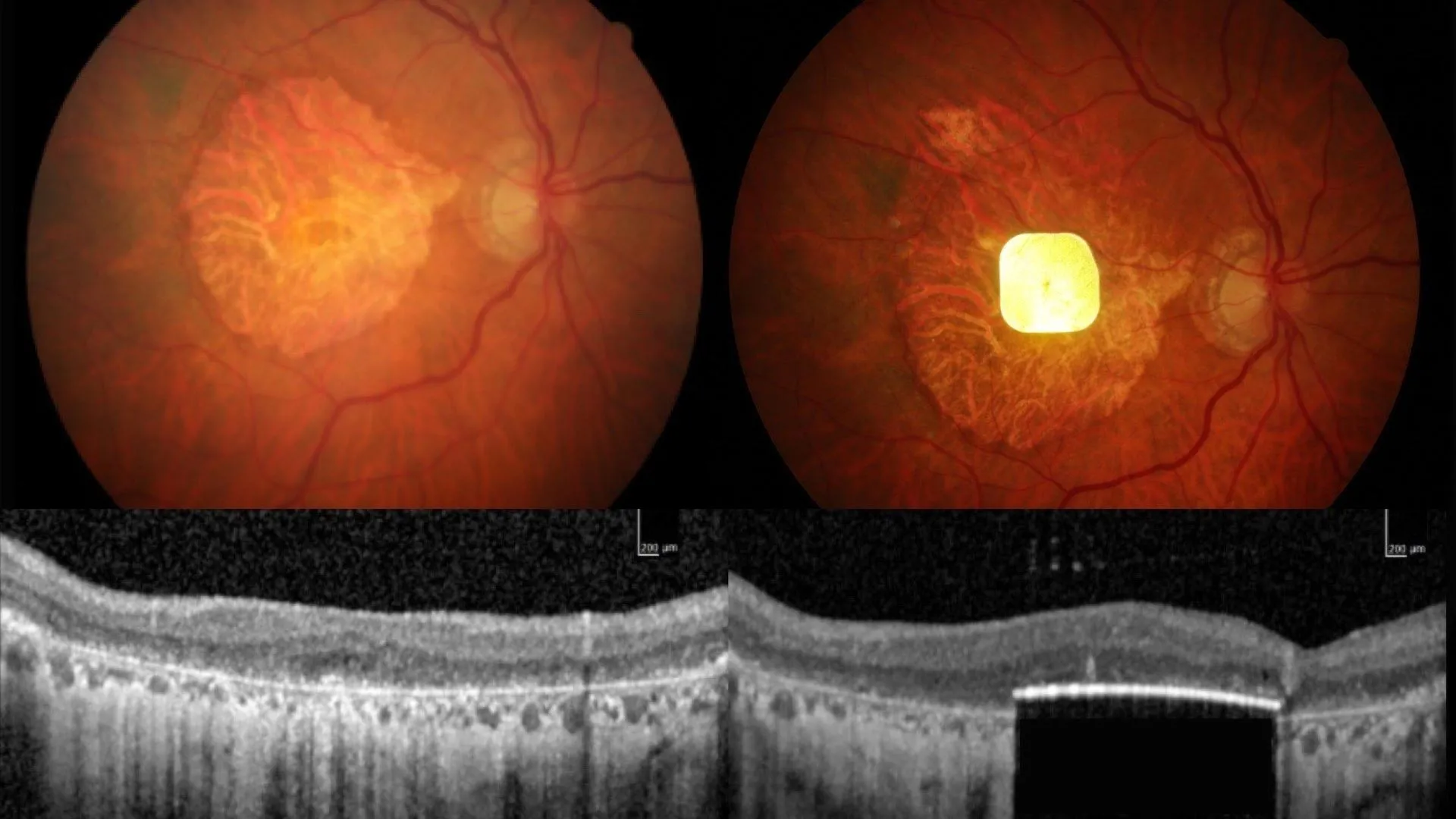
The procedure begins with a vitrectomy, where the eye’s vitreous gel is removed from between the lens and the retina. The surgeon then inserts a microchip shaped like a SIM card, only 2mm by 2mm, beneath the central retina through a small opening.
After surgery, patients use augmented-reality glasses equipped with a video camera linked to a pocket-sized computer with a zoom feature worn at the waist.

About a month after surgery, once the eye has healed, the implant is activated. The glasses’ camera captures visual scenes and projects them as an infra-red beam across the chip. Artificial intelligence (AI) algorithms in the computer process this data and convert it into electrical signals, which travel through retinal and optic nerve cells to the brain. The brain interprets these signals as images, allowing patients to perceive vision.
With training, patients learn to scan text using the glasses and adjust the zoom to read. Each participant underwent several months of rehabilitation to interpret these new signals and regain reading skills.
No participant experienced a decline in their remaining peripheral vision. The results pave the way for regulatory approval of the device and broader clinical use.
Patient Story: Seeing Words Again
Sheila Irvine, one of Moorfields’ patients on the trial who was diagnosed with age-related macular degeneration, said: “I wanted to take part in research to help future generations, and my optician suggested I get in touch with Moorfields. Before receiving the implant, it was like having two black discs in my eyes, with the outside distorted.
“I was an avid bookworm, and I wanted that back. I was nervous, excited, all those things. There was no pain during the operation, but you’re still aware of what’s happening. It’s a new way of looking through your eyes, and it was dead exciting when I began seeing a letter. It’s not simple, learning to read again, but the more hours I put in, the more I pick up.
“The team at Moorfields has given me challenges, like ‘Look at your prescription’, which is always tiny. I like stretching myself, trying to look at the little writing on tins, doing crosswords.
“It’s made a big difference. Reading takes you into another world, I’m definitely more optimistic now.”
The global trial was led by Dr. Frank Holz of the University of Bonn, with participants from the UK, France, Italy and the Netherlands.
The PRIMA System device used in this operation is being developed by Science Corporation (science.xyz), which develops brain-computer interfaces and neural engineering.
More About the Device
The device is a novel wireless subretinal photovoltaic implant paired with specialised glasses that project near-infrared light to the implant, which acts like a miniature solar panel.
It is 30 micrometers/microns (0.03mm) thick, about half the thickness of a human hair.
A zoom feature gives patients the ability to magnify letters. It is implanted in the subretinal layer, under the retinal cells that have died. Until the glasses and waistband computer are turned on, the implant has no visual stimulus or signal to pass through to the brain.
In addition to practicing their reading and attending regular training, patients on the trial were encouraged to explore ways of using the device. Sheila chose to learn to do puzzles and crosswords while one of the French patients used them to help navigate the Paris Metro — both tasks being more complex than reading alone.
Further quotes from Mr. Mahi Muqit, UCL and Moorfields clinical researcher:
“My feeling is that the door is open for medical devices in this area, because there is no treatment currently licensed for dry AMD — it doesn’t exist.
“I think it’s something that, in future, could be used to treat multiple eye conditions.
“The rehabilitation process is key to these devices. It’s not like you’re popping a chip in the eye and then you can see again. You need to learn to use this type of vision.
“These are elderly patients who were no longer able to read, write or recognize faces due to lost vision. They couldn’t even see the vision chart before. They’ve gone from being in darkness to being able to start using their vision again, and studies have shown that reading is one of the things patients with progressive vision loss miss most.”

Read more →

How just minutes of running can supercharge your health

Running offers a wide range of advantages for both body and mind. It can protect against disease, improve mood, and even slow down the body’s natural aging process.
Yet about 31% of adults still don’t get enough physical activity, including running. The most common reason people give is simple — they don’t have enough time.
That’s where interval running comes in. It promises nearly all the same benefits of regular running, but in a fraction of the time.
What Is Interval Running?
Interval running is based on the principles of high-intensity interval training (HIIT), a workout style that alternates short bursts of intense effort with brief recovery periods. Although HIIT has existed for nearly a century, it became widely popular in the 1990s and 2000s through training methods like Tabata (20 seconds of intense movement followed by ten seconds of rest) and CrossFit (a demanding combination of cardio, gymnastics, and weightlifting).
In a typical HIIT workout, you might do 30 seconds of burpees at full effort, rest for 30 seconds, and repeat the sequence several times. The alternating pattern challenges the body to adapt and recover quickly, leading to major fitness improvements in less time.
This same approach can easily be applied to running for those who want to see results faster without spending hours on the road.

How to Apply HIIT Principles to Running
One simple example is the “10-20-30 method.” This routine involves 30 seconds of walking or slow jogging, 20 seconds of running at a moderate pace, and a final 10 seconds of all-out sprinting.
Another option is the “fartlek” method (Swedish for “speed play”), which adds short sprints at random intervals during a jog. Instead of maintaining a steady pace, you mix in bursts of speed throughout your run, keeping things dynamic and fun.
The Science-Backed Benefits of Interval Running
Research consistently shows that HIIT-style running provides significant health improvements for the heart, metabolism, and body composition (how much fat the body stores and where it’s located).
In studies involving overweight or obese participants, sprint intervals led to greater gains in cardiovascular fitness than steady-paced runs. Those who sprinted showed higher improvements in their V̇O₂ max — the measure of how much oxygen the body can use during intense activity.

Even people who already run regularly can benefit. In one 12-week study, runners who added HIIT sessions to their endurance training saw greater improvements in V̇O₂ peak, another key measure of cardiovascular performance. Higher V̇O₂ values are linked not only to better athletic ability but also to a lower risk of premature death.
Improved Metabolism and Fat-Burning Power
Studies also show that alternating bursts of running and walking can improve metabolic health more effectively than continuous exercise. Specifically, interval training enhances blood glucose control, which helps reduce the risk of developing type 2 diabetes.
Workouts like the 10-20-30 method also stimulate the mitochondria, the energy-producing parts of cells, more than steady exercise does. This leads to improved stamina, better blood pressure, and healthier cholesterol levels. It even helps lower “bad” cholesterol while reducing overall cardiovascular risk.
Both continuous and interval running can decrease visceral fat (the harmful fat stored around organs), but HIIT accomplishes it in less time. Just 18 minutes of sprint intervals, three times a week, can lead to noticeable health benefits.
Getting Started With Interval Running
If you’re ready to try interval running, start small. When running outdoors, you can sprint between two lamp posts, then recover by walking or jogging to the next one. This simple approach is a classic form of fartlek training.
At the gym, you can replicate this using either a motorized or non-motorized treadmill. Many treadmills even have built-in interval programs that guide you through sprint and recovery periods. Walking intervals can also provide benefits for beginners or those easing back into exercise.
Sprints can last anywhere from a few seconds to two minutes. To maximize results, aim to push your heart rate to around 90% of your maximum effort during the intense intervals, then allow yourself enough time to recover before the next one.
Build Up Gradually and Stay Safe
Like any fitness plan, interval running should be introduced gradually. If you’ve been inactive for a long period, jumping straight into intense workouts can increase the risk of injury or strain.
It’s also wise to check with your doctor before beginning a new exercise routine, especially if you have health conditions. For example, people with diabetes should know that HIIT can cause temporary increases in blood sugar, so consulting a healthcare professional is important before starting.
A practical starting point is to include a few sprints in your next run — anywhere from several seconds to a full minute. Over two to three months, you’re likely to see measurable improvements in stamina and health.
If sprints feel too challenging at first, try the 10-20-30 method or “Jeffing” (alternating periods of running and walking). Even a small increase in running intensity now and then can lead to significant long-term fitness gains.
Small Effort, Big Results
Interval running proves that fitness doesn’t have to demand hours of dedication. By combining short bursts of effort with brief recovery periods, you can boost cardiovascular health, strengthen metabolism, and burn fat more efficiently — all in a fraction of the time.

Read more →

This common vitamin could cut your skin cancer risk in half

Since 2015, dermatologists have advised many patients with a history of skin cancer to consider taking nicotinamide, a form of vitamin B3. That recommendation was based on a clinical trial involving 386 participants, which found that those who took nicotinamide developed fewer new cases of skin cancer compared with those who did not.
Expanding the Evidence with a Larger Patient Group
Until now, confirming those earlier findings in a much larger population has been difficult. Because nicotinamide is available over the counter, its use is rarely documented in medical records. To overcome that obstacle, researchers turned to the Veterans Affairs (VA) Corporate Data Warehouse, where nicotinamide is listed on the VA’s official formulary.
Using these records, the research team reviewed the outcomes of 33,833 veterans who received baseline treatment with 500 milligrams of nicotinamide twice daily for more than 30 days. They tracked each patient’s next diagnosis of basal cell carcinoma or cutaneous squamous cell carcinoma.
Large Study Reveals Significant Risk Reduction
Among those studied, 12,287 patients had taken nicotinamide, while 21,479 had not. The researchers found a 14% overall decrease in skin cancer risk among nicotinamide users. For those who began taking the supplement after experiencing their first skin cancer, the risk reduction jumped to 54%. However, this benefit lessened when treatment began after multiple skin cancers had already developed. The effect was strongest for squamous cell carcinoma, one of the most common nonmelanoma skin cancers.
Early Use Could Shift Prevention Strategies
“There are no guidelines for when to start treatment with nicotinamide for skin cancer prevention in the general population. These results would really shift our practice from starting it once patients have developed numerous skin cancers to starting it earlier. We still need to do a better job of identifying who will actually benefit, as roughly only half of patients will develop multiple skin cancers,”

said the study’s corresponding author, Lee Wheless, MD, PhD, assistant professor of Dermatology and Medicine at Vanderbilt University Medical Center and staff physician at the VA Tennessee Valley Healthcare System.
Findings in Transplant Patients
The team also evaluated 1,334 patients who were immunocompromised due to solid organ transplants. In this group, overall risk reduction was not statistically significant, although early use of nicotinamide was linked to fewer cases of cutaneous squamous cell carcinoma.
Research Collaboration and Support
Wheless’s work was supported by a Department of Veterans Affairs grant (IK2CX002452). Co-authors from Vanderbilt University included Katyln Knox, Rachel Weiss, Siwei Zhang, PhD, Lydia Yao, MS, Yaomin Xu, PhD, and Kyle Maas.

Read more →

How this odd-looking animal outsmarted aging

Scientists believe that the remarkable longevity of naked mole-rats may be tied to subtle changes in just four amino acids. A recent study found that small evolutionary mutations in cGAS — an enzyme in the innate immune system that detects DNA and triggers immune defenses — could make these animals better at repairing age-related genetic damage. In contrast, the same enzyme in mice and humans can actually hinder DNA repair.
Despite their wrinkled, almost alien appearance, naked mole-rats (Heterocephalus glaber) are extraordinary among rodents, living up to 40 years, or about ten times longer than most animals their size. Surprisingly, their genetic blueprint is closer to humans than to mice, making them an important species for studying how organisms maintain health over long lifespans. One crucial element of longevity is the ability to preserve genome stability. Yet how naked mole-rats manage to keep their DNA so resilient has remained largely unclear.
How Naked Mole-Rats Protect Their DNA
A key process involved in maintaining DNA integrity is homologous recombination (HR), a pathway that repairs genetic breaks. When this process fails, it can accelerate aging and increase the risk of cancer. In humans and mice, the DNA-sensing enzyme cGAS (cyclic guanosine monophosphate-adenosine monophosphate synthase) is known to interfere with HR repair, which can lead to genome instability and a shorter lifespan.
To understand whether naked mole-rats have found a way around this limitation, Yu Chen and colleagues examined how their version of cGAS behaves. The researchers discovered that four amino acid substitutions in the naked mole-rat’s cGAS reduce the protein’s tagging for degradation, allowing it to remain active longer and accumulate after DNA damage. This stability enables the enzyme to interact more effectively with important DNA repair proteins, including FANCI and RAD50, strengthening the HR repair process.
Evolutionary Mutations That Extend Life
When the scientists removed cGAS from naked mole-rat cells, the amount of DNA damage sharply increased, confirming the enzyme’s protective role. Even more strikingly, fruit flies genetically engineered to carry the same four naked mole-rat-specific mutations in human cGAS lived longer than flies with the normal version of the enzyme.
These findings suggest that just a few precise molecular tweaks may help explain why naked mole-rats live so long. The mutations appear to turn cGAS from a potential inhibitor into a powerful ally of DNA repair, offering the animals a natural mechanism to combat the effects of aging.
A New Clue in the Search for Longevity
“The findings from Chen et al. describe an unexpected role for naked mole-rat cGAS in the nucleus that influences longevity,” write John Martinez and colleagues in a related Perspective. “Further research will be required to establish the roles that cGAS may play in the nucleus in other organisms, both short- and long-lived, but the answer may be substantially more complex than originally predicted.”

Read more →