One in ten have long-term effects 8 months following mild COVID-19

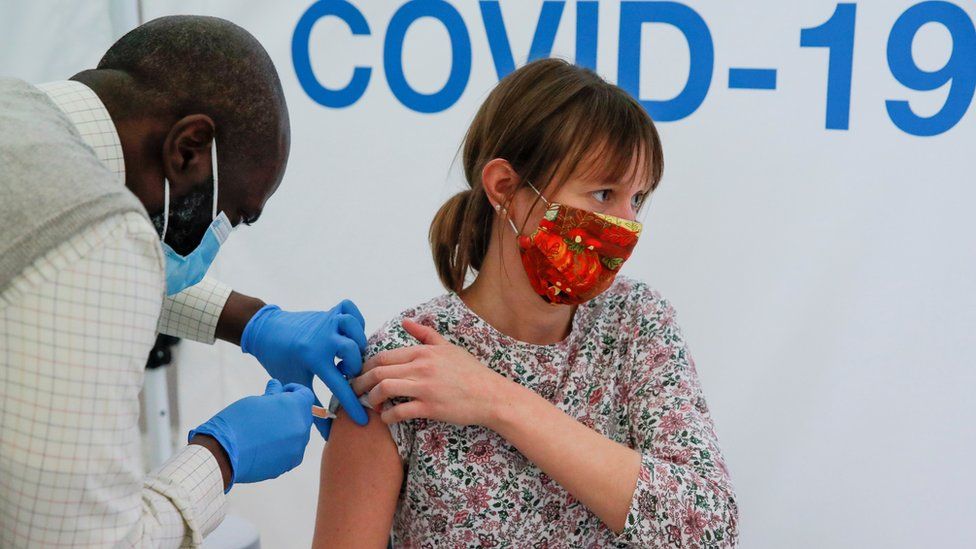
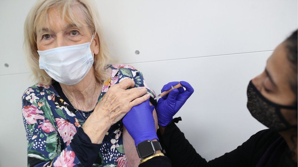
Eight months after mild COVID-19, one in ten people still has at least one moderate to severe symptom that is perceived as having a negative impact on their work, social or home life. The most common long-term symptoms are a loss of smell and taste and fatigue. This is according to a study published in the journal JAMA, conducted by researchers at Danderyd Hospital and Karolinska Institutet in Sweden.
Since spring 2020, researchers at Danderyd Hospital and Karolinska Institutet have conducted the so-called COMMUNITY study, with the main purpose of examining immunity after COVID-19. In the first phase of the study in spring 2020, blood samples were collected from 2,149 employees at Danderyd Hospital, of whom about 19 percent had antibodies against SARS-CoV-2. Blood samples have since then been collected every four months, and study participants have responded to questionnaires regarding long-term symptoms and their impact on the quality of life.
In the third follow-up in January 2021, the research team examined self-reported presence of long-term symptoms and their impact on work, social and home life for participants who had had mild COVID-19 at least eight months earlier. This group consisted of 323 healthcare workers (83 percent women, median age 43 years) and was compared with 1,072 healthcare workers (86 percent women, median age 47 years) who did not have COVID-19 throughout the study period.
The results show that 26 percent of those who had COVID-19 previously, compared to 9 percent in the control group, had at least one moderate to severe symptom that lasted more than two months and that 11 percent, compared to 2 percent in the control group, had a minimum of one symptom with negative impact on work, social or home life that lasted at least eight months. The most common long-term symptoms were loss of smell and taste, fatigue, and respiratory problems.
“We investigated the presence of long-term symptoms after mild COVID-19 in a relatively young and healthy group of working individuals, and we found that the predominant long-term symptoms are loss of smell and taste. Fatigue and respiratory problems are also more common among participants who have had COVID-19 but do not occur to the same extent,” says Charlotte Thålin, specialist physician, Ph.D. and lead researcher for the COMMUNITY study at Danderyd Hospital and Karolinska Institutet. “However, we do not see an increased prevalence of cognitive symptoms such as brain fatigue, memory and concentration problems or physical disorders such as muscle and joint pain, heart palpitations or long-term fever.”
“Despite the fact that the study participants had a mild COVID-19 infection, a relatively large proportion report long-term symptoms with an impact on quality of life. In light of this, we believe that young and healthy individuals, as well as other groups in society, should have great respect for the virus that seems to be able to significantly impair quality of life, even for a long time after the infection,” says Sebastian Havervall, deputy chief physician at Danderyd Hospital and PhD student in the project at Karolinska Institutet.
The COMMUNITY study will now continue, with the next follow-up taking place in May when a large proportion of study participants are expected to be vaccinated. In addition to monitoring immunity and the occurrence of re-infection, several projects regarding post- COVID are planned.
“We will, among other things, be studying COVID-19-associated loss of smell and taste more closely, and investigate whether the immune system, including autoimmunity, plays a role in post-COVID,” says Charlotte Thålin.
Story Source:
Materials provided by Karolinska Institutet. Note: Content may be edited for style and length.

Read more →

Surgical sutures inspired by human tendons

Sutures are used to close wounds and speed up the natural healing process, but they can also complicate matters by causing damage to soft tissues with their stiff fibers. To remedy the problem, researchers from Montreal have developed innovative tough gel sheathed (TGS) sutures inspired by the human tendon.
These next-generation sutures contain a slippery, yet tough gel envelop, imitating the structure of soft connective tissues. In putting the TGS sutures to the test, the researchers found that the nearly frictionless gel surface mitigated the damage typically caused by traditional sutures.
Conventional sutures have been around for centuries and are used to hold wounds together until the healing process is complete. But they are far from ideal for tissue repair. The rough fibers can slice and damage already fragile tissues, leading to discomfort and post-surgery complications.
Part of the problem lies in the mismatch between our soft tissues and the rigid sutures that rub against contacting tissue, say the researchers from McGill University and the INRS Énergie Matériaux Télécommunications Research Centre.
Inspired by the tendon
To tackle the problem, the team developed a new technology that mimics the mechanics of tendons. “Our design is inspired by the human body, the endotenon sheath, which is both tough and strong due to its double-network structure. It binds collagen fibers together while its elastin network strengthens it,” says lead author Zhenwei Ma, a PhD student under the supervision of Assistant Professor Jianyu Li at McGill University.
The endotenon sheath not only forms a slippery surface to reduce friction with surrounding tissues in joints, but it also delivers necessary materials for tissue repair in a tendon injury. In the same way, TGS sutures can be engineered to provide personalized medicine based on a patient’s needs, say the researchers.
Personalized wound treatment
“This technology provides a versatile tool for advanced wound management. We believe it could be used to deliver drugs, prevent infections, or even monitor wounds with near-infrared imaging,” says Li of the Department of Mechanical Engineering.
“The ability to monitor wounds locally and adjust the treatment strategy for better healing is an exciting direction to explore,” says Li, who is also a Canada Research Chair in Biomaterials and Musculoskeletal Health.
Story Source:
Materials provided by McGill University. Note: Content may be edited for style and length.

Read more →

Clinical trial completion rates decline during COVID-19 pandemic

Social distancing and lockdowns may have reduced the spread of COVID-19, but researchers from Penn State College of Medicine also report those actions may have affected clinical researchers’ ability to finish trials. Study completion rates dropped worldwide between 13% and 23%, depending on the type of research sponsor and geographic location, between April and October 2020.
Researchers previously reported that more than 80% of clinical trials suspended between March 1 and April 26, 2020, noted the pandemic as their chief reason for halting activity. Patient enrollment in studies was lower in April 2020, compared to April 2019. Arthur Berg, associate professor of public health sciences, and Nour Hawila, a biostatistics doctoral candidate, investigated how these trends may have affected the completion of clinical trials.
The researchers examined more than 117,000 trials in the United States, Europe, Asia and other regions to study whether the pandemic affected clinical research. Their goal was to assess how the pandemic’s mitigation efforts and financial setbacks may have contributed to decreased clinical trial enrollment and completion.
“The pandemic has made it more difficult for researchers to recruit and follow up on patients in clinical trials,” said Hawila, a research assistant from the Department of Public Health Sciences. “This analysis revealed that the impact was substantial — particularly for trials funded by government, academic or medical entities.”
Hawila and Berg analyzed data from ClinicalTrials.gov, a website that contains information on the status of thousands of clinical trials in the U.S. Pre-COVID-19 enrollment and completion data was pulled from March 2017 to February 2020. The post-COVID-19 period was defined as April through October 2020.
According to researchers, the pandemic reduced the number of new interventional clinical trial submissions to ClinicalTrials.gov by about 10%. Completed trials were down 13% to 23%, depending on the sector and location of the trial source. Clinical trials sponsored by pharmaceutical, biotechnology and therapeutic companies were more likely to complete enrollment.
However, some regions fared better than others during the pandemic. Egypt experienced an increase in both submitted (69%) and completed (73%) clinical trials. Berg explained that the rise is likely in response to the country’s recent parliamentary bill governing medical research.
Berg and Hawila also noted that the pandemic caused a shift in research priorities — 472 (11%) of trials submitted during the post-COVID period were pandemic-related. The results were published in the journal Clinical and Translational Science.
“Clinical research response to the pandemic has been robust,” said Berg, a Penn State Cancer Institute researcher and biostatistics doctoral program director. “But the impact of the pandemic on other types of clinical trials will be felt for decades to come. However, as demonstrated in Egypt, timely governmental action may be able to make a difference in reversing the pandemic’s impact on research.”
Story Source:
Materials provided by Penn State. Original written by Zachary Sweger. Note: Content may be edited for style and length.

Read more →

A drug that can stop tumors from growing

Cancer doctors may soon have a new tool for treating melanoma and other types of cancer, thanks to work being done by researchers at the University of Colorado Cancer Center.
In a paper published in the journal PNAS last month, CU Cancer Center members Mayumi Fujita, MD, PhD, Angelo D’Alessandro, PhD, Morkos Henen, PhD, MS, Beat Vogeli, PhD, Eric Pietras, PhD, James DeGregori, PhD, Carlo Marchetti, PhD, and Charles Dinarello, MD, along with Isak Tengesdal, MS, a graduate student in the Division of Infectious Diseases at the University of Colorado School of Medicine, detail their work on NLRP3, an intracellular complex that has been found to participate in melanoma-mediated inflammation, leading to tumor growth and progression. By inhibiting NLRP3, the researchers found, they can reduce inflammation and the resultant tumor expansion.
Specifically, NLRP3 promotes inflammation by inducing the maturation and release of interleukin-1-beta, a cytokine that causes inflammation as part of the normal immune response to infection. In cancer, however, inflammation can cause tumors to grow and spread.
“NLRP3 is a member of a larger family that is involved in sensing danger signals,” Marchetti says. “It is a receptor that surveils the intercellular compartment of a cell, looking for danger molecules or pathogens. When NLRP3 recognizes these signals, it leads to the activation of caspase-1, a protein involved in the processing and maturation of interleukin-1-beta into its biological active form, causing an intense inflammatory response. We found that in melanoma, this process is dysregulated, resulting in tumor growth.”
The oral NLRP3 inhibitor used in their study (Dapansutrile) has already shown to be effective in clinical trials to treat gout and heart disease, and it is currently being tested in COVID-19 as well. The CU cancer researchers are now trying to find out if this NLRP3 inhibitor can be successfully used in melanoma patients who are resistant to checkpoint inhibitors.
“Checkpoint inhibitors increase the efficacy of the immune system to kill tumors, but sometimes tumors become resistant to this treatment,” Marchetti says. “A big part of cancer research now is to find therapies that can be combined with checkpoint inhibitors to improve their efficacy.”
With the hypothesis that an NLRP3 inhibitor is one of those therapies, CU Cancer Center researchers are studying the drug’s effects on melanoma, as well as breast cancer and pancreatic cancer. In addition to improving the immune response, the NLRP3 inhibitor can also help reduce the side effects of checkpoint inhibitors. Marchetti says this research can make a big difference for melanoma patients who don’t respond to checkpoint inhibitors alone.
“This was a very collaborative project that involved a lot of members of the university, and we are very excited about it,” he says. This project is important because it further shows that NLRP3-mediated inflammation plays a critical role in the progression of melanoma, and it opens new strategies to improve patient care.”
Story Source:
Materials provided by University of Colorado Anschutz Medical Campus. Original written by Greg Glasgow. Note: Content may be edited for style and length.

Read more →

Chain length determines molecular color

Around the world, a huge amount of research and development work is currently being done on carbon-containing, or organic, molecules that emit coloured light after appropriate excitation. This research field is driven by the display industry and the development of biomedical imaging techniques. While precise colour tuning in organic fluorescent dyes has so far usually been achieved by mixing different molecules, ETH researchers have now developed an approach that can generate a broad palette of colours by way of chemical adjustments within the molecules themselves.
Yinyin Bao, a group leader in the group of ETH professor Jean-Christophe Leroux, and his team of scientists turned to fluorescent organic polymers for this work. These polymers can best be thought of as moving chains of varying lengths. “The chains have a symmetrical structure, and two components within them contribute to the fluorescence,” Bao explains. “One component, called the fluorophore, sits in the middle of the chain, while the other component occurs once at each of the chain’s two ends.” Joining the fluorophore in the middle of the chain with each end of the chain are links whose number and structure scientists can adjust. If the polymer chain is bent so that one of its ends comes to lie near the fluorophore and the chain is simultaneously irradiated with UV light, it fluoresces.
Distance affects the interaction
The scientists have now been able to show that the fluorescence colour depends not only on the structure of the chain links and ends, but also on the number of chain links. “It’s the interaction of the chain end and the fluorophore that’s responsible for the fluorescence of these polymers,” Bao says: “The distance between the two components affects how they interact and thus the colour that’s emitted.”
Using a method called living polymerisation, the researchers can regulate the number of chain links. First, they gradually grow the chain by a slow process of attaching building blocks to the fluorophore. Once the desired length is reached, the scientists can terminate the process and simultaneously generate the chain end molecule. This is how the researchers produced polymers with different colours: with fewer than 18 building blocks, the molecules fluoresce yellow; with 25 chain links, green; and with 44 or more links, blue. “What’s special about this is that these differently luminescent polymers are all composed of the exact same components. The only difference is the chain length,” Bao says.
Wide colour range OLEDs
The research team, including scientists from the group of ETH Professor Chih-Jen Shih and from the Royal Melbourne Institute of Technology in Australia, published their work in the journal Science Advances. Currently, the researchers can produce fluorescent polymers in yellow, green and blue, but they are working on extending the principle to include other colours, including red.
These new fluorescent polymers can’t be used directly as OLEDs (organic LEDs) in displays because their electrical conductivity is not sufficiently high, Bao explains. However, it ought to be possible to combine the polymers with semiconducting molecules in order to produce wide colour range OLEDs in a simple way. Used in concentrated solar power plants, they could also collect sunlight more efficiently and thus increase the plants’ efficiency. Bao sees their main areas of application in laboratory diagnostic procedures that use fluorescence, for example in PCR, as well as in microscopy and imaging procedures in cell biology and medicine. Other potential uses would be as security features on banknotes and certificates or in passports.
Story Source:
Materials provided by ETH Zurich. Original written by Fabio Bergamin. Note: Content may be edited for style and length.

Read more →

Carbon dioxide levels reflect COVID-19 risk

Tracking carbon dioxide levels indoors is an inexpensive and powerful way to monitor the risk of people getting COVID-19, according to new research from the Cooperative Institute for Research in Environmental Sciences (CIRES) and the University of Colorado Boulder. In any given indoor environment, when excess CO2 levels double, the risk of transmission also roughly doubles, two scientists reported this week in Environmental Science & Technology Letters.
The chemists relied on a simple fact already put to use by other researchers more than a decade ago: Infectious people exhale airborne viruses at the same time as they exhale carbon dioxide. That means CO2 can serve as a “proxy” for the number of viruses in the air.
“You’re never safe indoors sharing air with others, but you can reduce the risk,” said Jose-Luis Jimenez, co-author of the new assessment, a CIRES Fellow and professor of chemistry at the University of Colorado Boulder.
“And CO2 monitoring is really the only low-cost and practical option we have for monitoring,” said Zhe Peng, a CIRES and chemistry researcher, and lead author of the new paper. “There is nothing else.”
For many months, researchers around the world have been searching for a way to continually monitor COVID-19 infection risk indoors, whether in churches or bars, buses or hospitals. Some are developing instruments that can detect viruses in the air continually, to warn of a spike or to indicate relative safety. Others tested existing laboratory-grade equipment that costs tens of thousands of dollars.
Jimenez and colleagues turned to commercially available carbon dioxide monitors, which can cost just a few hundred dollars. First, they confirmed in the laboratory that the detectors were accurate. Then, they created a mathematical “box model” of how an infected person exhales viruses and CO2, how others in the room inhaled and exhaled, and how the viruses and gas accumulate in the air of a room or are removed by ventilation. The model takes into consideration infection numbers in the local community, but it does not detail air flow through rooms — that kind of modeling requires expensive, custom analysis for each room.
It’s important to understand that there is no single CO2 level at which a person can assume a shared indoor space is “safe,” Peng emphasized. That’s partly because activity matters: Are people in the room singing and talking loudly or exercising, or are they sitting quietly and reading or resting? A CO2 level of 1,000 ppm, which is well above outside levels of about 400 ppm, could be relatively safe in a quiet library with masks but not in an active gym without masks.
But in each indoor space, the model can illuminate “relative” risk: If CO2 levels in a gym drop from 2,800 to 1,000 ppm (~2,400 above background levels to 600), the risk of COVID-19 transmission drops, too, to one-quarter of the original risk. In the library, if an influx of people makes CO2 jump from 800 to 1,600 (400 to 1,200 above background), COVID transmission risk triples.
In the new paper, Peng and Jimenez also shared a set of mathematical formulae and tools that experts in building systems and public health can use to pin down actual, not just relative, risk. But the most important conclusion is that to minimize risk, keep the CO2 levels in all the spaces where we share air as low as practically possible.
“Wherever you are sharing air, the lower the CO2, the lower risk of infection,” Jimenez said.

Read more →

Late night snacks may hurt your workplace performance, study finds

A recent study finds that unhealthy eating behaviors at night can make people less helpful and more withdrawn the next day at work.
“For the first time, we have shown that healthy eating immediately affects our workplace behaviors and performance,” says Seonghee “Sophia” Cho, corresponding author of the study and an assistant professor of psychology at North Carolina State University. “It is relatively well established that other health-related behaviors, such as sleep and exercise, affect our work. But nobody had looked at the short-term effects of unhealthy eating.”
Fundamentally, the researchers had two questions: Does unhealthy eating behavior affect you at work the next day? And, if so, why?
For the study, researchers had 97 full-time employees in the United States answer a series of questions three times a day for 10 consecutive workdays. Before work on each day, study participants answered questions related to their physical and emotional well-being. At the end of each workday, participants answered questions about what they did at work. In the evening, before bed, participants answered questions about their eating and drinking behaviors after work.
In the context of the study, researchers defined “unhealthy eating” as instances when study participants felt they’d eaten too much junk food; when participants felt they’d had too much to eat or drink; or when participants reporting having too many late-night snacks.
The researchers found that, when people engaged in unhealthy eating behaviors, they were more likely to report having physical problems the next morning. Problems included headaches, stomachaches and diarrhea. In addition, when people reported unhealthy eating behaviors, they were also more likely to report emotional strains the next morning — such as feeling guilty or ashamed about their diet choices. Those physical and emotional strains associated with unhealthy eating were, in turn, related to changes in how people behaved at work throughout the day.

Read more →

Novel hydrogel carriers for anti-cancer drugs offer new hope for cancer treatment

Cancer therapy in recent times relies on the use of several drugs derived from biological sources including different bacteria and viruses, among others. However, these bio-based drugs get easily degraded and therefore inactivated on administration into the body. Thus, effective delivery to and release of these drugs at target tumor sites are of paramount importance from the perspective of cancer therapy.
Recently, scientists have discovered unique three-dimensional, water-containing polymers, called hydrogels, as effective drug delivery systems (DDSs). Drugs loaded into these hydrogels remain relatively stable owing to the network-like structure and organic tissue-like consistency of these DDSs. Besides, drug release from hydrogels can be controlled by designing them to swell and shrink in response to certain stimuli, or minute changes in conditions, like temperature or pH (which determines the acidity of an environment). For instance, when conditions are just the right level of acidic in the tumor microenvironment, these DDSs either shrink or swell and release the drug.
However, there has been no method for the one-pot synthesis of hydrogels that respond to more than one such stimulus and degrade to release drugs at target tumor sites. Until now.
Now, a team of scientists, led by Professor Akihiko Kikuchi from Tokyo University of Science, reports the production of unique degradable hydrogels that respond to changes under multiple conditions in “reducing” environments mimicking the microenvironment of tumors. As Prof. Kikuchi observes, “In order to prepare degradable hydrogels that can release drugs in response to changes in the tumor microenvironment, we prepared hydrogels that respond to temperature, pH, and reducing environment, and analyzed their properties.”
In their study published in the Journal of Controlled Release, Prof. Kikuchi — along with his colleagues from Tokyo University of Science, Dr. Syuuhei Komatsu, Ms. Moeno Tago, and Ms. Yu Ando, and his collaborator on the study, Prof. Taka-Aki Asoh from Osaka University — details the steps of designing these novel hydrogels from the synthetic polymer poly(ethylene glycol) diglycidyl ether and the sulfur-containing organic compound cystamine. In response to low temperatures, these hydrogels swell up while they shrink at the physiological temperature. Additionally, the hydrogels respond to pH changes by virtue of possessing tertiary amino groups. It must be noted here that the pH of the tumor microenvironment fluctuates between 5.5 and 6.5 owing to glycolysis in the tumor cells. Under the reducing conditions of this environment, the hydrogels degrade because of the breakage of disulfide bonds and change into low molecular-weight water-soluble oligomers that are easily excreted from the body.
To further test their drug release properties, the scientists loaded these hydrogels with specific proteins by exploiting their temperature-dependent swelling-deswelling behavior and tested the controlled release of drugs under acidic or reducing conditions. It was found that the amount of drug loaded onto these hydrogels could be controlled by changing the mesh size of the hydrogel polymer network by changing temperature, suggesting the possibility of customizing these DDSs for specific drug delivery. Besides, the hydrogel network structure and electrostatic interactions in the network ensured that the proteins were preserved intact until delivery, unaffected by the swelling and shrinking of the hydrogels with pH changes in the surrounding environment. The scientists found that the loaded protein drugs were completely released only under reducing conditions.
Using these hydrogels and the tractability that they provide, doctors may soon be able to design “customized” hydrogels that are specific to patients, giving personalized medicine a big boost. In addition to that, this new DDS provides a way to kill cancer cells that are left behind after surgery. As Prof. Kikuchi states, “The implantation of this material in the affected area after cancer resection may eliminate residual cancer cells, making it a more powerful therapeutic tool.”
As cancer tightens its vise grip around the world, treatment options need to be varied and upgraded for customized and effective therapy. This unique and simple design technique to produce multi-stimuli-responsive hydrogels for effective drug delivery to target tumor sites may just be one among several such promising techniques to mount an answer to the challenge cancer poses to humanity.
Story Source:
Materials provided by Tokyo University of Science. Note: Content may be edited for style and length.

Read more →

Ant responses to social isolation resemble those of humans

Ants react to social isolation in a similar way as do humans and other social mammals. A study by an Israeli-German research team has revealed alterations to the social and hygienic behavior of ants that had been isolated from their group. The research team was particularly surprised by the fact that immune and stress genes were downregulated in the brains of the isolated ants. “This makes the immune system less efficient, a phenomenon that is also apparent in socially isolating humans — notably at present during the COVID-19 crisis,” said Professor Susanne Foitzik, who headed up the study at Johannes Gutenberg University Mainz (JGU). The study on a species of ant native to Germany has recently been published in Molecular Ecology.
Effects of isolation in social insects little studied so far
Humans and other social mammals experience isolation from their group as stressful, having a negative impact on their general well-being and physical health. “Isolated people become lonely, depressed, and anxious, develop addictions more easily, and suffer from a weakened immune system and impaired overall health,” added Professor Inon Scharf, lead author of the article and cooperation partner of the Mainz research group at Tel Aviv University in Israel. While the effects of isolation have been extensively studied in social mammals such as humans and mice, less is known about how social insects respond in comparable situations — even though they live in highly evolved social systems. Ants, for instance, live their entire lives as members of the same colony and are dependent on their colony mates. The worker ants relinquish their own reproductive potential and devote themselves to feeding the larvae, cleaning and defending the nest, and searching for food, while the queen does little more than just lay eggs.
The research team looked at the consequences of social isolation in the case of ants of the species Temnothorax nylanderi. These ants inhabit cavities in acorns and sticks on the ground in European forests, forming colonies of a few dozen workers. Young workers engaged in brood care were taken singly from 14 colonies and kept in isolation for varying lengths of time, from one hour to a maximum of 28 days. The study was conducted between January and March 2019 and highlighted three particular aspects in which changes were observed. After the end of their isolation, the workers were less interested in their adult colony mates, but the length of time they spent in brood contact increased; they also spent less time grooming themselves. “This reduction in hygienic behavior may make the ants more susceptible to parasites, but it is also a feature typical of social deprivation in other social organisms,” explained Professor Susanne Foitzik.
Stress due to isolation adversely affects the immune system
While the study revealed significant changes in the behaviors of the isolated insects, its findings with regard to gene activity were even more striking: Many genes related to immune system function and stress response were downregulated. In other words, these genes were less active. “This finding is consistent with studies on other social animals that demonstrated a weakening of the immune system after isolation,” said Professor Inon Scharf.
The discovery by the team of biologists led by Professor Susanne Foitzik is the first of its kind, combining behavioral and genetic analyses on the effects of isolation in social insects. “Our study shows that ants are as affected by isolation as social mammals are and suggests a general link between social well-being, stress tolerance, and immunocompetence in social animals,” concluded Foitzik, summarizing the results of the Israeli-German study. Foitzik is also collaborating with her Israeli partner Professor Inon Scharf and with co-author and group leader Dr. Romain Libbrecht of JGU on a new joint project on the fitness benefits and the molecular basis of spatial learning in ants, funded by the German Research Foundation (DFG).
Story Source:
Materials provided by Johannes Gutenberg Universitaet Mainz. Note: Content may be edited for style and length.

Read more →

Inheriting acquired traits requires trailblazer modifications to unfertilized eggs

An epigenetic study at the RIKEN Center for Integrative Medical Sciences shows that in mouse egg cells, modifications to histone H2A at lysine 119 lay the groundwork for inherited DNA functional modifications from the mother.
In books and the movies, a group of people on a special mission always sends out a scout to do reconnaissance before they proceed. Sometimes, the scouts leave signs or markers that allow the group to know where there should go. Researchers led by Azusa Inoue at the RIKEN Center for Integrative Medical Sciences in Japan have discovered a mark left behind in unfertilized egg cells that determine which DNA modifications will be inherited if the egg is fertilized. Specifically, they found that without initial modifications to histone H2A at lysine 119 — technically called H2AK119ub1 — later inheritable modifications would not occur. When allowed to develop, one consequence of this deficit was an enlarged placenta after embryo implantation. This study was published in Nature Genetics on April 5.
For many years we were taught in school that acquired traits were not inherited. In some sense this was correct; stretching your neck a lot to get food will not result in children with longer necks. However, your DNA function can be modified throughout your life. For example, DNA structure in chromosomes is supported by proteins called histones. When histones are modified, they can change how genes are expressed in the body. This is epigenetics, and a previous study by Inoue and colleagues showed that acquired tri-methylation of histone H3 at lysine 27 (thankfully abbreviate to H3K27me3) in mammalian egg cells can be inherited. In the new study, the team used technology called low-input CUT&RUN to begin answering the question of how this happens.
First, the researchers examined the timing of the two different histone modifications. They found that every gene exhibiting H3K27me3 also showed H2AK119ub1 in mouse egg cells. Suspecting its importance, the researchers knocked out two proteins that make up H2AK119ub1 in egg cells. Low-input CUT&RUN showed that the knock-out egg cells had much less H3K27me3 than controls at a subset of genes that normally bring H3K27me3 into the next generation. Thus, H2AK119ub1 acts like a kind of marker left by a scout, identifying where subsequent H3K27me3 should follow. “We discovered that H2AK119ub1 is necessary for maternal inheritance of H3K27me3, making the H2AK119ub1-H3K27me3 pathway a major player in transgenerational epigenetic inheritance in mammals,” says Inoue.
The researchers then found something they didn’t expect. Testing showed that the loss of H3K27me3 was itself inherited by fertilized embryos, and could not be reversed. Furthermore, this deficiency led increased lethality — miscarriages — and enlarged placentas. “It was surprising to find that defects in an egg’s histone modification are irreversibly inherited by embryos and cause long term consequences in development,” says Inoue.
The results thus showed that despite normal DNA in the mouse egg cell, if the proper instructions — first H2AK119ub1 and then H3K27me3 modifications — were missing, miscarriages and enlarged placentas could occur. These findings have clinical implications, especially for reproductive medicine and placental defects. “The next step,” says Inoue, “is to see whether any diseases or surrounding environments can affect the heritable histone modification.”
Story Source:
Materials provided by RIKEN. Note: Content may be edited for style and length.

Read more →