Covishield: India seeks EU travel approval for its main vaccine

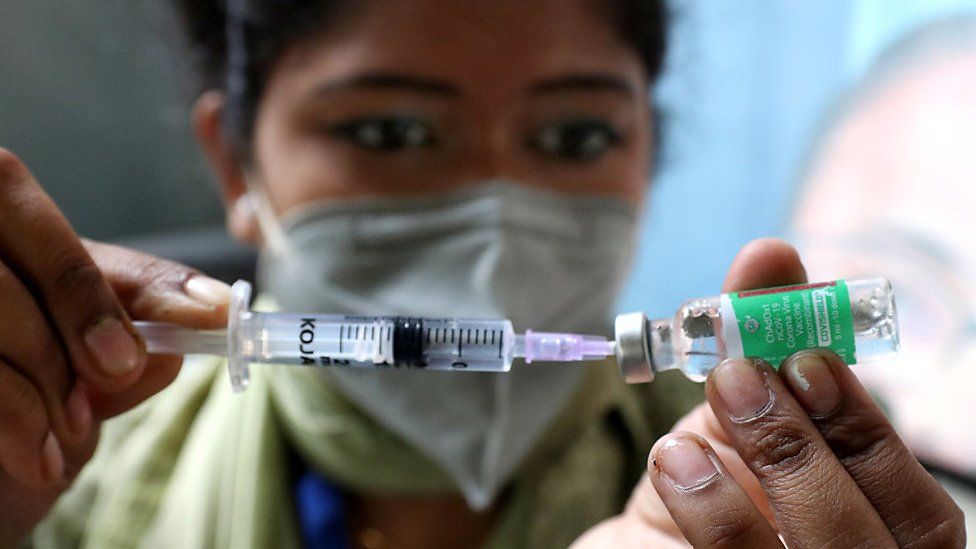
SharecloseShare pageCopy linkAbout sharingimage copyrightGetty ImagesIndia’s Serum Institute is seeking emergency authorisation in the European Union for its Covishield jab, sources told the BBC.The move comes amid reports that Covishield is not yet eligible for the digital green certificate, an EU-wide travel pass, set to launch on 1 July. The certificate is currently for EU citizens only. Covishield is the Indian-made version of AstraZeneca’s Vaxzevria jab, which has been authorised in the EU.It’s manufactured in India by Serum, the world’s largest vaccine maker. The vaccines currently eligible for the green pass have all been approved by the European Medicines Agency (EMA). Serum’s CEO, Adar Poonawalla, said on Monday that his company hopes to “resolve this matter soon”, referring to the fact that Covishield had not yet been authorised in the EU. I realise that a lot of Indians who have taken COVISHIELD are facing issues with travel to the E.U., I assure everyone, I have taken this up at the highest levels and hope to resolve this matter soon, both with regulators and at a diplomatic level with countries.— Adar Poonawalla (@adarpoonawalla) June 28, 2021
The BBC is not responsible for the content of external sites.View original tweet on TwitterThe EMA told the BBC on Monday that the Serum Institute had not yet applied for authorisation.The European Commission has left it to individual member states to decide whether to allow travellers who have received vaccines “that have been authorised at the national level or by the World Health Organization (WHO)”. Covishield was listed for emergency use by the WHO in February. It’s unclear yet if the same rules will also apply to international travellers visiting the EU. India has so far overwhelmingly administered Covishield jabs – they account for more than 284 million of the 323 or so million vaccinations given so far. Covaxin, an Indian homegrown vaccine, which has not yet received WHO approval, has also not applied for EMA authorisation. Sputnik V, which is the third vaccine approved for use in India and by the WHO, is on the EMA’s list of vaccines currently under review. But it has not been rolled out yet in India due to supply delays.At a recent meeting of G7 countries to which India was invited, India’s health minister Dr Harsh Vardhan said that India was “strongly opposed to a ‘vaccine passport’ at this juncture”.Expressed India’s concern & strong opposition to ‘Vaccine Passport’ at this juncture of the #pandemic With vaccine coverage as a % of population in developing countries still low compared to developed countries, such an initiative could prove to be highly discriminatory.@G7 pic.twitter.com/zh6nhkEfbv— Dr Harsh Vardhan (@drharshvardhan) June 4, 2021
The BBC is not responsible for the content of external sites.View original tweet on TwitterThe UK’s Equality and Human Rights Commission recently said that introducing Covid status certificates would be discriminating against some groups saying they could create a “two-tier society whereby only certain groups are able to fully enjoy their rights”.

Read more →

Covid: Masks upgrade cuts infection risk, research finds

SharecloseShare pageCopy linkAbout sharingThe quality of face masks healthcare workers wear makes a huge difference to their risk of coronavirus infection, Cambridge University Hospitals NHS Foundation Trust research has found.Wearing a high grade mask known as an FFP3 can provide up to 100% protection. By contrast, there is a far greater chance of staff wearing standard issue surgical masks catching the virus. Professional bodies have long campaigned for staff to be given better personal protective equipment.The data was gathered during a programme of regular testing for Covid at the trust. The results are published in a pre-print paper that has not been peer-reviewed. Which kind of mask is best?Covid PPE: How healthcare workers came to feel ‘expendable’For most of last year, the hospital followed national guidance which specifies that healthcare workers should wear surgical masks, except in a few limited situations.Though fluid resistant, these masks are relatively flimsy and loose-fitting and are not meant to screen out infectious aerosols – tiny virus particles that can linger in the air and are now widely accepted as a source of coronavirus infection. The study found that staff caring for Covid patients on “red” wards faced a risk that was up to 47 times higher than those on “green” or non-Covid wards. Lead researcher Dr Mark Ferris, a specialist in occupational health at the hospital, said staff were getting Covid despite doing everything they were asked to in terms of infection control.So as the second wave of the pandemic started to hit last December, managers in Cambridge made a local decision to upgrade the protection on red wards. “The only thing left to try that could make a difference was FFP3 respirators, and they did,” Dr Ferris said. FFP3 masks have a close fit and are specifically designed to filter out aerosols.Huge impactIn the weeks following this move, the rate of infections among healthcare workers on red wards dropped spectacularly, quickly falling to the level experienced by staff on green wards where there were no Covid patients.The study concludes that “cases attributed to ward-based exposure fell significantly, with FFP3 respirators providing 31-100% protection (and most likely 100%) against infection from patients with Covid-19”.Any remaining cases were likely to be caused by spread in the community, rather than in the hospital. The paper says fluid-resistant surgical masks were “insufficient” to protect healthcare workers. Dr Mike Weekes, of Cambridge University NHS Hospitals Foundation Trust, who also worked on the study, said the work gives “some real world evidence that FFP3 masks are actually effective and more effective than the surgical masks”. He added: “Clearly, it’s a relatively small study in one trust and so we need to see these findings replicated elsewhere. “But given the difference in the results that we’ve seen, as a sort of precautionary principle effect, what we should be thinking about is changing to use FFP3 masks for anyone caring for a patient with coronavirus.”Campaign intensifiesThe Cambridge trust is among 17 across the UK known to have decided to upgrade PPE regardless of national policy.The call for FFP3 masks to be issued more widely is in line with a long-standing demand by the British Medical Association, the Royal College of Nursing (RCN) and many other professional bodies. They have repeatedly appealed for higher standards of protection to face the threat of airborne spread of the disease, most recently in a high-level meeting with health officials earlier this month.In an open letter to the new Health Secretary Sajid Javid, the Fresh Air NHS group of consultants and doctors says the new study provides yet more evidence of why the policy needs to change.”This has important implications for healthcare worker protection as the UK copes with what is hopefully an ‘exit wave’, as well as trying to reduce the massive backlog of other work whilst coping with inevitable staff sickness and isolation,” the group wrote.It says supply chains have now been largely restored after the shortages of the early days of the pandemic, and that although FFP3 masks are less comfortable than surgical ones, surveys show staff generally prefer a higher level of protection.Rose Gallagher, the RCN’s lead for infection prevention and control, also welcomed the research, telling the BBC: “This important study adds even further weight to the RCN’s continuing call for nursing staff to be better protected from Covid-19 and given routine access to the highest levels of respiratory protective equipment whenever they need it.”We are still seeing cases of Covid-19, even from some who have been vaccinated, and it is vital staff are fully protected and there are no attempts to restrict or ease off on measures to further reduce the risk of infection.”A Department of Health and Social Care spokesperson said guidance on PPE standards was regularly updated to reflect the latest science.”The safety of the NHS and social care staff has always been our top priority and we continue to work round the clock to deliver PPE to protect those on the frontline.”Emerging evidence and data are continually monitored and reviewed and guidance will be amended accordingly if appropriate.”

Read more →

Inside India's deadly mucormycosis fungus epidemic

After India’s devastating second Covid wave, the country is now grappling with tens of thousands of cases of a rare fungal infection called mucormycosis. The disease has been declared an epidemic in the nation. Thousands have died of it. Doctors say Covid, and the treatment being given for it, has led to this surge. From a public hospital in Pune, one of the worst affected districts, the BBC’s Yogita Limaye reports.Filmed by Sanjay Ganguly. Produced and edited by Aakriti Thapar

Read more →

Maternal diets rich in Omega-3 fatty acids may protect offspring from breast cancer, study suggests

According to researchers at Marshall University, a maternal diet rich in Omega-3 fatty acids protects from breast cancer development in offspring. In a new study recently published by Frontiers in Cell and Developmental Biology, researchers noted a significant difference in mice from mothers that were fed a diet rich in canola oil, compared with mothers fed a diet rich in corn oil. A maternal Omega 3-rich diet affected genome-wide epigenetic landscape changes in offspring and potentially modulated gene expression patterns.
Dr. Ata Abbas, a former postdoctoral research fellow in Marshall’s Department of Biological Sciences, headed a research team under the leadership of Dr. Philippe Georgel in the College of Science. Research was done in the Cell Differentiation and Development Center at Marshall as part of a collaborative effort with the Joan C. Edwards School of Medicine’s Department of Biochemistry and Microbiology, under the leadership of Dr. W. Elaine Hardman.
Researchers noticed a three-week delay in mortality in mice whose mothers were fed canola oil versus corn oil. The early delay in mortality was significantly different, but the ultimate overall survival rate was not. Eventually, all the mice developed tumors, but the ones fed canola oil had tumors that were slower-growing and smaller than the mice fed corn oil. Translated to human time scale, the duration of the protective effect linked to the maternal diet would be equivalent to several months (Sengupta et al., 2016).
This study is among a body of work done by Marshall University scientists and others looking at the link between Omega-3 fatty acids and reduced incidence of various types of cancer including, but not restricted to, Chronic Lymphocytic Leukemia and Diffuse Large B-Cell Lymphoma.
“The issue of parental diet and inter-generational transmission has become an important field of research; however, the mode of action often remains partially elusive,” said Georgel, a professor in the Department of Biological Sciences at Marshall. “The MU research group focused on ‘epigenetic’ aspects of trans-generational transmission to explain the reported role of Omega-3 fatty acids. Epigenetics involves changes in gene expression which are not linked to changes in genetic sequences. These results have the potential to promote the design of simple changes in diet which would allow for reduced onset of various types of cancer, not only for the individuals using that diet but also for their offspring.”
The study was conducted with funding from the Department of Defense Health Program and the National Institutes of Health.
Story Source:
Materials provided by Marshall University Joan C. Edwards School of Medicine. Note: Content may be edited for style and length.

Read more →

Researchers question prevailing Alzheimer's theory with new discovery

Experts estimate more than 6 million Americans are living with Alzheimer’s dementia. But a recent study, led by the University of Cincinnati, sheds new light on the disease and a highly debated new drug therapy.
The UC-led study, conducted in collaboration with the Karolinska Institute in Sweden, claims that the treatment of Alzheimer’s disease might lie in normalizing the levels of a specific brain protein called amyloid-beta peptide. This protein is needed in its original, soluble form to keep the brain healthy, but sometimes it hardens into “brain stones” or clumps, called amyloid plaques.
The study, which appears in the journal EClinicalMedicine (published by the Lancet), comes on the heels of the FDA’s conditional approval of a new medicine, aducanumab, that treats the amyloid plaques.
“It’s not the plaques that are causing impaired cognition,” says Alberto Espay, the new study’s senior author and professor of neurology at UC. “Amyloid plaques are a consequence, not a cause,” of Alzheimer’s disease, says Espay, who is also a member of the UC Gardner Neuroscience Institute.
Alzheimer’s disease became widely known as “the long goodbye” in the late 20th century due to the disease’s slow deterioration of brain function and memory. It was over 100 years ago, however, that scientist Alois Alzheimer first identified plaques in the brain of patients suffering from the disease.
Since then, Espay says that scientists have focused on treatments to eliminate the plaques. But the UC team, he says, saw it differently: Cognitive impairment could be due to a decline in soluble amyloid-beta peptide instead of the corresponding accumulation of amyloid plaques. To test their hypothesis, they analyzed the brain scans and spinal fluid from 600 individuals enrolled in the Alzheimer’s Disease Neuroimaging Initiative study, who all had amyloid plaques. From there, they compared the amount of plaques and levels of the peptide in the individuals with normal cognition to those with cognitive impairment. They found that, regardless of the amount of plaques in the brain, the individuals with high levels of the peptide were cognitively normal.

Read more →

Mouse brain imaged from the microscopic to the macroscopic level

Researchers at the University of Chicago and the U.S. Department of Energy’s (DOE) Argonne National Laboratory have leveraged existing advanced X-ray microscopy techniques to bridge the gap between MRI (magnetic resonance imaging) and electron microscopy imaging, providing a viable pipeline for multiscale whole brain imaging within the same brain. The proof-of-concept demonstration involved imaging an entire mouse brain across five orders of magnitude of resolution, a step which researchers say will better connect existing imaging approaches and uncover new details about the structure of the brain.
The advance, which was published on June 9 in NeuroImage, will allow scientists to connect biomarkers at the microscopic and macroscopic level, improving the resolution of MRI imaging and providing greater context for electron microscopy.
“Our lab is really interested in mapping brains at multiple scales to get an unbiased description of what brains look like,” said senior author Narayanan “Bobby” Kasthuri, MD, Assistant Professor of Neurobiology at UChicago and neuroscience researcher at Argonne. “When I joined the faculty here, one of the first things I learned was that Argonne had this extremely powerful X-ray microscope, and it hadn’t been used for brain mapping yet, so we decided to try it out.”
The microscope uses a type of imaging called synchrotron-based X-ray tomography, which can be likened to a “micro-CT,” or micro-computerized tomography scan. Thanks to the powerful X-rays produced by the synchrotron particle accelerator at Argonne, the researchers were able to image the entire mouse brain — roughly one cubic centimeter — at the resolution of a micron, 1/10,000 of a centimeter. It took roughly six hours to collect images of the entire brain, adding up to around 2 terabytes (TB) of data. This is one of the fastest approaches for whole brain imaging at this level of resolution.
MRI can quickly image the whole brain to trace neuronal tracts, but the resolution isn’t sufficient to observe individual neurons or their connections. On the other end of the scale, electron microscopy (EM) can reveal the details of individual synapses, but generates an enormous amount of data, making it computationally challenging to look at pieces of brain tissue larger than a few micrometers in volume. Existing techniques for studying neuroanatomy at the micrometer resolution typically are either merely two-dimensional or use protocols that are incompatible with MRI or EM imaging, making it impossible to use the same brain tissue for imaging at all scales.
The researchers quickly realized that their new micro-CT, or μCT, approach could help bridge this existing resolution gap. “There have been a lot of imaging studies where people use MRI to look at the whole brain level and then try to validate those results using EM, but there’s a discontinuity in the resolutions,” said first author Sean Foxley, PhD, Research Assistant Professor at UChicago. “It’s hard to say anything about the large volume of tissue you see with an MRI when you’re looking at an EM dataset, and the X-ray can bridge that gap. Now we finally have something that can let us look across all levels of resolution seamlessly.”
Combining their expertise in MRI and EM, Foxley, Kasthuri, and the rest of their team opted to attempt mapping a single mouse brain using these three approaches. “Why did we choose the mouse brain? Because it fits in the microscope,” Kasthuri said with a laugh. “But also, the mouse is the workhorse of neuroscience; they’re very useful for analyzing different experimental conditions in the brain.”

Read more →

COVID-19 vaccine generates immune structures critical for lasting immunity

The first two COVID-19 vaccines authorized for emergency use by the Food and Drug Administration (FDA) employed a technology that had never before been used in FDA-approved vaccines. Both vaccines performed well in clinical trials, and both have been widely credited with reducing disease, but concerns remain over how long immunity induced by the new vaccine technology will last.
Now, a study from researchers at Washington University School of Medicine in St. Louis, published June 28 in the journal Nature, has found evidence that the immune response to such vaccines is both strong and potentially long-lasting. Nearly four months after the first dose, people who received the Pfizer vaccine still had so-called germinal centers in their lymph nodes churning out immune cells directed against SARS-CoV-2, the virus that causes COVID-19. Germinal centers, which form as the result of natural infection or vaccination, are boot camps for immune cells, a place where inexperienced cells are trained to better recognize the enemy and weapons are sharpened. A better germinal center response may equal a better vaccine.
Moreover, vaccination led to high levels of neutralizing antibodies effective against three variants of the virus, including the Beta variant from South Africa that has shown some resistance to vaccines. Vaccination induced stronger antibody responses in people who had recovered from SARS-CoV-2 infection compared to those who had never been infected.
In April, both Pfizer and Moderna reported that their vaccines provided at least six months of protection. Their reports were based on tracking whether vaccinated people came down with COVID-19. Other groups have monitored antibody levels in the blood and concluded that the vaccine provides at least months of protection. But nobody had looked to see how the immune response was developing in the body, which could provide important clues to the strength and persistence of the immune response without requiring years of follow-up.
“Germinal centers are the key to a persistent, protective immune response,” said senior author Ali Ellebedy, PhD, an associate professor of pathology & immunology, of medicine and of molecular microbiology. “Germinal centers are where our immune memories are formed. And the longer we have a germinal center, the stronger and more durable our immunity will be because there’s a fierce selection process happening there, and only the best immune cells survive. We found that germinal centers were still going strong 15 weeks after the vaccine’s first dose. We’re still monitoring the germinal centers, and they’re not declining; in some people, they’re still ongoing. This is truly remarkable.”
Scientists don’t fully understand why some vaccines, such as the one for smallpox, induce strong protection that lasts a lifetime, while others, such as the vaccine for whooping cough, require regular boosters. But many suspect that the difference lies in the quality of the germinal centers induced by different vaccines.

Read more →

Baby reef fishes swim for gold

A new study has found baby coral reef fishes can outpace all other baby fishes in the ocean.
Lead author Adam Downie is a PhD candidate at the ARC Centre of Excellence for Coral Reef Studies at James Cook University (Coral CoE at JCU).
Mr Downie said when considering aquatic athletes, young coral reef fishes shine: they are some of the fastest babies, swimming around 15-40 body lengths per second.
As a comparison, herring babies swim up to two body lengths per second, and the fastest human in the water, Olympic gold medalist Michael Phelps, can only swim 1.4 body lengths per second.
“We found the swimming performance in baby fishes relates to whether they ultimately associate with a reef or not,” Mr Downie said.
“When they’re a baby searching for a new reef to call home, a reef fish has to navigate the open ocean and its currents. To be successful at this they need a higher swimming capacity than other non-reef fishes.”
“We think that, over evolutionary time, habitat association shaped the swimming performance in the early life stages of these marine fishes,” said co-author Dr Peter Cowman, also from Coral CoE at JCU and Senior Curator of Biosystematics from Queensland Museum’s Project DIG.

Read more →

New molecule found in chestnut leaves disarms dangerous staph bacteria

Scientists isolated a molecule, extracted from the leaves of the European chestnut tree, with the power to neutralize dangerous, drug-resistant staph bacteria. Frontiers in Pharmacology published the finding, led by scientists at Emory University.
The researchers dubbed the molecule Castaneroxy A, after the genus of the European chestnut, Castanea. The use of chestnut leaves in traditional folk remedies in rural Italy inspired the research.
“We were able to isolate this molecule, new to science, that occurs only in very tiny quantities in the chestnut leaves,” says Cassandra Quave, senior author of the paper and associate professor in Emory’s Center for the Study of Human Health and the School of Medicine’s Department of Dermatology. “We also showed how it disarms Methicillin-resistant Staphylococcus aureus by knocking out the bacteria’s ability to produce toxins.”
Methicillin-resistant Staphylococcus aureus (MRSA) causes infections that are difficult to treat due to its resistance to antibiotics. It is one of the most serious infectious disease concerns worldwide, labeled as a “serious threat” by the Centers for the Disease Control and Prevention. In the United States alone, nearly 3 million antibiotic-resistant infections occur in the U.S. each year, killing more than 35,000 people.
Antibiotics work by killing staph bacteria, which can lead to greater resistance among those few bacteria that survive, spawning “super bugs.” The Quave lab has identified compounds from the Brazilian peppertree, in addition to the European chestnut tree, that simply neutralize the harmful effects of MRSA, allowing cells and tissue to naturally heal from an infection without boosting resistance.
“We’re trying to fill the pipeline for antimicrobial drug discovery with compounds that work differently from traditional antibiotics,” Quave says. “We urgently need these new strategies.” She notes that antimicrobial infections kill an estimated 700,000 globally each year, and that number is expected to grow exponentially if new methods of treatment are not found.

Read more →

Pulling wisdom teeth can improve long-term taste function, research finds

Patients who had their wisdom teeth extracted had improved tasting abilities decades after having the surgery, a new Penn Medicine study published in the journal Chemical Senses found. The findings challenge the notion that removal of wisdom teeth, known as third molars, only has the potential for negative effects on taste, and represent one of the first studies to analyze the long-term effects of extraction on taste.
“Prior studies have only pointed to adverse effects on taste after extraction and it has been generally believed that those effects dissipate over time,” said senior author Richard L. Doty, PhD, director of the Smell and Taste Center at the University of Pennsylvania. “This new study shows us that taste function can actually slightly improve between the time patients have surgery and up to 20 years later. It’s a surprising but fascinating finding that deserves further investigation to better understand why it’s enhanced and what it may mean clinically.”
Doty and co-author Dane Kim, a third-year student in the University of Pennsylvania School of Dental Medicine, evaluated data from 1,255 patients who had undergone a chemosensory evaluation at Penn’s Smell and Taste Center over the course of 20 years. Among that group, 891 patients had received third molar extractions and 364 had not.
The “whole-mouth identification” test incorporates five different concentrations of sucrose, sodium chloride, citric acid, and caffeine. Each solution is sipped, swished in the mouth, and then spit out. Subjects then indicate whether the solution tastes sweet, salty, sour, or bitter.
The extraction group outperformed the control group for each of the four tastes, and in all cases, women outperformed men. The study suggests, for the first time, that people who have received extractions in the distant past experience, on average, an enhancement (typically a three to 10 percent improvement) in their ability to taste.
“The study strongly suggests that extraction of the third molar has a positive long-term, albeit subtle, effect on the function of the lingual taste pathways of some people,” Kim said.
Two possibilities, the authors said, could explain the enhancement. First, extraction damage to the nerves that innervate the taste buds on the front of the mouth can release inhibition on nerves that supply the taste buds at the rear of the mouth, increasing whole-mouth sensitivity. Second, hypersensitivity after peripheral nerve injury from a surgery like an extraction has been well documented in other contexts. There is evidence, for example, from animal studies that repetitive light touch, which might occur during chewing, gradually accentuates neural responses from irritated tissue that can lead to progressive long-term tactile hypersensitivity. Whether this occurs for taste, however, is not known.
“Further studies are needed to determine the mechanism or mechanisms behind the extraction-related improvement in taste function,” Doty said. “The effects are subtle but may provide insight into how long-term improvement in neural function can result from altering the environment in which nerves propagate.”
The study was supported in part by the National Institute on Deafness and Other Communication Disorders (PO1 DC 00161).

Read more →