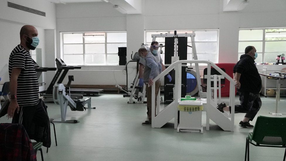
Thomas Brock, Whose Discovery Paved the Way for PCR Tests, Dies at 94

In 1966, he found heat-resistant bacteria in a hot spring at Yellowstone National Park. That led to the development of the chemical process behind the test for Covid-19.Thomas Brock, a microbiologist, was driving west to a laboratory in Washington State in 1964 when he stopped off at Yellowstone National Park.“I’d never seen Yellowstone before,” he said in an interview in 2017. “I came in the south entrance, got out of my car, and there were all these thermal areas spreading out from the hot springs into the lake. I was stunned by the microbes that were living in the hot springs, and nobody seemed to know anything about them.”What fascinated him, on what would be the first of many trips to Yellowstone, were the blue-green algae living in a hot spring — proof that some life could tolerate temperatures above the boiling point of water.It was the beginning of research that led to a revolutionary find in 1966: a species of bacteria that he called Thermus aquaticus, which thrived at 70 degrees Celsius (158 degrees Fahrenheit) or more.The yellow bacteria — discovered by Dr. Brock and Hudson Freeze, his undergraduate assistant at Indiana University — survived because all their enzymes are stable at very high temperatures, including one, Taq polymerase, that replicates its own DNA and was essential to the invention of the process behind the gold standard in Covid-19 testing.Dr. Brock died on April 4 at his home in Madison, Wis. He was 94.His wife, Katherine (Middleton) Brock, known as Kathie, said the cause was complications of a fall.Thermus aquaticus was used in the 1980s by the biochemist Kary B. Mullis to help create the polymerase chain reaction, or PCR, which earned him a share of the 1993 Nobel Prize in Chemistry.Dr. Brock made his discovery after he became fascinated by the blue-green algae living in a hot spring. They offered proof that some life tolerated temperatures above the boiling point of water.Thomas Brock“I remember running into Mullis at a meeting,” Dr. Freeze, now the director of the human genetics program at Sanford-Burnham Prebys Medical Discovery Institute in San Diego, said in an interview. “And I said, ‘I’m the guy who found Thermus aquaticus with Tom Brock,’ and he said that he used the very strain that we isolated in Yellowstone.” (Dr. Brock had deposited cultures at the American Type Culture Collection in Gaithersburg, Md.)The PCR technology, which requires cycles of extreme heating and cooling, can multiply small segments of DNA millions or even billions of times in a short period. It has proved crucial in many ways, including the identification of DNA at a crime scene and, more recently, detecting whether someone has Covid-19.“PCR is fundamental to everything we do in molecular biology today,” said Yuka Manabe, a professor of medicine in the division of infectious diseases at the Johns Hopkins University School of Medicine. “Mullis couldn’t have done PCR without a rock-stable enzyme.”Thomas Dale Brock was born on Sept. 10, 1926, in Cleveland. His father, Thomas, an engineer who ran the boiler room at a hospital, died when Tom was 15, pushing him and his mother, Helen (Ringwald) Brock, a nurse, into poverty. Tom, an only child, took jobs in stores to help her.When he was a teenager, his interest in chemistry led him to set up a small laboratory with a friend in the loft of a barn behind his house in Chillicothe, Ohio, where he and his mother lived after his father’s death. They experimented there with explosives and toxic gas.After serving in the Navy’s electronics training program, Dr. Brock earned three degrees at Ohio State University: a bachelor’s in botany and a master’s and Ph.D. in mycology, the study of fungi.With faculty jobs in short supply, Dr. Brock spent five years as a research microbiologist at the Upjohn Company before he was hired as an assistant professor of biology at Western Reserve University (now Case Western Reserve University) in Cleveland. After two years, he became a postdoctoral fellow in the university’s medical school. In 1960, he joined the department of bacteriology at Indiana University, Bloomington, where he taught medical microbiology.When he arrived at Yellowstone, he did not have grandiose ambitions.A view of a Thermus aquaticus bacterium. It is able to survive hot temperatures because of an enzyme called Taq polymerase that protects it from heat.UW-Madison News & Public Affairs“I was just looking for a nice, simple ecosystem where I could study microbial ecology,” he said in an interview for the website of the University of Wisconsin, Madison, where he was a professor of natural sciences in the department of bacteriology from 1971 to 1990. “At higher temperatures, you don’t have the complications of having animals that eat all the microbes.”Stephen Zinder worked with Dr. Brock as a student from 1974 to 1977, a period that included Dr. Brock’s last summer of work at Yellowstone and his research into the ecology of Wisconsin’s lakes, including Lake Mendota in Madison.“He had an encyclopedic knowledge of microbiology and science in general,” said Dr. Zinder, now a professor of microbiology at Cornell University. “He was always learning and picking up new things.” He added, “I think his real ability was to see things simply and to figure out simple techniques to find out what the organisms were doing in their environment.”Dr. Brock wrote or edited numerous books, including “Milestones in Microbiology” (1961); “Biology of Microorganisms” (1970), now in its 16th edition; and “A Eutrophic Lake: Lake Mendota, Wisconsin” (1985).After retiring from the University of Wisconsin, Dr. Brock focused on ecological strategies to restore oak savanna, prairie and marshland on 140 acres that he and his wife had purchased in Black Earth, Wis., about 35 minutes from Madison.Dr. Brock in 2013 at the Pleasant Valley Conservancy in Black Earth, Wis. He and his wife, the microbiologist Katherine (Middleton) Brock, restored 140 acres of oak savanna, prairie and marshland there.Jeff Miller/UW-MadisonThe land, initially intended as a place for their two children to play, later became the Pleasant Valley Conservancy.“It was less expensive than land nearer Madison, and it turned out to be more interesting,” said Mrs. Brock, who is also a microbiologist.In addition to his wife, Dr. Brock is survived by their daughter, Emily Brock, and their son, Brian. His first marriage, to Mary Louise Louden, ended in divorceTo Dr. Brock, the discovery of Thermus aquaticus exemplified the benefits of being given the freedom to perform fundamental research without fixating on practical results.“It’s kind of an interesting story,” he told Wyoming Public Radio in 2020, “how research that was being done for just basic research, trying to find out what kind of weird critters might be living in boiling water in Yellowstone,” would eventually lead to “extremely widespread practical applications.”

Read more →

Genetic effects of Chernobyl radiation

In two landmark studies, researchers have used cutting-edge genomic tools to investigate the potential health effects of exposure to ionizing radiation, a known carcinogen, from the 1986 accident at the Chernobyl nuclear power plant in northern Ukraine. One study found no evidence that radiation exposure to parents resulted in new genetic changes being passed from parent to child. The second study documented the genetic changes in the tumors of people who developed thyroid cancer after being exposed as children or fetuses to the radiation released by the accident.
The findings, published around the 35th anniversary of the disaster, are from international teams of investigators led by researchers at the National Cancer Institute (NCI), part of the National Institutes of Health. The studies were published online in Science on April 22.
“Scientific questions about the effects of radiation on human health have been investigated since the atomic bombings of Hiroshima and Nagasaki and have been raised again by Chernobyl and by the nuclear accident that followed the tsunami in Fukushima, Japan,” said Stephen J. Chanock, M.D., director of NCI’s Division of Cancer Epidemiology and Genetics (DCEG). “In recent years, advances in DNA sequencing technology have enabled us to begin to address some of the important questions, in part through comprehensive genomic analyses carried out in well-designed epidemiological studies.”
The Chernobyl accident exposed millions of people in the surrounding region to radioactive contaminants. Studies have provided much of today’s knowledge about cancers caused by radiation exposures from nuclear power plant accidents. The new research builds on this foundation using next-generation DNA sequencing and other genomic characterization tools to analyze biospecimens from people in Ukraine who were affected by the disaster.
The first study investigated the long-standing question of whether radiation exposure results in genetic changes that can be passed from parent to offspring, as has been suggested by some studies in animals. To answer this question, Dr. Chanock and his colleagues analyzed the complete genomes of 130 people born between 1987 and 2002 and their 105 mother-father pairs.
One or both of the parents had been workers who helped clean up from the accident or had been evacuated because they lived in close proximity to the accident site. Each parent was evaluated for protracted exposure to ionizing radiation, which may have occurred through the consumption of contaminated milk (that is, milk from cows that grazed on pastures that had been contaminated by radioactive fallout). The mothers and fathers experienced a range of radiation doses.

Read more →

Immune system, not COVID virus, may pose greatest risk to pregnant women

For reasons not yet clear, pregnant women infected with the virus that causes COVID-19 are more likely to experience preterm births, pre-eclampsia, and other neonatal problems than non-infected women.
A team of Yale scientists decided to investigate whether the virus could be affecting placental tissue of infected expectant mothers. Their analysis found that while evidence of the virus in the placenta is rare, the placenta in infected mothers tended to exhibit a much higher level of immune system activity than those of non-infected pregnant women, they report April 22 in the journal Med.
“The good news is the placenta is mounting a robust defense against an infection that is far distant, in lungs or nasal tissue,” said Shelli Farhadian, assistant professor of internal medicine (infectious diseases) and neurology at Yale and co-corresponding author. “On the other hand, the high level of immune system activity might be leading to other deleterious effects on the pregnancy.”
The team headed by Farhadian and Akiko Iwasaki, the Waldemar Von Zedtwitz Professor of Immunobiology at Yale, analyzed blood and placental tissue in 39 infected and as well as COVID-free expectant mothers at different stages of pregnancy. While they found evidence of the virus in only two samples of placental tissue, they did find ACE2 receptors — which the SARS-CoV-2 virus uses to enter cells — in the placentas of most women during the first trimester of pregnancy. Those receptors had largely disappeared in healthy women at later stages of pregnancy.
“It is very important to closely monitor expectant mothers who become infected early in pregnancy,” Farhadian said.
Immune system activity in the placenta during infections like COVID-19 has not been extensively studied and it is not known whether other types of infections would behave similarly to SARS-CoV-2, she said.
Alice Lu-Culligan is lead author of the study, which was primarily funded by the National Institutes of Health and the Emergent Ventures Fund at the Mercatus Center at George Mason University.
Story Source:
Materials provided by Yale University. Original written by Bill Hathaway. Note: Content may be edited for style and length.

Read more →

A new method for fighting 'cold' tumors

Not all cancerous tumors are created equal. Some tumors, known as “hot” tumors, show signs of inflammation, which means they are infiltrated with T cells working to fight the cancer. Those tumors are easier to treat, as immunotherapy drugs can then amp up the immune response.
“Cold” tumors, on the other hand, have no T-cell infiltration, which means the immune system is not stepping in to help. With these tumors, immunotherapy is of little use.
It’s the latter type of tumor that researchers Michael Knitz and radiation oncologist and University of Colorado Cancer Center member Sana Karam, MD, PhD, address in new research published this week in the Journal for ImmunoTherapy of Cancer. Working with mouse models in Karam’s specialty area of head and neck cancers, Knitz and Karam studied the role of T cells in tumor treatment.
“What we found is that the cells that normally tell the T cell, ‘Hey, here’s a tumor — come and attack it,’ are being silenced,” Karam says.
She and her team found that regulatory T cells (Tregs), a specialized T cell type that suppresses immune response, are essentially telling the T cells to stop fighting the cancer.
“Tregs normally serve as an important balance in a healthy immune system,” Knitz says. “They prevent autoimmune disease and put the brakes on the T cells when needed. However, in many tumors, Tregs are too numerous or overly suppressive, bringing the T cell response to a halt.”
Using medication that deactivates the Tregs can help boost the immune response in patients with cold tumors, the researchers found, as can radiation treatment that causes enough injury that the immune cells known as dendritic cells work to put the regular T cells into fight mode.
But this is only part of the story. The T cells need to know what to attack. “You need the radiation to create injury and bring in the immune cells so that the tumor can be recognized and targeted,” says Karam, also an associate professor of radiation oncology at the University of Colorado School of Medicine. “That way, the dendritic cells trigger the immune system to produce a lot of T cells, similar to what a vaccine does. Those T cells then go back to the tumor to kill cancer cells. The pieces are already in place; they just need the proper signals. Activating the dendritic cells is a crucial step in allowing radiation to heat up these cold tumors.”
Importantly, Karam and her team, which includes post-doctorate fellow Thomas Bickett, found that the radiation must be administered in a specific way.
“A specific dosing is needed,” Karam says. “You have to pulse it. You can’t just give one dose. You have to give it again and combine it with things that remove the suppression — the Tregs — while simultaneously keeping those antigen-presenting dendritic cells active and on board.”
Karam says the next step in her research is clinical trials she hopes will eventually change the treatment paradigm from surgery and weeks of chemotherapy and radiation to just three sessions of radiation and immunotherapy, then surgery. She is driven to change the standard of care for cold tumors, she explains, because of the horrendous effects they have on patients.
“These tumors resemble those in patients who are heavy smokers,” she says. “They’re very destructive to bone and muscle, infiltrating the tongue, jaw, gum, and lymph nodes. It’s horrible. We have very high failure rates with them, and the treatment often involves removing the tongue and weeks of radiation and chemotherapy, only for the patient to fail. I’m confident that we can do better for our patients.”

Read more →

Newly-discovered molecule provides dual protection against vascular inflammation

A multidisciplinary team of researchers from Duke-NUS Medical School and the Agency for Science, Technology and Research (A*STAR) in Singapore discovered a new mitochondrial peptide called MOCCI that plays an important role in regulating inflammation of blood vessel and immunity. The study, published in the journal Nature Communications, revealed how one gene encoded two molecules that provide two-pronged protection following viral infection.
Chronic and excessive inflammation of the blood vessels, known as vascular inflammation, can lead to tissue damage and cardiovascular diseases such as atherosclerosis and fibrosis. Although some therapies have shown promising results in clinical trials, they have considerable side effects, such as immunosuppression leading to increased risk of infection, and limited efficacy. Therefore, more effective treatments are urgently needed.
“In this study, we aimed to identify new targets to combat inflammation in the lining of blood vessels. Specifically, we wanted to target small naturally-produced peptides that have not been studied before,” explained Assistant Professor Lena Ho, from the Cardiovascular and Metabolic Diseases Programme at Duke-NUS, who led the team that included Associate Professor Ashley St John, Assistant Professor Owen Rackham and Senior Research Fellow Dr Cheryl Lee.
The Duke-NUS team, in collaboration with colleagues from the Institute of Molecular and Cell Biology at A*STAR Singapore, investigated a group of peptides called Mito-SEPs that localise in mitochondria, the cellular organelles well-known for their role in cellular energy production. After observing that Mito-SEPs appear to be involved in regulating inflammation, they screened cells from the lining of human aortic blood vessels to uncover peptides involved in this process.
They found a new peptide, which they named MOCCI — short for Modulator of Cytochrome C oxidase during Inflammation — that is made only when cells undergo inflammation and infection.
To their surprise, they discovered that MOCCI is a hitherto unknown component of Complex IV, a part of a series of enzymes in the mitochondria responsible for energy production, called the electron transport chain. During inflammation, MOCCI incorporates into Complex IV to dampen its activity. Collaborating with Assoc Prof St John at Duke-NUS, the researchers found that this dampening is required to reduce inflammation following viral infection.
“Our finding that the composition of the electron transport chain changes in response to inflammation is novel. MOCCI in essence repurposes part of the energy production centre in the cell to regulate inflammation,” said Dr Lee, the lead author of this study.
The researchers also discovered that MOCCI is made together with a micro-RNA molecule called miR-147b. The two molecules are made from different sections of the same gene. MOCCI originates from the sequence of the gene that codes for proteins, while miR-147b is made from the non-coding section.
While the miR-147b molecule also exerts anti-inflammatory effects, it actively prevents viruses from replicating at the same time. This implies that MOCCI and miR-147b function in tandem to help to control viral infection and suppress inflammation.
“This dual-pronged strategy is an elegant mechanism that the body has put in place to prevent excessive and potentially tissue-damaging inflammation during infection, such as the cytokine storm seen in COVID-19 infection, and colitis” said Asst Prof Ho. “The gene encoding MOCCI is one of the first genes described to have both coding and non-coding functions. The fact these dual functions are coordinated to achieve a concerted biological outcome is a significant finding in cell biology.”
Professor Patrick Casey, Senior Vice-Dean for Research at Duke-NUS, commented, “Medicine and healthcare advance with the aid of new discoveries in fundamental research. This study by Asst Prof Ho and her collaborators provides valuable insight on inflammation and immunity — a topic that has become even more important in the context of COVID-19.”
The researchers say the next step is to explore how to develop targeted pharmacological treatments that can mimic the anti-inflammatory effects of MOCCI and miR147b. They also plan to investigate the role of MOCCI in common chronic inflammatory diseases such as colitis and psoriasis.
Story Source:
Materials provided by Duke-NUS Medical School. Note: Content may be edited for style and length.

Read more →

Experimental drug shows potential against Alzheimer's disease

Researchers at Albert Einstein College of Medicine have designed an experimental drug that reversed key symptoms of Alzheimer’s disease in mice. The drug works by reinvigorating a cellular cleaning mechanism that gets rid of unwanted proteins by digesting and recycling them. The study was published online today in the journal Cell.
“Discoveries in mice don’t always translate to humans, especially in Alzheimer’s disease,” said co-study leader Ana Maria Cuervo, M.D., Ph.D., the Robert and Renée Belfer Chair for the Study of Neurodegenerative Diseases, professor of developmental and molecular biology, and co-director of the Institute for Aging Research at Einstein. “But we were encouraged to find in our study that the drop-off in cellular cleaning that contributes to Alzheimer’s in mice also occurs in people with the disease, suggesting that our drug may also work in humans.” In the 1990s, Dr. Cuervo discovered the existence of this cell-cleaning process, known as chaperone-mediated autophagy (CMA) and has published 200 papers on its role in health and disease.
CMA becomes less efficient as people age, increasing the risk that unwanted proteins will accumulate into insoluble clumps that damage cells. In fact, Alzheimer’s and all other neurodegenerative diseases are characterized by the presence of toxic protein aggregates in patients’ brains. The Cell paper reveals a dynamic interplay between CMA and Alzheimer’s disease, with loss of CMA in neurons contributing to Alzheimer’s and vice versa. The findings suggest that drugs for revving up CMA may offer hope for treating neurodegenerative diseases.
Establishing CMA’s Link to Alzheimer’s
Dr. Cuervo’s team first looked at whether impaired CMA contributes to Alzheimer’s. To do so, they genetically engineered a mouse to have excitatory brain neurons that lacked CMA. The absence of CMA in one type of brain cell was enough to cause short-term memory loss, impaired walking, and other problems often found in rodent models of Alzheimer’s disease. In addition, the absence of CMA profoundly disrupted proteostasis — the cells’ ability to regulate the proteins they contain. Normally soluble proteins had shifted to being insoluble and at risk for clumping into toxic aggregates.
Dr. Cuervo suspected the converse was also true: that early Alzheimer’s impairs CMA. So she and her colleagues studied a mouse model of early Alzheimer’s in which brain neurons were made to produce defective copies of the protein tau. Evidence indicates that abnormal copies of tau clump together to form neurofibrillary tangles that contribute to Alzheimer’s. The research team focused on CMA activity within neurons of the hippocampus — the brain region crucial for memory and learning. They found that CMA activity in those neurons was significantly reduced compared to control animals.

Read more →

Anti-aging compound improves muscle glucose metabolism in people

A natural compound previously demonstrated to counteract aspects of aging and improve metabolic health in mice has clinically relevant effects in people, according to new research at Washington University School of Medicine in St. Louis.
A small clinical trial of postmenopausal women with prediabetes shows that the compound NMN (nicotinamide mononucleotide) improved the ability of insulin to increase glucose uptake in skeletal muscle, which often is abnormal in people with obesity, prediabetes or Type 2 diabetes. NMN also improved expression of genes that are involved in muscle structure and remodeling. However, the treatment did not lower blood glucose or blood pressure, improve blood lipid profile, increase insulin sensitivity in the liver, reduce fat in the liver or decrease circulating markers of inflammation as seen in mice.
The study, published online April 22 in the journal Science, is the first randomized clinical trial to look at the metabolic effects of NMN administration in people.
Among the women in the study, 13 received 250 mg of NMN orally every day for 10 weeks, and 12 were given an inactive placebo every day over the same period.
“Although our study shows a beneficial effect of NMN in skeletal muscle, it is premature to make any clinical recommendations based on the results from our study,” said senior investigator Samuel Klein, MD, the William H. Danforth Professor of Medicine and Nutritional Science and director of the Center for Human Nutrition. “Normally, when a treatment improves insulin sensitivity in skeletal muscle, as is observed with weight loss or some diabetes medications, there also are related improvements in other markers of metabolic health, which we did not detect in our study participants.”
The remarkable beneficial effects of NMN in rodents have led several companies in Japan, China and in the U.S. to market the compound as a dietary supplement or a neutraceutical. The U.S. Food and Drug Administration is not authorized to review dietary supplement products for safety and effectiveness before they are marketed, and many people in the U.S. and around the world now take NMN despite the lack of evidence to show clinical benefits in people.
The researchers studied 25 postmenopausal women who had prediabetes, meaning they had higher than normal blood sugar levels, but the levels were not high enough to be diagnosed as having diabetes. Women were enrolled in this trial because mouse studies showed NMN had the greatest effects in female mice.
NMN is involved in producing an important compound in all cells, called nicotinamide adenine dinucleotide (NAD). NAD plays a vital role in keeping animals healthy. Levels of NAD decline with age in a broad range of animals, including humans, and the compound has been shown to contribute to a variety of aging-associated problems, including insulin resistance in studies conducted in mice. Supplementing animals with NMN slows and ameliorates age-related decline in the function of many tissues in the body.
Co-investigator Shin-ichiro Imai, MD, PhD, a professor of developmental biology and of medicine who has been studying NMN for almost two decades and first reported on its benefits in mice said, “This is one step toward the development of an anti-aging intervention, though more research is needed to fully understand the cellular mechanisms responsible for the effects observed in skeletal muscle in people.”
Insulin enhances glucose uptake and storage in muscle, so people who are resistant to insulin are at increased risk for developing Type 2 diabetes. But the researchers caution that more studies are needed to determine whether NMN has beneficial effects in the prevention or management of prediabetes or diabetes in people. Klein and Imai are continuing to evaluate NMN in another trial involving men as well as women.
Story Source:
Materials provided by Washington University School of Medicine. Original written by Jim Dryden. Note: Content may be edited for style and length.

Read more →

Researchers trace spinal neuron family tree

Spinal cord nerve cells branching through the body resemble trees with limbs fanning out in every direction. But this image can also be used to tell the story of how these neurons, their jobs becoming more specialized over time, arose through developmental and evolutionary history. Salk researchers have, for the first time, traced the development of spinal cord neurons using genetic signatures and revealed how different subtypes of the cells may have evolved and ultimately function to regulate our body movements.
The findings, published in the journal Science on April 23, 2021, offer researchers new ways of classifying and tagging subsets of spinal cord cells for further study, using genetic markers that differentiate branches of the cells’ family tree.
“A study like this provides the first molecular handles for scientists to go in and study the function of spinal cord neurons in a much more precise way than they ever have before,” says senior author of the study Samuel Pfaff, Salk Professor and the Benjamin H. Lewis Chair. “This also has implications for treating spinal cord injuries.”
Spinal neurons are responsible for transmitting messages between the spinal cord and the rest of the body. Researchers studying spinal neurons have typically classified the cells into “cardinal classes,” which describe where in the spinal cord each type of neuron first appears during fetal development. But, in an adult, neurons within any one cardinal class have varied functions and molecular characteristics. Studying small subsets of these cells to tease apart their diversity has been difficult. However, understanding these subset distinctions is crucial to helping researchers understand how the spinal cord neurons control movements and what goes awry in neurogenerative diseases or spinal cord injury.
“It’s been known for a long time that the cardinal classes, as useful as they are, are incomplete in describing the diversity of neurons in the spinal cord,” says Peter Osseward, a graduate student in the Pfaff lab and co-first author of the new paper, along with former graduate student Marito Hayashi, now a postdoctoral fellow at Harvard University.
Pfaff, Osseward and Hayashi turned to single-cell RNA sequencing technologies to analyze differences in what genes were being activated in almost 7,000 different spinal neurons from mice. They used this data to group cells into closely related clusters in the same way that scientists might group related organisms into a family tree.

Read more →

Salad or cheeseburger? Your co-workers shape your food choices

The foods people buy at a workplace cafeteria may not always be chosen to satisfy an individual craving or taste for a particular food. When co-workers are eating together, individuals are more likely to select foods that are as healthy — or unhealthy — as the food selections on their fellow employees’ trays. “We found that individuals tend to mirror the food choices of others in their social circles, which may explain one way obesity spreads through social networks,” says Douglas Levy, PhD, an investigator at the Mongan Institute Health Policy Research Center at Massachusetts General Hospital (MGH) and first author of new research published in Nature Human Behaviour. Levy and his co-investigators discovered that individuals’ eating patterns can be shaped even by casual acquaintances, evidence that corroborates several multi-decade observational studies showing the influence of people’s social ties on weight gain, alcohol consumption and eating behavior.
Previous research on social influence upon food choice had been primarily limited to highly controlled settings like studies of college students eating a single meal together, making it difficult to generalize findings to other age groups and to real-world environments. The study by Levy and his co-authors examined the cumulative social influence of food choices among approximately 6,000 MGH employees of diverse ages and socioeconomic status as they ate at the hospital system’s seven cafeterias over two years. The healthfulness of employees’ food purchases was determined using the hospital cafeterias’ “traffic light” labeling system designating all food and beverages as green (healthy), yellow (less healthy) or red (unhealthy).
MGH employees may use their ID cards to pay at the hospitals’ cafeterias, which allowed the researchers to collect data on individuals’ specific food purchases, and when and where they purchased the food. The researchers inferred the participants’ social networks by examining how many minutes apart two people made food purchases, how often those two people ate at the same time over many weeks, and whether two people visited a different cafeteria at the same time. “Two people who make purchases within two minutes of each other, for example, are more likely to know each other than those who make purchases 30 minutes apart,” says Levy. And to validate the social network model, the researchers surveyed more than 1,000 employees, asking them to confirm the names of the people the investigators had identified as their dining partners.
“A novel aspect of our study was to combine complementary types of data and to borrow tools from social network analysis to examine how the eating behaviors of a large group of employees were socially connected over a long period of time,” says co-author Mark Pachucki, PhD, associate professor of Sociology at the University of Massachusetts, Amherst.
Based on cross-sectional and longitudinal assessments of three million encounters between pairs of employees making cafeteria purchases together, the researchers found that food purchases by people who were connected to each other were consistently more alike than they were different. “The effect size was a bit stronger for healthy foods than for unhealthy foods,” says Levy.
A key component of the research was to determine whether social networks truly influence eating behavior, or whether people with similar lifestyles and food preferences are more likely to become friends and eat together, a phenomenon known as homophily. “We controlled for characteristics that people had in common and analyzed the data from numerous perspectives, consistently finding results that supported social influence rather than homophily explanations,” says Levy.
Why do people who are socially connected choose similar foods? Peer pressure is one explanation. “People may change their behavior to cement the relationship with someone in their social circle,” says Levy. Co-workers may also implicitly or explicitly give each other license to choose unhealthy foods or exert pressure to make a healthier choice.
The study’s findings have several broader implications for public health interventions to prevent obesity. One option may be to target pairs of people making food choices and offer two-for-one sales on salads and other healthful foods but no discounts on cheeseburgers. Another approach might be to have an influential person in a particular social circle model more healthful food choices, which will affect others in the network. The research also demonstrates to policymakers that an intervention that improves healthy eating in a particular group will also be of value to individuals socially connected to that group.
“As we emerge from the pandemic and transition back to in-person work, we have an opportunity to eat together in a more healthful way than we did before,” says Pachucki. “If your eating habits shape how your co-workers eat — even just a little — then changing your food choices for the better might benefit your co-workers as well.”
Story Source:
Materials provided by Massachusetts General Hospital. Note: Content may be edited for style and length.

Read more →