People given 'friendly' bacteria in nose drops protected against meningitis, study suggests

Led by Professor Robert Read and Dr Jay Laver from the NIHR Southampton Biomedical Research Centre and the University of Southampton, the work is the first of its kind.
Together they inserted a gene into a harmless type of a bacteria, that allows it to remain in the nose and trigger an immune response. They then introduced these bacteria into the noses of healthy volunteers via nose drops.
The results, published in the journal Science Translational Medicine, showed a strong immune response against bacteria that cause meningitis. Published in Science Translational Medicine, those data also show long-lasting protection.
Meningitis occurs in people of all age groups but affects mainly infants, young children and the elderly. Meningococcal meningitis, is a bacterial form of the disease, causing 1,500 cases a year in the UK. It can lead to death in as little as four hours after symptoms start.
Around 10% of adults carry N. meningitidis in the back of their nose and throat with no signs or symptoms. However, in some people it can invade the bloodstream. That can lead to life-threatening conditions including meningitis and blood poisoning (‘septicaemia).
The ‘friendly’ bacteria Neisseria lactamica (N. lactamica) also lives in some people’s noses naturally. By occupying the nose, it protects from a severe type of meningitis. It does so by denying a foothold to its close cousin Neisseria meningitidis (N. meningitidis).

Read more →

Sensing 'junk' RNA after chemotherapy enhances blood regeneration

Chemotherapy is widely used to treat cancer patients. During the treatment, chemotherapeutic agents affect various biochemical processes to kill or reduce the growth of cancer cells, which divide uncontrollably in patients. However, the cell-damaging effect of chemotherapy affects cancer cells but also in principle many other cell types, including cycling blood cells. This puts the hematopoietic system under severe stress and pushes hematopoietic stem cells (HSCs) in the bone marrow to produce fresh cells and replenish the stable pool of differentiated blood cells in the body.
Researchers from the MPI of Immunobiology and Epigenetics, together with colleagues from the University of Freiburg, Lyon, Oxford, and St Jude Children’s Research Hospital in Memphis, now discovered that hematopoietic stem cells make use of RNA molecules from junk DNA sections to enhance their activation after chemotherapy.
Wake-up inflammation for HSC
Hematopoietic stem cells lie on the top of the hematopoietic hierarchy and can give rise to the majority of blood cells including immune cells. Under normal conditions, HSCs kept dormant in the bone marrow to preserve their long-term self-renewal potential and prevent stem cell exhaustion. However, upon chemotherapy, they are “forced” to exit quiescence and start cycling. “Hematopoietic stem cells respond to chemotherapy by starting proliferating. We know that inflammatory signaling is pivotal for HSC activation but we still don’t understand completely how this happens,” says Eirini Trompouki, group leader at the MPI of Immunobiology and Epigenetics in Freiburg.
A link between chemotherapy-induced inflammation and junk RNA
Interestingly, she and her team observed that other RNA molecules besides the RNAs of “classic” coding genes are transcribed in HSCs after chemotherapy. A part of these RNAs stems from active or inactive transposable elements. Transposable elements are remnants of pathogens such as viruses or bacteria that have been integrated into the genome through millions of years of evolution. Researchers often considered these extensive strands of genetic material that dominate the human and mouse genome by more than one-third but seem to lack specific functions, as “junk DNA.”
Once the team noticed that RNA from these elements is increased after chemotherapy, the question then became: “Is there a link between transposable element RNA and the increased inflammatory signals observed after chemotherapy?” explains Thomas Clapes, lead author in the study. Indeed, HSCs express some receptors that could induce inflammation but they are primarily associated with immune cells and their role is to sense viral RNA. “We hypothesized that these receptors could also bind to transposable element RNA,” says Aikaterini Polyzou. The data of the scientists show that transposable element RNA can bind to the immune receptor MDA5 and trigger an inflammatory response that results in HSCs exiting quiescence and starting to proliferate. “Without these interactions, HSC activation becomes slower and less efficient. This indicates that RNA sensing is probably not necessary for hematopoietic regeneration but helps to enhance blood regeneration after chemotherapy,” say Thomas Clapes, Aikaterini Polyzou and Pia Prater.
Mechanism or adaptation?
These findings help to better understand the molecular underpinnings of hematopoietic regeneration, especially after chemotherapy. However, the results also pinpoint that transposable element RNA is used by the cells during developmental transitions. The transition of a cell from an inactive-quiescent to an active proliferative state means a massive reorganization of the genome. For example, the cell needs to switch off genes responsible for the energy-saving mode and turns on genes essential for increased metabolism or cell cycling. “It is interesting to think that cells make use of transposable elements or other repetitive RNAs to finetune and adapt whenever they need to change their state for example after stress, like chemotherapy or even after physiological stress signals like development or aging,” says Eirini Trompouki. The scientists assume that the usage of RNA is a way for the cell to sense and buffer transcription. “We have many more things to find out to be able to understand if RNA sensing is an evolutionary adaptation used in cases of high cellular plasticity to finetune cell fate decisions,” says Eirini Trompouki.
Story Source:
Materials provided by Max Planck Institute of Immunobiology and Epigenetics. Note: Content may be edited for style and length.

Read more →

Every spot of green space counts

The city park may be an artificial ecosystem but it plays a key role in the environment and our health, the first global assessment of the microbiome in city parks has found.
The study, published in Science Advances, found that even roadside verges contribute a range of important microbial communities that are critical for sustaining productive ecosystem services, such as filtering pollutants and sequestering carbon dioxide.
“Parks are not the homogenised ecological deserts that we think they are — they are living ecosystems that do amazing things,” study co-author, Professor David Eldridge from the Centre for Ecosystem Science in UNSW Science’s School of Biological, Earth and Environmental Sciences says.
“Urban greenspaces harbour important microbes, so if you want to sustain a bunch of ecosystem services, you need to have plenty of parks and green spaces.”
The study took soil samples from different type of urban green spaces and comparable neighbouring natural ecosystems in 56 cities from 17 countries across six continents.
The urban greenspaces studied included Olympic Park in Beijing, the University of Queensland campus in Brisbane, Retiro in Madrid Spain, and the park surrounding Uppsala Castle in Uppsala, Sweden.

Read more →

Remarkable new insights into the pathology of Usher syndrome

Human Usher syndrome (USH) is the most common form of hereditary deaf-blindness. Sufferers can be deaf from birth, suffer from balance disorders, and eventually lose their eyesight as the disease progresses. For some 25 years now, the research group led by Professor Uwe Wolfrum of the Institute of Molecular Physiology at Johannes Gutenberg University Mainz (JGU) has been conducting research into Usher syndrome. Working in cooperation with the group headed up by Professor Reinhard Lührmann at the Max Planck Institute for Biophysical Chemistry in Göttingen, his team has now identified a novel pathomechanism leading to Usher syndrome. They have discovered that the Usher syndrome type 1G protein SANS plays a crucial role in regulating splicing process. Furthermore, the researchers have been able to demonstrate that defects in the SANS protein can lead to errors in the splicing of genes related to the Usher syndrome, which may provoke the disease.
Further research on how the SANS protein contributes to the development of blindness needed
“We are aiming to elucidate the molecular basis that leads to the degeneration of the light-sensitive photoreceptor cells in the eye in cases of Usher syndrome,” said Professor Uwe Wolfrum. For sufferers with USH, cochlea implants can be used to compensate for hearing loss. However, there are currently no existing treatments for the associated blindness. The current investigation is focusing on one of the Usher syndrome proteins, the USH1G protein, known as SANS. Previous research undertaken by Wolfrum’s team established that SANS acts as a scaffold protein. SANS has multiple domains to which other proteins can dock, thus ensuring correct cellular function. Mutations in the USH1G/SANS gene lead to malfunctions of the auditory and vestibular hair cells in the inner ear and of the photoreceptor cells of the retina, which are responsible for the sensory defects experienced by Usher syndrome patients.
It remains unclear how SANS contributes to pathogenic processes in the eye. Encoded by the USH1G gene, the protein is expressed in the photoreceptors of the retina and glia cells. “So far, we had thought of SANS simply as a scaffold molecule that participates in transport processes in the cytoplasm associated with ciliary extensions,” said Wolfrum. “But recently, Adem Yildirim in his PhD thesis conducted in the International PhD Program (IPP) in Mainz discovered that SANS interacts with splicing factors to regulate pre-mRNA splicing.”
SANS regulates the splicing of pre-mRNA
Splicing is an important process in path from the coding gene to the biosynthesis of proteins. What happens during splicing is that non-coding introns are removed from initially transcribed pre-mRNA or, in the case of alternative splicing, exons that are not required for the subsequent protein variant are excluded. The resulting mRNA is then used for protein biosynthesis. The splicing process is catalyzed in the nucleus by the spliceosome, a dynamic, highly complex molecular machine that is successively assembled during the splicing process from a number of subcomplexes of protein and RNA components.
“We were surprised by our finding that SANS is not only a component of the transport to cilia at the surface of the cell but also active in the nucleus and can modulate the splicing process there too,” said Wolfrum, referring to their results published in Nucleic Acids Research. In the cell nucleus, SANS is responsible for transferring tri-snRNP complexes, or components of spliceosome subcomplexes, from the Cajal bodies, a kind of molecular assembly line, to the so-called nuclear speckles. In this compartment, tri-snRNP complexes bind to the spliceosome assembly to subsequently activate it. SANS is also likely to be involved in recycling the tri-snRNP components back to the Cajal bodies.
The absence of SANS and also pathogenic mutations of the USH1G/SANS gene prevent the spliceosome being correctly assembled and sequentially activated. This, in turn, suppresses the correct splicing of other Usher syndrome-related genes, ultimately leading to their dysfunction and therefore to the development of the disorder. “Thus, we provide the first evidence that splicing dysregulation may participate in the pathophysiology of Usher syndrome,” is how the authors sum up their results in their article. And Professor Uwe Wolfrum added: “In addition to the new findings relating to the splicing mechanism, we have also identified new aspects that we aim to investigate with regard to developing concepts for the treatment and therapy of the Usher syndrome in future.”
Story Source:
Materials provided by Johannes Gutenberg Universitaet Mainz. Note: Content may be edited for style and length.

Read more →

A step toward advancing precision hormone therapies to reduce Alzheimer's risk

A new University of Arizona Health Sciences study found women on hormone therapy were up to 58% less likely to develop neurodegenerative diseases including Alzheimer’s disease, and reduction of risk varied by type and route of hormone therapy and duration of use. The findings could lead to the development of a precision medicine approach to preventing neurodegenerative diseases.
The study, published in Alzheimer’s & Dementia: Translational Research & Clinical Interventions, found that women who underwent menopausal hormone therapy for six years or greater were 79% less likely to develop Alzheimer’s and 77% less likely to develop any neurodegenerative disease.
“This is not the first study on the impact of hormone therapies on neurodegenerative disease reduction,” said Roberta Diaz Brinton, PhD, director of the UArizona Center for Innovation in Brain Science and senior author on the paper. “But what is important about this study is that it advances the use of precision hormone therapies in the prevention of neurodegenerative disease, including Alzheimer’s.”
Hormone therapy is the most effective treatment for the symptoms of menopause, which can include hot flashes, night sweats, insomnia, weight gain and depression. During the study, Dr. Brinton and the research team examined the insurance claims of nearly 400,000 women aged 45 and older who were in menopause.
They focused on the effects of individual U.S. Food and Drug Administration-approved hormone therapy medications, including estrogens and progestins, and combination therapies on neurodegenerative diseases. Additionally, they evaluated the impacts of the type of hormone therapy, the route of administration — oral vs. through the skin — and the duration of therapy on the risk of developing disease.
They found that using the natural steroids estradiol or progesterone resulted in greater risk reduction than the use of synthetic hormones. Oral hormone therapies resulted in a reduced risk for combined neurodegenerative diseases, while hormone therapies administered through the skin reduced the risk of developing dementia. Overall risk was reduced the most in patients 65 years or older.
Additionally, the protective effect of long-term therapy lasting longer than one year on Alzheimer’s, Parkinson’s disease and dementia was greater than with short-term therapy of less than one year.
“With this study, we are gaining mechanistic knowledge. This reduction in risk for Alzheimer’s disease, Parkinson’s and dementia means these diseases share a common driver regulated by estrogen, and if there are common drivers, there can be common therapies,” said Dr. Brinton, who has researched neurodegenerative diseases and the aging female brain for more than 25 years. “The key is that hormone therapy is not a treatment, but it’s keeping the brain and this whole system functioning, leading to prevention. It’s not reversing disease; it’s preventing disease by keeping the brain healthy.”
Dr. Brinton’s co-authors include first author Gregory L. Branigan, PhD, an MD-PhD student in the UArizona College of Medicine — Tucson; Kathleen Rodgers, PhD, associate director of translational neuroscience at the Center for Innovation in Brain Science and professor of pharmacology in the UArizona College of Medicine — Tucson; postdoctoral research associate Yu Jin Kim, PhD, in the Center for Innovation in Brian Sciences; and former postdoctoral research associate Maira Soto, PhD.
Dr. Brinton recently co-authored another paper led by researchers at Weill Cornell Medicine and published in Scientific Reports. Those findings demonstrated that the menopausal transition stage has pronounced effects on the brain’s structure, connectivity and energy metabolism, and provides a neurological framework for both vulnerability and resilience.
Neurodegenerative diseases associated with aging are a major public health concern as the proportion of populations aged 65 and older increase. There is no known cure for Alzheimer’s disease, which affects more than 5.5 million people in the United States.

Read more →

Innovative gene therapy 'reprograms' cells to reverse neurological deficiencies

A novel method of gene therapy is helping children born with a rare genetic disorder called AADC deficiency that causes severe physical and developmental disabilities. The study, led by researchers at The Ohio State University Wexner Medical Center and The Ohio State University College of Medicine, offers new hope to those living with incurable genetic and neurodegenerative diseases.
Research findings are published online in the journal Nature Communications.
This study describes the findings from the targeted delivery of gene therapy to midbrain to treat a rare deadly neurodevelopmental disorder in children with a neurogenetic disease, aromatic L-amino acid decarboxylase (AADC) deficiency characterized by deficient synthesis of dopamine and serotonin.
Only about 135 children worldwide are known to be missing the enzyme that produces dopamine in the central nervous system, which fuels pathways in the brain responsible for motor function and emotions. Without this enzyme, children lack muscle control, and are usually unable to speak, feed themselves or even hold up their head. They also suffer from seizure-like episodes called oculogyric crises that can last for hours.
“Remarkably, these episodes are the first symptom to disappear after gene therapy surgery, and they never return,” said study co-author Dr. Krystof Bankiewicz, professor of neurological surgery at Ohio State College of Medicine who leads the Bankiewicz Lab. “In the months that follow, many patients experience life-changing improvements. Not only do they begin laughing and have improved mood, but many are able to begin speaking and even walking. They are making up for the time they lost during their abnormal development.”
The directed gene therapy in seven children ages 4 to 9 who were infused with the viral vector resulted in dramatic improvement of symptoms, motor function and quality of life. Six children were treated at UCSF Benioff Children’s Hospital in San Francisco and one at Ohio State Wexner Medical Center. This therapeutic modality promises to transform the treatment of AADC deficiency and other similar disorders of the brain in the future, Bankiewicz said.
During the gene therapy surgery, physicians infuse a benign virus programmed with specific DNA into precisely targeted areas of the brain. The infusion is delivered extremely slowly as surgeons monitor exactly how it spreads within the brain using real-time MRI imaging.
“Really, what we’re doing is introducing a different code to the cell,” said Dr. James “Brad” Elder, director of neurosurgical oncology at Ohio State Wexner Medical Center’s Neurological Institute. “And we’re watching the whole thing happen live. So we continuously repeat the MRI and we can see the infusion blossom within the desired nucleus.”
Researchers believe this same method of gene therapy can be used to treat other genetic disorders as well as common neurodegenerative diseases, such as Parkinson’s and Alzheimer’s disease. Clinical trials are underway to test this procedure in others living with debilitating and incurable neurological conditions.
The directed gene therapy, in these patients, resulted in dramatic improvement of symptoms, motor function and quality of life. This therapeutic modality promises to transform the treatment of AADC deficiency and other similar disorders of the brain in the future.
Story Source:
Materials provided by The Ohio State University Wexner Medical Center. Note: Content may be edited for style and length.

Read more →

Scientists blueprint bacterial enzyme believed to 'stealthily' suppress immune response

Scientists have produced the first fine-detail molecular blueprints of a bacterial enzyme known as Lit, which is suspected to play a “stealthy” role in the progression of infection by reducing the immune response.
Blueprints such as these allow drug designers to uncover potential weaknesses in bacterial arsenals as they seek to develop new therapeutics that may help us win the war against antibiotic resistance.
The study, led by scientists from the School of Biochemistry and Immunology and the Trinity Biomedical Sciences Institute (TBSI) at Trinity College Dublin, has just been published by leading international journal Nature Communications.
Lipoproteins and their role in bacterial infection
Lipoproteins serve diverse functions in the bacterial cell. Some are essential for survival while some play an important role by engaging with the innate immune response of the host.
The growing list of enzymes responsible for building lipoproteins includes the recently discovered Lit (lipoprotein intramolecular transacylase), which creates a specific lipoprotein that “cools the immune response” — raising the likelihood that Lit enables the bacteria to gain a foothold in the host by stealth.
With a view to understanding how Lit functions at the molecular level, the Trinity-led team has just produced Lit’s all-important, high-resolution crystal structure from Bacillus cereus — a common bacteria found in soil and food.
Combined with other analytical techniques, molecular dynamics simulations and quantum mechanics approaches, the team now has a detailed understanding of how it functions.
Professor Martin Caffrey, Fellow Emeritus (Trinity and TBSI), is the senior author of the research. Underlining its significance and potential societal impact, he said:
“We believe Lit is very likely a virulence factor, negatively impacting host immune response to infection. As such, it could well turn out to be an important target for the development of critically needed antibiotics against which resistance is much less likely to evolve. And it is no exaggeration to say that antibiotic resistance poses a genuine, growing threat to our society.
“With a high-resolution crystal structure and a strong foundation for understanding how it functions in bacterial cells, we are in a similar position to where we were four years ago when we published similar work concerning a related lipoprotein processing enzyme, known as lipoprotein signal peptidase II (or LspA). LspA is currently under intense scrutiny as an antibiotic target by several research groups — including ours in TBSI — and by a number of pharmaceutical companies.”
Story Source:
Materials provided by Trinity College Dublin. Note: Content may be edited for style and length.

Read more →

A Trojan horse could help get drugs past our brain's tough border patrol

Sclerosis, Parkinson’s Disease, Alzheimer’s and epilepsy are but a few of the central nervous system disorders. They are also very difficult to treat, since the brain is protected by the blood-brain barrier.
The blood-brain barrier works as a border wall between the blood and the brain, allowing just certain molecules to enter the brain. Although certain substances, such as water, oxygen and alcohol can penetrate the blood-brain barrier, 99 percent of potentially neuroprotective compounds are blocked from reaching their targets in the brain.
Now, in a study conducted in living, including awake mice, a team of researchers from the University of Copenhagen provides direct insight on how to trick the blood-brain barrier’s impermeable walls to allow drug delivery to the brain.
They investigated so-called nanoparticle liposome drug carriers and delivered them past the blood-brain barrier while tracking and monitoring them all the way through the system.
“Before this study, the community had no insight what was happening in the blood-brain barrier in the living brain, and why some nanoparticles crossed and others wouldn’t. In this regard, the blood-brain barrier was a ‘black-box’ where the events between drug administration and detection in the brain remaind obscures. It was even doubted whether nanoparticle entry to the brain was possible at all. With our paper, we now provide a direct proof of nanoparticle entry to the brain and describe why, when, and where it happens,” explains Assistant Professor Krzysztof Kucharz from the Department of Neuroscience.
The researchers, aided by colleagues at the Technical University of Denmark and Aalborg University, used two-photon imaging to deconstruct the blood-brain barrier in order to understand how the nanoparticle drug carriers travel past the blood-brain barrier in a living organism.

Read more →

Protein appears to prevent tumor cells from spreading via blood vessels

Researchers have identified a specialized protein that appears to help prevent tumor cells from entering the bloodstream and spreading to other parts of the body.
“We have discovered that this protein, TRPM7, senses the pressure of fluid flowing in the circulation and stops the cells from spreading through the vascular system,” said Kaustav Bera, a Johns Hopkins University PhD candidate in chemical and biomolecular engineering and a lead author of the study, which was done with colleagues at the University of Alberta and Universitat Pompeu Fabra.
“We found that metastatic tumor cells have markedly reduced levels of this sensor protein, and that is why they efficiently enter into the circulation rather than turning away from fluid flow,” said Bera.
The findings, published in Science Advances, help shed light on a little-understood part of metastasis called intravasation, when cancer cells that have separated from a primary tumor enter the circulation in order to travel to other parts of the body and establish colonies.
The researchers further show that artificially increasing the expression of TRPM7 in tumor cells may help stop intravasation — and ultimately metastasis — in its tracks.
TRPM7 has long been known to regulate calcium in cells, but this new insight into its role in cell migration is exciting, according to the researchers. “The process is akin to what happens when you touch a hot kettle, feel it’s hot, and remove your hand,” said senior study author Konstantinos Konstantopoulos, a professor of chemical and biomolecular engineering and member of the Johns Hopkins Kimmel Cancer Center.

Read more →

BioBonds Use Wall Street Tools to Fund Medical Research

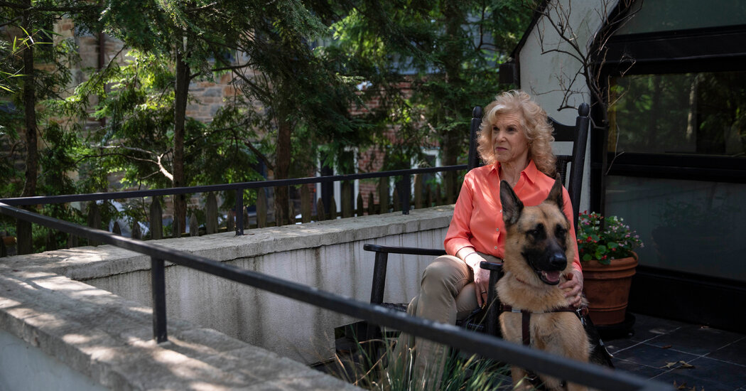
Karen Petrou invented a new funding model for curing blindness. Proposed legislation aims to apply it to medical research more generally.In the development of disease treatments, the stage between basic research and advanced clinical trials is known as “the valley of death.”While ample public grants fund early-stage research and pharmaceutical companies are willing to fund studies on proven solutions, research at the “translational” stage, when basic findings are applied to potential treatments, is notoriously difficult to finance. Some promising treatments are never pursued as a result.The pandemic made this perilous valley “a whole lot deeper,” said Karen Petrou, the co-founder and managing partner of Federal Financial Analytics, a financial services consulting firm in Washington that created a new financial instrument designed to help solve this problem.During the pandemic, clinical trials were halted, resources were diverted from labs, attention was focused on immediate needs, and much funding dried up. New research projects were difficult to kick-start.At the same time, the value of funding scientific research became even clearer: Without the initial efforts of academic labs, it would have been impossible for big pharmaceutical companies to fast-track vaccine development.Ms. Petrou’s proposed solution, known as BioBonds, gained traction.The program would create low-interest, government-backed loans for translational research. These would be packaged into a bond, similarly to how mortgages are, and sold into the secondary market for risk-averse institutional investors like pension funds.In May, Representative Bobby Rush, Democrat of Illinois, and Representative Brian Fitzpatrick, Republican of Pennsylvania, introduced legislation that, if passed, would create $30 billion worth of these loans over three years.Ms. Petrou, who was diagnosed with retinal degeneration as a teen and went blind in her 40s, first stumbled upon the “valley of death” in 2013. She was raising funding for studies to speed up treatment for retinal degeneration, but potential investors told her translational projects were too speculative — they needed results that show a potential idea works, preferably involving a large population that will rely on pills.She refused to accept that as a final answer. Many countries support private-sector funding for biomedical research and each does it differently, Ms. Petrou said: “We needed an American model.”Ms. Petrou and her husband, Basil, had been advising Wall Street executives and regulators for decades. (She recently wrote a book on monetary policy driving inequality.) They had thought a lot about mixed public-private markets during the mortgage finance crisis. Inspired by green bonds — publicly-backed loans that since 2007 have created a $750 billion private market in sustainability projects — they started working on the idea that became BioBonds.“It’s a lifeline,” Attila Seyhan, the director of translational oncology operations at Brown University and a former Pfizer scientist, said of the idea. He said his colleagues were similarly intrigued.Unlike with grants, researchers would need to repay BioBonds loans. Still, getting no-strings funding is a “constant struggle,” Dr. Seyhan said, and “there is an enormous amount of frustration about lack of alternatives.”He believes university business units will get “creative” to make BioBonds work. “There will be losses,” he said. “But if 1 percent succeeds, you pay off the losses. This is how drug development works.”Many schools already encourage scientists to find money outside of grants with which to pursue their ideas. Increasingly, scientists say they have to think like venture capitalists, keeping commercialization in mind when they design clinical trials so that they are able to raise money from private companies to fund them. “There’s a recognition now that even if we discover something, universities now have to help researchers transition to commercialization,” says Dr. Richard Burkhart, a surgeon and researcher at The Johns Hopkins University School of Medicine. Currently, his work is funded by the National Institutes of Health, but he is working with the Technology Ventures team at his institution on trying to commercialize his work.While grants are preferable, they aren’t abundant. Dr. Burkhart believes BioBonds bonds may help scientists and institutions navigate the difficult translational space.When the Petrous first came up with the BioBond concept, they proposed a modest pilot program targeting blindness research. The legislation was introduced in the House in 2018 session and again in a new session in 2019. Then everything changed. “Covid hit and U.S. biomedicine just shut down,” Ms. Petrou recalled.Ms. Petrou is determined to see a BioBonds bill passed, to pay tribute to her husband and business partner.Sarah Silbiger for The New York TimesMeanwhile, the couple’s understanding of the need for more translational research evolved, tragically. Mr. Petrou was diagnosed with pancreatic cancer in 2018. After undergoing surgery in 2019 as part of a clinical trial run by Dr. Burkhart, Mr. Petrou was believed to be cancer-free. But in April of last year, a routine screening revealed the disease had reappeared.The Petrous were determined to find another trial, but thousands of them were being halted because of the pandemic. Stuck at home in lockdown, they decided to revisit their BioBonds idea but think bigger. They repurposed their first proposal, expanding it to address added stress on the already ailing translational space.“When we began to hear about devastation in the clinical trial context, I was quickly able to pivot,” said Valerie White, a recently retired financial services lobbyist, formerly at Akin Gump. She had helped shepherd the original bond concept and immediately began talking to contacts in Congress about BioBonds.The legislation that Mr. Rush and Mr. Fitzpatrick introduced in May, called the “Long-term Opportunities for Advancing New Studies for Biomedical Research Act,” or LOANS for Biomedical Research, would require the secretary of health and human services to guarantee $10 billion a year for three years to fund loans for universities and other labs to conduct F.D.A.-approved clinical trials. The bill has 14 co-sponsors and support from about 20 organizations, including the Alliance for Aging Research, the Alzheimer’s Drug Discovery Association, the Blinded Veterans Association, and the Juvenile Diabetes Research Foundation.“This should, quite frankly, capture the attention of a lot of different sectors in Congress,” said Ms. White. From her perspective, more biomedical research won’t just save lives but will also lead to increased military readiness and economic viability, among other things.She has volunteered four years to the project and said she would keep going for as long as it takes for the BioBonds bill to become law.Mr. Petrou will not be there to celebrate if that day comes. He died in March. Ms. Petrou believes that the surgery he underwent as part of the clinical trial would have saved his life but for other complications.Ms. Petrou is determined to see the LOANS Act passed, to pay tribute to her partner of more than a quarter-century. She thinks a lot about all the pain people go through now, anguish that might be avoided in the future if there were more work being done on cures of all kinds, including for cancer and for blindness.“This was their baby from inception,” said Ms. White, who was present at the couple’s wedding and remained friends with them over the years. “It’s almost ironic that this whole project started with eye bonds that could have helped Karen, but in the end, it was Basil who could have benefited if this idea had existed before.”

Read more →