Nanoparticles create heat from light to manipulate electrical activity in neurons

Nanomaterials have been used in a variety of emerging applications, such as in targeted pharmaceuticals or to bolster other materials and products such as sensors and energy harvesting and storage devices. A team in the McKelvey School of Engineering at Washington University in St. Louis is using nanoparticles as heaters to manipulate the electrical activity of neurons in the brain and of cardiomyocytes in the heart.
The findings, published July 3, 2021, in Advanced Materials, have the potential to be translated to other types of excitable cells and serve as a valuable tool in nano-neuroengineering.
Srikanth Singamaneni, a materials scientist, and Barani Raman, a biomedical engineer, and their teams collaborated to develop a noninvasive technology that inhibits the electrical activity of neurons using polydopamine (PDA) nanoparticles and near-infrared light. The negatively charged PDA nanoparticles, which selectively bind to neurons, absorb near-infrared light that creates heat, which is then transferred to the neurons, inhibiting their electrical activity.
“We showed we can inhibit the activity of these neurons and stop their firing, not just on and off, but in a graded manner,” said Singamaneni, the Lilyan & E. Lisle Hughes Professor in the Department of Mechanical Engineering & Materials Science. “By controlling the light intensity, we can control the electrical activity of the neurons. Once we stopped the light, we can completely bring them back again without any damage.”
In addition to their ability to efficiently convert light into heat, the PDA nanoparticles are highly biocompatible and biodegradable. The nanoparticles eventually degrade, making them a convenient tool for use in in vitro and in vivo experiments in the future.
“When you pour cream into hot coffee, it dissolves and becomes creamed coffee through the process of diffusion,” he explained. “It is similar to the process that controls which ions flow in and out of the neurons. Diffusion depends on temperature, so if you have a good handle on the heat, you control the rate of diffusion close to the neurons. This would in turn impact the electrical activity of the cell. This study demonstrates the concept that the photothermal effect, converting light into heat, near the vicinity of nanoparticles tagged neurons can be used as a way to control specific neurons remotely.”
To continue the coffee analogy, the team has designed a photothermal foam that is similar to a sugar cube, forming a dense population of nanoparticles in tight packaging that acts more quickly than individual sugar crystals that disperse, Raman said.
“With so many of them packed in a small volume, the foam is quicker in transducing light to heat and give more efficient control to only the neurons we want,” he said. “You don’t have to use high-intensity power to generate the same effect.”
In addition, the team, which includes Jon Silva, associate professor of biomedical engineering, applied the PDA nanoparticles to cardiomyocytes, or heart muscle cells. Interestingly, the photothermal process excited the cardiomyocytes, showing that the process can increase or decrease the excitability in cells depending on their type.
“The excitability of a cell or tissue, whether it be cardiomyocytes or muscle cells, depends to a certain extent on diffusion,” Raman said. “While cardiomyocytes have a different set of rules, the principle that controls the sensitivity to temperature can be expected to be similar.”
Now, the team is looking at how different types of neurons respond to the stimulation process. They will be targeting particular neurons by selectively binding the nanoparticles to provide more selective control.

Read more →

Effectiveness of mRNA vaccines against the Alpha and Beta variants in France

Over the past six months, the World Health Organization has categorized four SARS-CoV-2 variants as being “of concern” because they are more transmissible or may escape the immune response. They have been termed the Alpha, Beta, Gamma and Delta variants.
Scientists from the Institut Pasteur, in collaboration with the French National Health Insurance Fund (CNAM), Ipsos and Santé publique France, conducted a nationwide case-control study to evaluate the effectiveness of mRNA vaccines against symptomatic forms of SARS-CoV-2 infection, be that non-variant virus or the Alpha and Beta variants. The results show that the two-dose vaccination regimen of mRNA vaccines provides 88% protection against non-variant virus, 86% against the Alpha variant and 77% against the Beta variant. The results of this study were published in The Lancet Regional Health Europe on July 14, 2021.
In late 2020, two new variants of SARS-CoV-2 were reported in the United Kingdom and in South Africa, respectively. This occurred at a time when the United Kingdom was experiencing a resurgence in the incidence of SARS-CoV-2 infections. This was later attributed to the emergence of the Alpha variant, which has now demonstrated increased transmissibility as compared to the original SARS-CoV-2. The Beta variant was first reported in South Africa and later identified to carry a mutation known as E484K and which is associated with immune evasion. The reporting of the first SARS-CoV-2 variants occurred as mass COVID-19 vaccination campaigns were commencing at global level. In France, both Alpha and Beta were circulating in the first months of 2021. It was therefore important to analyze the effectiveness of the messenger RNA (mRNA) COVID-19 vaccines against these SARS-CoV-2 variants of concern.
In October 2020, the Institut Pasteur, in collaboration with the French National Health Insurance Fund (CNAM), Ipsos and Santé publique France, launched the ComCor study, a case-control study at national level to analyze the sociodemographic, behavioral and practical factors associated with SARS-CoV-2 infection. All those infected with SARS-CoV-2 were invited to take part in the study by the CNAM, while non-infected control were identified through an Ipsos representative panel. The first results of the study were published in The Lancet Regional Health on June 7, 2021.
In February 2021, the scientists adapted the ComCor questionnaire to add information about vaccination against COVID-19, previous SARS-CoV-2 infection and information on the variants responsible for infection, provided as part of test results. This information was used to assess the effectiveness of two doses of mRNA vaccine against the Alpha and Beta variants, and to evaluate the protection conferred by previous SARS-CoV-2 infection.
In this study, 7,288 people infected with the original strain, 31,313 people infected with the Alpha variant, 2,550 people infected with the Beta variant and 3,644 non-infected controls were included between February and May 2021. The study enabled the scientists to demonstrate that two doses of mRNA vaccine conferred 88% (81-92) effectiveness against infection with original virus, 86% (81-90) effectiveness against infection with the Alpha variant and 77% (71-90) effectiveness against infection with the Beta variant, as measured seven days after the second dose. No difference in vaccine effectiveness was found between different categories of age, sex or occupational exposure.
“There was much expectation surrounding the (vaccine effectiveness) results for the Beta variant, known for its E484K mutation associated with immune evasion. Our estimate of 77% protection is very close to the estimate of 75% reached by the only other study worldwide to have evaluated the effectiveness of mRNA vaccines against this variant. These analyses confirm the effectiveness of the COVID-19 vaccines and the key role that they have to play in tackling the epidemic,” explains Arnaud Fontanet, Head of the Epidemiology of Emerging Diseases Unit at the Institut Pasteur and Professor at the Conservatoire National des Arts et Métiers (CNAM).
Another important finding of the study related to previous SARS-CoV-2 infection. The scientists demonstrated that recent infection (in the past 2 to 6 months) confers similar protection to that observed with mRNA vaccines.
The analyses will now be extended to estimate vaccine effectiveness against the Delta variant, which has been the dominant variant in France since early July 2021.
The ComCor project is funded by REACTing, the Fondation de France via the “All United Against Coronavirus” alliance, and the Institut Pasteur. It was recently awarded the CAPNET “national research priority” label.
Story Source:
Materials provided by Institut Pasteur. Note: Content may be edited for style and length.

Read more →

Cancer: Information theory to fight resistance to treatments

One of the major challenges in modern cancer therapy is the adaptive response of cancer cells to targeted therapies: initially, these therapies are very often effective, then adaptive resistance occurs, allowing the tumor cells to proliferate again. Although this adaptive response is theoretically reversible, such a reversal is hampered by numerous molecular mechanisms that allow the cancer cells to adapt to the treatment. The analysis of these mechanisms is limited by the complexity of cause and effect relationships that are extremely difficult to observe in vivo in tumor samples. In order to overcome this challenge, a team from the University of Geneva (UNIGE) and the University Hospitals of Geneva (HUG), Switzerland, has used information theory for the first time, in order to objectify in vivo the molecular regulations at play in the mechanisms of the adaptive response and their modulation by a therapeutic combination. These results are published in the journal Neoplasia.
Adaptive response limits the efficiency of targeted therapies used to fight the development of tumors: after an effective treatment phase that reduces the tumor size, an adaptation to the used molecule occurs that allows the tumor cells to proliferate again. “We now know that this resistance to treatment has a large reversible component that does not involve mutations, which are an irreversible process,” explains Rastine Merat, a researcher in the Department of Pathology and Immunology at the UNIGE Faculty of Medicine, the head of the Onco-Dermatology Unit at the HUG and the principal investigator of the study.
Research confronted with the complexity of biological regulations
In order to prevent resistance to targeted therapies, scientists need to understand the molecular mechanisms of the adaptive response. “These mechanisms may involve variations in gene expression, for example,” explains Rastine Merat. It is then necessary to modify or prevent these variations by means of a therapeutic combination that blocks the consequences or even prevents them. One challenge remains: the description of these mechanisms and their modulation under the effect of a therapeutic combination is very often carried out on isolated cultured cells and not validated in tumor tissue in the body. “This is essentially due to the difficulty of objectifying these mechanisms, which may occur in a transient manner and only in a minority of cells in tumor tissues, and above all which involve non-linear cause and effect relationships,” explains the Geneva researcher.
Applying information theory to tumors
To counter these difficulties, the UNIGE and HUG team came up with the idea of using information theory, more specifically by quantifying mutual information. This approach has previously been used in biology, mainly to quantify cell signaling and understand genetic regulation networks. “This statistical method makes it possible to link two parameters involved in a mechanism by measuring the reduction in the uncertainty of one of the parameters when the value of the other parameter is known,” simplifies Rastine Merat.
Practically, the scientists proceed step by step: they take biopsies of tumors (in this case melanomas) in a mouse model at different stages of their development during therapy. Using immunohistochemical analyses — i.e. tumor sections — they measure, using an automated approach, the expression of proteins involved in the mechanism at play in the adaptive response. “The proposed mathematical approach is easily applicable to routine techniques such as immunohistochemistry and makes it possible to validate in vivo the relevance of the mechanisms under study, even if they occur in a minority of cells and in a transient manner,” the Geneva researcher explains. Thus, scientists can not only validate in the organism the molecular mechanisms they are studying, but also the impact of innovative therapeutic combinations that result from the understanding of these mechanisms. “Similarly, we could use this approach in therapeutic trials as a predictive marker of response to therapeutic combinations that seek to prevent adaptive resistance,” he continues.
A method suitable for all types of cancer
“This method, developed in a melanoma model, could be applied to other types of cancer for which the same issues of adaptive resistance to targeted therapies occur and for which combination therapy approaches based on an understanding of the mechanisms involved are under development,” concludes Rastine Merat.
Story Source:
Materials provided by Université de Genève. Note: Content may be edited for style and length.

Read more →

Many ICU staff have experienced mental health conditions in COVID-19 pandemic

A high proportion of staff working in intensive care units during the COVID-19 pandemic have experienced mental health conditions, according to a new study.
In a study of 515 healthcare staff working in intensive care units (ICUs) across seven countries, the researchers found that on average 48 percent of participants showed signs of mental health conditions — depression, insomnia and post-traumatic stress disorder (PTSD). Their mental health was assessed using a detailed questionnaire and a clinical scoring system.
The team also found a 40 per cent increase in the conditions for those who spent more than six hours in personal protective equipment (PPE) over a course of a day, compared to those who didn’t.
The study, led by researchers at Imperial College London, is published in the British Journal of Nursing and is the first to evaluate ICU workers’ mental health during the COVID-19 pandemic. In line with the UK Government’s report on burnout in NHS staff published in June 2021, the researchers suggest that the high level of mental health conditions found among the ICU staff surveyed should inform local and national wellbeing policies.
Dr Ahmed Ezzat, lead author of the study and Honorary Clinical Fellow at Imperial College London, said:
“This is a timely study which acts as a stark reminder of the personal challenges healthcare staff are facing as a result of COVID-19. As within wider society, mental illness of healthcare staff still remains a taboo subject for some.

Read more →

SARS-CoV-2 variant B.1.617 gives the immune system a hard time

SARS-CoV-2 still poses major challenges to mankind. The frequent emergence of mutant forms makes the threat posed by the virus difficult to predict. The SARS-CoV-2 variant B.1.617 circulated in India and gave rise to the Delta variant, B.1.617.2, which is now becoming dominant in many countries.
Infection researchers from the German Primate Center (DPZ) – Leibniz Institute for Primate Research in Göttingen have investigated the B.1.617 variant in detail. In cell culture studies, they found that this variant can infect certain lung and intestinal cell lines more efficiently than the original virus. The researchers also demonstrated that B.1.617 is less sensitive to inhibition by antibodies present in the blood of convalescent or vaccinated individuals and resistant to a therapeutic antibody used for COVID-19 treatment. These properties may enable B.1.617 and its subtypes to rapidly spread in the human population, thereby increasing the risk of incompletely vaccinated individuals and individuals with declining immune protection to become infected (Cell Reports).
The spike protein is embedded in the viral envelope and facilitates SARS-CoV-2 entry into host cells. Without the activity of the spike protein the virus cannot replicate in the human body. The currently known virus variants harbor different mutations in the spike protein, some of which make it easier for them to infect host cells and evade the immune system of infected individuals. The B.1.617 variant carries eight different mutations in the spike protein, including two within the receptor binding domain, which is essential for viral attachment to cells and represents the main target for neutralizing antibodies.
A team led by Markus Hoffmann and Stefan Pöhlmann, infection researchers at the German Primate Center, and including scientists from the University Hospital of Göttingen, the University of Erlangen and the Hannover Medical School, investigated how the mutations impact the ability of the B.1.617 variant to enter host cells and how efficiently the antibody response in vaccinated and recovered individuals inhibits this variant.
First, the researchers analyzed entry of the B.1.617 variant into different human cell lines. In two cell lines derived from lung and colon, respectively, they detected a 50 percent increase in the virus’ entry efficiency.
The researchers also investigated the efficacy of four different therapeutic antibodies that have been approved for the treatment of COVID-19 patients. They found that the B.1.617 variant was completely resistant against one of these antibodies and slightly less inhibited by another antibody.
In a third step, the scientists tested the efficacy of antibodies from the blood of recovered and vaccinated individuals. Here, they found a two- to threefold reduction in protection against the B.1.617 variant.
“Our study shows that this virus variant can infect lung and intestinal cells more efficiently, suggesting increased viral fitness,” Markus Hoffmann, lead author of the study, summarizes the results. “In addition, the protective effect of antibodies is limited because they block the cell entry of B.1.617 less efficiently than that of the original virus. As a result, individuals who are not fully vaccinated or were infected a long time ago and thus produce low amounts of antibodies, may be poorly protected against infection with the B.1.617 variant.
Stefan Pöhlmann, Head of the Infection Biology Unit at the DPZ adds: “In order to prevent further spread of the B.1.617 variant, especially its subtype Delta, and to prevent the emergence of new virus variants it is advisable to quickly achieve full immunization of all people that are willing to be vaccinated. It is also necessary to investigate whether booster vaccinations with existing vaccines or vaccines optimized to target variants provide long-lasting and broad protection.” 

Read more →

In an ICU, a Photographer's View of a Desperate Covid Fight

After documenting a last-resort treatment at one hospital, a photographer who spent hours in patients’ rooms describes capturing the humanity of the struggle.Times Insider explains who we are and what we do, and delivers behind-the-scenes insights into how our journalism comes together.As I photographed people in Covid-19 intensive care units early this year, I was protected by four sets of plastic: glasses, goggles, face shield and viewfinder. But there is no protection for the pain one takes in.I captured images for a recent Times article about a last-resort Covid treatment called ECMO, documenting coronavirus patients and the medical professionals caring for them at Providence Saint John’s Health Center in Santa Monica, Calif. The families allowed me to share in the darkest moments of their lives.I felt privileged to be let into these sacred spaces. As a journalist, I feel it is my responsibility to have the emotional bandwidth to be with people in moments that most of society cannot handle. Despite safety guidelines that advised against spending long periods inside ICU rooms, I spent hours with each patient, lingering for an extended amount of time to be able to get a sense of the person and bring forth an emotional spectrum of moments.José Cervantes plays a song by Nipsey Hussle for his son, Alfred Sablan, in the ICU at Saint John’s.Isadora Kosofsky for The New York TimesDr. Terese Hammond watches a procedure to take Dr. David Gutierrez off of ECMO in the ICU. He was released in June.Isadora Kosofsky for The New York TimesVerbal interaction helps me connect with those I photograph. On this assignment, some people were not awake or couldn’t speak, and the most powerful connection was often silent.I would stand next to the bed of Alfred Sablan, 25, and imagine the sound of his voice, trying to sense the gentle manner his mother described. I would lean over the bed of Dr. David Gutierrez, 62, a physician who had become a patient himself, and remind him who I was. He would look back, unable to respond with words, but I felt our connection over the classic rock playing on his iPad.Ruby Acosta looking at her son, Alfred Sablan. He died in March. Isadora Kosofsky for The New York TimesPeriodically, a staff member would enter to check on Mr. Sablan or Dr. Gutierrez. “Are you OK?” asked a nurse as she cracked the door of Dr. Gutierrez’s room. He nodded “yes.”Amid all the pain, there were reminders of grace.

Read more →

A Personal History of the C-Section

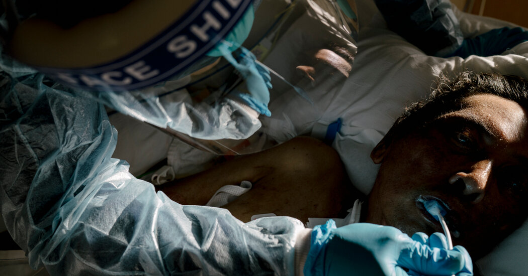
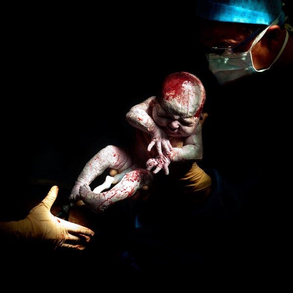
I didn’t realize that I cared about getting a cesarean until I listened to myself telling the story of my cesarean. Describing my daughter’s birth during the first months of her life, I often stumbled over my words. “When she was born,” I would say, then correct myself: “When they got her out.” This was less poetic, but it seemed more accurate. I mean, of course she’d been born. She was here, wasn’t she? But it never felt quite right to say that I gave birth to her. The literal truth was something else: A doctor opened me up, parted my skin and reached inside to pull my baby into the world. Whenever I told my birth story, I noticed myself stressing that it was an emergency C-section — wanting people to know that there wasn’t any other option, that I didn’t choose to forgo labor, wasn’t coerced into the procedure by an intervention-happy, efficiency-obsessed, liability-avoidant medical establishment. At first, I was mainly just relieved that my daughter survived the delivery, that I could wonder at her little burrito of a body in the swaddling blanket or her impossibly tiny fingernails. My C-section was simply the intervention that had been necessary; now it was just a set of physical inconveniences. When I laughed or coughed, I felt as though I was going to split open along my new seam. When I searched online for “C-section shelf?” it was only because I didn’t know what other word to use for the bulge of numb skin that hung like a rock formation over my scar. (Apparently “shelf” was good enough, yielding pages of message-board entries: Is it fat? Is it skin? How do I make it go away?)Listen to This ArticleAudio Recording by AudmTo hear more audio stories from publications like The New York Times, download Audm for iPhone or Android.When I talked about the days after my daughter’s birth, I found myself emphasizing how much I held her, how I never wanted to put her down. It was as if I felt the need to compensate narratively for that first hour, when I wasn’t able to hold her at all — to insist that we bonded just as much anyway. I found myself exaggerating the part about not caring if I was numb before they cut me open, when in fact I did care; I told the doctors that I would actually love some more anesthesia in my epidural if they had a split second to spare. My impulse to exaggerate my stoicism felt like another shameful compensation — as if I were trying to make up for other kinds of pain I didn’t experience, unwittingly obeying the cultural script that insisted on suffering and sacrifice as the primary measures of maternal love. Louann, April 12, 2013; 14 seconds old.Artwork by Christian BerthelotEven now, three and a half years later, I still feel a pang when I hear women use the phrase “natural childbirth” or describe pushing out their babies after 40 hours of labor. Imagining all that effort inspires a deep awe but also a splinter of shame — as if my own birth story wasn’t one that merited pride or celebration but was instead a kind of blemish, a beginning from which my daughter and I must recover. “That operation is called Caesarean by which any way is opened for the child” other “than that destined for it by nature,” wrote a late-18th-century French obstetrician named Jean Louis Baudelocque. Since ancient history, the “unnatural” quality of the cesarean has made it both miraculous and suspect, simultaneously a deus ex machina and a tyrannical intervention. It’s an apocryphal story that Julius Caesar was born by cesarean — his mother survived his birth to bear more children, and at that point, the C-section was impossible to survive — most likely spun to grant more drama to the story of his birth. In his 1925 history of the operation, Herbert Spencer, a professor of obstetrics at University College London, speculates that it “was called Caesarean as being too grand to have been first performed on ordinary mortals” and calls it “the greatest of all operations, in that it directly affects two lives.” For most of its history, however, it saved only one of them. Mothers didn’t routinely survive the procedure until the 20th century. Before then, it was generally deployed as a last-ditch measure to save the baby once the mother was dying or already dead. In many languages, the name for the procedure invokes its ostensibly regal lineage: The Danish, Dutch and Swedish terms are all variations of “the imperial cut.” In German, it’s kaiserschnitt; in Slovenian, it’s cesarski rez. (Hardly surprising that even though it’s the woman who is cut open, the procedure is named for a man.) A friend who grew up in Belgium told me his grandmother believed that all royalty were delivered by cesarean. It has retained an enduring association with privilege or indulgence: too posh to push.Even before it was imperial, the C-section was associated with divinity. The Greek god of medicine, Asklepios, was born by cesarean, rescued from his mother’s body as she burned on a funeral pyre. In Shakespeare’s “Macbeth,” the cesarean-born Macduff famously arrives as the answer to a riddle: Although the witches have promised that “none of woman born shall harm Macbeth,” Macduff turns out to be exempt from the prophecy because he “was from his mother’s womb untimely ripped.” Macduff’s exceptional birth grants him a singular power, but its exceptionality also carries a whiff of monstrosity: “Untimely ripped” doesn’t exactly summon the epidural and the blue tarp.Why do we want so much from our birth stories, anyway?During the medieval era, some babies born by cesarean were called “the fortunate” or “the unborn,” deemed miraculous not despite being born from corpses but because of it. They were proof of hope and possibility salvaged from the jaws of death, emblems of life plucked from wombs growing cold. Cesareans were understood as both miraculous interventions from saints — the so-called apertura mirabilis, or “wondrous opening” — and unholy abominations. The birth of the Antichrist was sometimes depicted as a cesarean; in one 15th-century woodcut, a winged demon clutches the new baby by his wrist while the mother looks away with her head cocked from exhaustion, or horror, a gaping wound still furrowing her stomach. As one widely circulated medieval account of the birth of the Antichrist put it: “The devil will go down into the womb of Antichrist’s mother and fill her completely, possess her completely inside and out, so that she will conceive by man with the devil’s assistance, and what is born will be completely foul, completely evil, completely ruined.”Now, 500 years later, the “greatest of all operations” has become one of the most common surgeries in America. By 2019, almost one-third of American births happened by C-section, more than double the share that the World Health Organization considers the ideal rate to reduce maternal and infant mortality (10 to 15 percent). In some countries, the rate is even higher: In the Dominican Republic, about 60 percent of all babies are born by cesarean, and in Brazil, the so-called C-section capital of the world, cesareans account for almost 85 percent of all births in private hospitals, where women throw parties around their planned C-sections. One “presidential suite” in a São Paulo maternity ward includes a balcony and a minibar; another ward has a videography wing where women can get blowouts, manicures and makeup before being filmed with their newborns.But the rise of the C-section has brought with it a powerful backlash, in which legitimate arguments against the procedure’s ubiquity have become Trojan horses, carrying within them age-old ideals of motherhood that fetishize sacrifice and pain. The dismissive, often unspoken critique of the C-section understands it as birth without labor, birth without pain, birth without sacrifice. If a mother is supposed to do anything, she is supposed to sacrifice herself for her children, and pain in childbirth is the earliest barometer of that sacrifice, the punishment God bestows upon Eve in the Book of Genesis: “I will greatly multiply thy sorrow and thy conception; in sorrow thou shalt bring forth children.” A cesarean often involves pain, but it’s unnatural pain, and it’s typically medicated away. Even when a C-section isn’t elective, it still means a woman doesn’t undergo that supreme, heroic effort of pushing a baby through the birth canal. Although the cesarean backlash arose from an impulse to empower women, it has perversely also become another way to shame mothers, or make them feel inadequate, as soon as they’ve given birth. When the British doctor Grantly Dick-Read coined the term “natural birth” in his 1933 book “Natural Childbirth,” he meant childbirth without any intervention that would disrupt or change the process of labor. In “Childbirth Without Fear,” his internationally best-selling 1942 manifesto, he wrote that childbirth is “nature’s first hard lesson in the two greatest assets of good motherhood. Children will always mean hard work and will always demand self-control.” The woman who has a C-section is a woman who doesn’t learn those lessons. I can still remember the sheer awe I felt in birth class when a lovely woman holding a plastic pelvis explained the interlocking stages of the process: how the pressure of contractions pushing my baby’s head against my cervix would stretch it, prompting my body to produce more prostaglandins, making it more receptive to oxytocin, which would thin the cervix and help it dilate; how my endorphins would carry me through the pain and my adrenaline would surge for that final push. It struck me as almost beautiful, how all these parts fit together like jigsaw-puzzle pieces. It was less like the hydraulics of a machine and more like the choreography of a dance.Although I was never ferociously attached to the idea of a natural birth, in that class I finally got it: the primal drive to let your body do its work without intervention. It wasn’t just a sense of wonder at what my body was capable of; it was also about how my baby and I could be joined together by this shared labor. Before my daughter was born, a friend sent me an email describing her own recent birth as utterly collaborative. “For the final two hours we were working together totally,” she wrote. “Once she transitioned into the birth canal, I was just following her lead. I was aware of it, and of her and of my body, totally.” I was viscerally compelled toward this collaboration: not just the idea of my body delivering this new body into the world, but the idea that it would be the first thing my baby and I ever did together. My birth ended up being a different kind of collaboration, one between two doctors, an anesthesiologist, a team of nurses and a pediatrician. For those last moments, I was something more like an extension of the operating theater. In this sense, the intervention of a C-section not only disrupts the ideal of a “natural” birth but also the ideal of autonomy: the fantasy of a mother’s body as an unassisted miracle worker, a self-contained ecosystem of fertility and capacity. “Basically it has made me feel less than a total woman,” one woman, quoted in an anti-cesarean book from the early 1980s, said after her C-section. “I felt like I had failed.”My birth tableau was a far cry from what childbirth looked like for most of human history. Before the 1900s, women rarely even labored in hospitals. Increasing levels of intervention across the course of the 20th century — of which the C-section was the most extreme manifestation — were fueled by a radical shift in our understanding of labor itself. It ceased to be seen as a natural process that required largely passive oversight and became a dangerous predicament from which mother and baby each needed to be protected. Joseph DeLee, an early-20th-century obstetrician, understood childbirth as a process riddled with inherent risks: “So frequent are these bad effects that I often wonder whether nature did not deliberately intend women to be used up in the process of reproduction, in a manner analogous to that of salmon, which dies after spawning.” By 1964, an article in Harper’s warned that “a soldier in wartime has a better chance for survival than a baby during birth.”Sarah, April 1, 2016; 17 seconds old.Artwork by Christian BerthelotAs the medical historian Jacqueline H. Wolf recounts in her 2018 history, “Cesarean Section: An American History of Risk, Technology and Consequence,” the cesarean became perceived as “an avenue to perfection” within an increasingly quantitative approach to childbirth in the 1950s and ’60s: the Friedman curve (measuring length of labor), the Bishop score (measuring readiness for induction) and the Apgar score (measuring health of newborn). The C-section rate rose by 455 percent between 1965 and 1987. Many doctors were frustrated by mothers who, inspired by the natural-birth movement of the 1970s and early ’80s, fought for births without intervention. As Wolf recounts, one obstetrician used to press his fetoscope to women’s abdomens and pretend to listen to the voices of their unborn children: “You know, I can hear your baby!” he would tell them. “Your baby is telling me: ‘Don’t listen to my mother! She doesn’t know anything!’” The same medical paternalism that judged women for resisting C-sections also judged women — just a few decades earlier — for having them. In a 1921 medical analysis of the procedure, Franklin Newell, a doctor affiliated with Harvard Medical School, describes an ideal candidate as a woman with “poor nervous equipment.” Even though this type of woman doesn’t have a deformed pelvis or a medical condition, Newell argues, she is suited to a C-section because she is so averse to pain and so constitutionally weak. “Such women are very prone to respond badly to the strain of labor, and to them pain is a real evil,” he writes. “What they cannot recover from is a long strain, particularly if much pain accompanies it, and all pain is exaggerated to them.” Newell cobbles together a set of enduring female stereotypes: a woman who likes to play victim, hates to work and constantly inflates her own discomfort. She is incapable of the self-sacrifice that is the hallmark of virtuous motherhood. In fact, he argues, centuries ago she most likely would have been killed off by natural selection. “These patients are the abnormal product of an overcivilization and are much like hothouse plants,” he writes. “They represent in our civilized communities a type which would have been largely eliminated, if medical care had not interfered with the law of the survival of the fittest.” Rising from the natural-birth movement of the 1970s and consolidated by the 1977 publication of Ina May Gaskin’s canonical text, “Spiritual Midwifery,” the strong cesarean backlash found its stride in the 1980s. In the digital era, it eventually bloomed into a proliferation of websites and forums, including the popular “Unnecesarean” blog. A slew of recent books has criticized the overabundance of C-sections, including “Cut It Out: The C-Section Epidemic in America” (ha!). The International Cesarean Awareness Network offers meetings for women looking for a “safe space to process cesarean experience and/or getting support for a VBAC” (vaginal birth after cesarean). In one cesarean memoir, a woman describes herself murmuring “VBAC” like a mantra during her third pregnancy: “My soul craved natural birth the way a lover’s very being calls to her mate.” It’s surprisingly intuitive to frame the longing for natural childbirth in terms of romantic desire. Both kinds of intimacy promise to deliver consummation. They promise to let you become — by giving birth to another human being, or making life with another human being — the fullest version of yourself. When I first encountered the taxonomy of “cesarean mothers” created by the cesarean-prevention movement in the early 1980s, paraphrased in Wolf’s book, I couldn’t help wondering where I fell: “The ‘cesarean mourner’ had never made peace with not having a vaginal birth. The ‘cesarean victim’ suspected her surgery had been unnecessary. The ‘cesarean learner’ was now empowered to seek a vaginal birth the next time around. The ‘cesarean surrender’ had given up the fight. The ‘cesarean gratitude’ was thankful for the surgery that had saved her and her baby. The ‘cesarean activist’ was determined that no woman ever have unnecessary surgery again. The ‘cesarean phoenix’ rose ‘victorious from bitter ashes!’”Some part of me yearned for natural childbirth as a threshold of redemption.The truth is, before I started reading books by and about women who felt traumatized by their C-sections, I never felt particularly traumatized by my own. After enough reading, however, I started to think: Maybe I shouldn’t feel like a real woman either? Was I a “cesarean surrender” for having capitulated so easily to the narrative that my surgery was necessary? Wolf’s history of the American C-section made me think of my cesarean as part of a long history of intervention. Bolstered by technological triumphalism, the procedure became commonplace as an attempt to standardize an essentially variable experience so that it would hew as closely as possible to an “ideal” birth and an ideal baby. Of course, it is now the very thing an “ideal” birth seeks to avoid. This was the uncomfortable truth of my labor experience, of anyone’s labor experience: It feels deeply personal but has in fact been shaped by impersonal societal forces. Wolf describes the drastic increase in the use of electronic fetal monitors, which offered a technological peephole into the well-being of the unborn and, in so doing, effectively created more situations where a C-section was deemed necessary — as one doctor put it, “dropping the knife with each drop in the fetal heart rate.” I started to wonder if the surgery I understood as lifesaving had in fact been a mere symptom of risk-averse medical culture. But I could still hear those nurses calling out my baby’s falling heart rate — “It’s in the 60s! It’s in the 50s!” — and the memory of their voices, their utter panic, still lifted the hair on my arms. Maybe my cesarean was necessary. But what did it say about the cultural ideals of motherhood I had internalized that I felt such a frantic desire to insist that it was an emergency? It was as if I needed to prove I wasn’t a bad mother for having given birth by C-section, or as if I still believed, in some sense, that I hadn’t given birth at all. Was I another “cesarean victim” who was emotionally blackmailed into surgery? Or had my baby been saved from death? These were very different stories to live inside. They were very different ways to understand the start of my daughter’s life. Owen, March 27, 2015; 9 seconds old.Artwork by Christian BerthelotIt would be a lie — or at least an incomplete truth — to deny that some part of me yearned for natural childbirth as a threshold of redemption. I had never fully treated my body as an ally. I had starved myself to whittle it down and spent years drinking myself to blackout and various other perils. Pregnancy already felt like a more redemptive chapter in this fraught relationship between body and spirit: I was taking care of another tiny body inside my own! Everything my body ate was feeding hers. All the blood pumping through my heart was flowing through hers. Giving birth to her would not only be the culmination of her nine-month incubation but would also be a refutation of all the ways I abused or punished my body over the years, all the ways I treated it as an encumbrance rather than a collaborator. My mind resisted this logic, but I could feel — on a visceral, cellular, hormonal level — its gravitational pull. “Silent Knife: Cesarean Prevention & Vaginal Birth After Cesarean,” an influential anti-cesarean manifesto published by the writers Nancy Wainer Cohen and Lois Estner in 1983, insists that what it calls a “purebirth” is “not a cry or demand for perfection,” though the definition ends up sounding a little … demanding: “Birth that is completely free of medical intervention. It is self-determined, self-assured and self-sufficient.” The unstated tension of the entire book is also the unstated tension embedded in the broader backlash against C-sections: between recognizing the trauma of a C-section and reinforcing or creating that trauma by framing the C-section as a compromised or lesser birth. A section called “Voices of the Victims” quotes women traumatized by their C-sections: “It felt as if I was being raped,” one woman says. “I couldn’t do anything but wait until it was over.” A father says: “A c-sec is one of the worst mutilations that can be perpetrated on a woman as well as a denial of a fundamental right of a woman to experience childbirth.” Inspired by Ina May Gaskin’s famous pronouncement that “you can fix the body by working on the mind,” Cohen and Estner argue that our wombs are cluttered with “unaddressed stresses or fears” that obstruct the birth process, but that they can be swept aside through self-awareness to “clear a passageway for normal birth.” The implication is that, conversely, emotional baggage could be “blamed” for a cesarean. Reading the book 38 years after it was written, I immediately dismissed this notion. But another part of me — the part that had been conditioned for my entire life to feel accountable to impossible ideals of motherhood — wasn’t immune to this magical thinking. In secret, I had indulged my own pet theories about the possible psychological causes of my C-section: my eating disorder, my abortion, my maternal ambivalence. Had I mistreated my body so much that it refused to give birth naturally as an act of retaliation? Had I been more attached to the idea of being a mother than I was prepared for the actuality of being a mother? Was my labor stalling out — as my baby’s heart rate dropped — a sign of this subconscious unwillingness? If “Silent Knife” was written to restore agency to women by pushing back against the tyrannical paternalism of C-sections, then there’s a different tyranny embedded in its ostensible restoration of agency, a tyranny that abides today: a script of self-possession that can become another straitjacket, another iteration of the claustrophobic maternal ideals. Expressing compassion for a woman who feels like an inadequate mother because she hasn’t given birth “naturally” can easily slide into implying that she should feel that way. Many of the ideas that “Silent Knife” made explicit years ago are still deep forces shaping childbirth today, even if people might be less likely to confess to them: the notion that birth by C-section is less “real,” that it might imply some lack of willpower or failure of spirit.Motherhood is instinctual, but it’s also inherited: a set of circulating ideals we encounter and absorb. The fact that we are constantly shaped by external models of an internal impulse makes women intensely vulnerable to narratives of “right” or “real” motherhood, and all the more susceptible to feeling scolded or excluded by them. A woman’s right to state her preferences during the birth process is increasingly prioritized, and rightly so, but it’s easy to fetishize these preferences as the ultimate proof of female empowerment, when they are, of course, shaped by societal forces too. It’s a kind of partial vision to hold up a woman’s desire for natural birth as a badge of unpolluted female agency, when that desire has been shaped by all the voices extolling natural birth as the consummation of a woman’s feminine identity. As my daughter has grown from newborn to infant to toddler, I have been daydreaming about getting a tattoo on my abdominal scar. There are entire Pinterest boards full of C-section-scar tattoos and Instagram hashtags devoted to them (#csectionscarsarebeautiful): angel wings, diamonds, draping pearls, blazing guns. Ganesh, the remover of obstacles. A blue rose unfurling into cursive: “Imperfection is beautiful.” Bolder Gothic script: “MAN’S RUIN.” A “Star Wars” scene of two snub fighters approaching the Death Star. A zipper partly unzipped to show an eye lurking inside. A pair of scissors poised to cut along a dotted line, inked beside the scar itself. A trompe l’oeil of a paper clip piercing the skin, as if it were holding the abdomen together across the line of its rupture. My favorites are the ones in which the scar is intentionally incorporated into the design itself. A low transverse cut becomes the spine of a feather or a branch bursting with cherry blossoms. These tattoos don’t try to hide the scar from view but instead put it to work as part of a larger vision. I have started to imagine, on my skin, a row of songbirds on a wire. The fantasy of this tattoo has been part of a deeper reckoning with the question of whether I want to narrate the birth — to myself, to others — as miraculous, traumatic or simply banal, a commonplace necessity. Around the time I started to consider a tattoo, I read a memoir by an Oregon writer named Roanna Rosewood called “Cut, Stapled and Mended: When One Woman Reclaimed Her Body and Gave Birth on Her Own Terms After Cesarean.” My inner Sontag (“Illness is no metaphor!”) bristled at the endorsement from a mother on the front flap: “I blamed my midwife for my failure to progress but secretly knew it was me; my lack of confidence led to my failure.” Though I resented what I interpreted as the book’s veneration of vaginal birth as the only “real” kind, I could recognize — if I was honest with myself — that my resistance also rose from the fear that I had missed out on an extraordinarily powerful experience. When I read Rosewood’s declaration that a “clean and passive birth resembles an empowered one in the same way that an annual exam resembles making love,” it made me feel deeply foolish — as if understanding my daughter’s birth as the most powerful experience of my life (which I did) was somehow akin to mistaking a Pap smear for an orgasm. Partway through Rosewood’s memoir, however, I encountered a moment that resonated so strongly that I had to put the book down. When she describes her body shaking uncontrollably after her C-section and her anguish at being unable to hold her baby, I was taken back to the flurry of my own post-op desire, my arms twitching beneath their gurney straps while the doctors carried my daughter away from me. Rosewood and I each found ourselves — in the first moments after our births — strangers to our own bodies and separated from the bodies of our children. The difference between us didn’t live in those moments of fear and loss but in what we wanted to do with them afterward. When I read about Rosewood’s desire to “write over” the story of her eldest son’s birth, it made me feel defensive on my daughter’s behalf. I don’t want to write over her birth story. I don’t want anyone else to, either. Maybe it wasn’t ideal, but it was ours. Why do we want so much from our birth stories, anyway? It’s tempting to understand life in terms of pivotal moments, when it is actually composed of ongoing processes: not the single day of birth but the daily care that follows, the labor of diapers and midnight crying, playground tears and homework tantrums, speeding tickets and long-distance phone calls — all that work of sustenance and reinvention. If we’re lucky, birth is just the beginning. The labor isn’t done. It has only just begun.Leslie Jamison is the author, most recently, of “Make It Scream, Make It Burn.” She last wrote for the magazine about visiting Istanbul’s public baths weeks before the lockdown.

Read more →

We Need to Know How Menopause Changes Women’s Brains

This might turn out to be a crucial window to try to prevent Alzheimer’s and other chronic diseases that often accompany older age.During menopause, which marks the end of a woman’s menstrual cycles, her ovaries stop producing the hormones estrogen and progesterone, bringing an end to her natural childbearing years. But those hormones also regulate how the brain functions, and the brain governs their release — meaning that menopause is a neurological process as well. “Many of the symptoms of menopause cannot possibly be directly produced by the ovaries, if you think about the hot flashes, the night sweats, the anxiety, the depression, the insomnia, the brain fog,” says Lisa Mosconi, an associate professor of neurology at Weill Cornell Medicine and director of its Women’s Brain Initiative. “Those are brain symptoms, and we should look at the brain as something that is impacted by menopause at least as much as your ovaries are.”In June, Mosconi and her colleagues published in the journal Scientific Reports one of the few studies to observe in detail what happens to the brain throughout the menopause transition, not just before and after. Using various neuroimaging techniques, they scanned the brains of more than 160 women between the ages of 40 and 65 who were in different stages of the transition to examine the organ’s structure, blood flow, metabolism and function; they did many of the same scans two years later. They also imaged the brains of men in the same age range. “What we found in women and not in men is that the brain changes quite a lot,” Mosconi says. “The transition of menopause really leads to a whole remodeling.”On average, women in the United States enter the menopause transition — defined as the first 12 consecutive months without a period — at around 50; once diagnosed, they are in postmenopause. But they may begin to have hormonal fluctuations in their 40s. (For some women, this happens in their 30s, and surgical removal of the ovaries causes immediate menopause, as do some cancer treatments.) Those fluctuations cause irregular periods and potentially a wide variety of symptoms, including hot flashes, insomnia, mood swings, trouble concentrating and changes in sexual arousal. During this phase, known as perimenopause, which averages four years in length (but can last from several months to a decade), Mosconi and colleagues observed that their female subjects experienced a loss of both gray matter (the brain cells that process information) and white matter (the fibers that connect those cells). Postmenopause, however, that loss stopped, and in some cases brain volume increased, though not to its premenopausal size. The researchers also detected corresponding shifts in how the brain metabolized energy, but these did not affect performance on tests of memory, higher-order processing and language. This suggests that the female brain “goes through this process, and it recoups,” says Jill M. Goldstein, a professor of psychiatry and medicine at Harvard Medical School and founder and executive director of the Innovation Center on Sex Differences in Medicine at Massachusetts General Hospital. “It adapts to a new normal.”Understanding what happens in the brain around the time of the menopause transition could inform when and how doctors treat a given woman’s symptoms. Hormone therapy — whether estrogen alone or in combination with a progestogen — is not ordinarily prescribed until postmenopause, and carries risks; on the other hand, it can help treat hot flashes, bone loss or undesirable urinary or vaginal changes for women under 60 (or who have begun menopause within the past 10 years), according to the North American Menopause Society. Some women who receive hormone therapy might also gain cognitive benefits, but more evidence is needed to identify who should be treated. Randomized control trials of postmenopausal women have tried to assess whether hormone therapy decreased the risk of Alzheimer’s disease or other cognitive declines, but these have returned mixed results so far.Illustration by Ori ToorYet Mosconi and colleagues found that women in their study who had a particular genetic risk factor for Alzheimer’s disease began to develop amyloid plaques, which are linked to the disease, during perimenopause in their late 40s and early 50s — earlier than previously thought. If the brain changes significantly during perimenopause, that might turn out to be a crucial window during which to try to prevent Alzheimer’s and other chronic diseases that often accompany older age. (Because hormone therapy is not generally prescribed for perimenopausal women, clinical trials on its potential cognitive benefits have not been done for them.) Several major chronic diseases, including Alzheimer’s, appear to afflict women disproportionately. As Goldstein and her colleagues noted in a January opinion column in JAMA Psychiatry, more than two-thirds of those diagnosed with Alzheimer’s are women (only in part because they live longer, and older people are at greater risk). Women, too, are at twice the risk of developing a major depressive disorder, and they do so in tandem with cardiovascular disease at twice the rate men do — a combination, the authors point out, that increases their risk of death from cardiovascular causes as much as fivefold. Heart disease is also a risk factor for Alzheimer’s.Figuring out why those health disparities exist and what to do about them will require researchers to consider sex and gender specifically as variables, which science has been slow to do. Over the past 30 years, for example, researchers hoping to understand age-related cognitive decline have generally analyzed data from men and women collectively, obscuring differences between the sexes as far as when deficits tend to appear and how to diagnose them. “We need to think about designing studies from the outset in a way that’s relevant for women and men,” says Janine Austin Clayton, director of the Office of Research on Women’s Health at the National Institutes of Health. “Men and women both undergo chronological aging and reproductive aging, but in distinct ways,” she says. “Not looking at those separately masks findings and is a missed opportunity.”Another challenge is separating the impacts on health caused by aging versus those caused by the hormonal changes that accompany menopause. Ideally, you would compare a large number of women who are experiencing them to women of the same age who are not. But by their 50s, most women have reached perimenopause; by their 60s, almost all are postmenopausal. Mosconi and her colleagues accounted for this by comparing women with age-matched men. But, as Stephanie Faubion, director of the Mayo Clinic Center for Women’s Health and medical director of the NAMS, points out, “Men’s brains are going to be different than women’s.”The fact that women can experience significant brain changes around menopause also raises questions about how commonly this happens and the extent to which it affects women’s daily lives, says Pauline Maki, a professor of psychiatry, psychology and obstetrics & gynecology at the University of Illinois at Chicago College of Medicine. It’s crucial to note, she says, that women frequently report cognitive deficits around menopause, and that such symptoms are usually temporary. But her work has shown that they are more likely to have a lasting impact on low-income women of color — probably, she says, because those women have higher rates of stress, disrupted sleep and other mental-health burdens that “make the brain more vulnerable.”Conversely, there are a number of possible preventive measures to protect cognitive health before and after the menopause transition. Abstaining from tobacco, being physically active, eating a plant-rich diet, reducing stress and getting enough sleep — these are all ways to support brain function. “Menopause is a critical window, when a woman might begin to develop the first signs of chronic disease,” Clayton says. As such, it’s an important time for her to check in with her health care provider and discuss her reproductive history and menopause status, each of which can influence her disease risk and treatment options. In turn, providers of all kinds need to be prepared to care for women throughout their transition: “It’s not just in the realm of gynecology,” Faubion says, “and we have to stop thinking of it that way.”Kim Tingley is a contributing writer for the magazine.

Read more →

Lifting Weights? Your Fat Cells Would Like to Have a Word

A cellular chat after your workout may explain in part why weight training burns fat.We all know that lifting weights can build up our muscles. But by changing the inner workings of cells, weight training may also shrink fat, according to an enlightening new study of the molecular underpinnings of resistance exercise. The study, which involved mice and people, found that after weight training, muscles create and release little bubbles of genetic material that can flow to fat cells, jump-starting processes there related to fat burning. The results add to mounting scientific evidence that resistance exercise has unique benefits for fat loss. They also underscore how extensive and interconnected the internal effects of exercise can be.Many of us pigeonhole resistance training as muscle building, and with good reason. Lifting weights — or working against our body weight as we bob through push-ups, squats or chair dips — will noticeably boost our muscles’ size and strength. But a growing number of studies suggest weight training also reshapes our metabolisms and waistlines. In recent experiments, weight workouts goosed energy expenditure and fat burning for at least 24 hours afterward in young women, overweight men and athletes. Likewise, in a study I covered earlier this month, people who occasionally lifted weights were far less likely to become obese than those who never lifted. But how weight training revamps body fat remains murky. Part of the effect occurs because muscle is metabolically active and burns calories, so adding muscle mass by lifting should increase energy expenditure and resting metabolic rates. After six months of heavy lifting, for example, muscles will burn more calories just because they are larger. But that doesn’t fully explain the effect, because adding muscle mass requires time and repetition, while some of the metabolic effects of weight training on fat stores seem to occur immediately after exercise.Perhaps, then, something happens at a molecular level right after resistance workouts that targets fat cells, a hypothesis that a group of scientists at the University of Kentucky in Lexington and other institutions recently decided to investigate. The researchers had been studying muscle health for years, but had grown increasingly interested in other tissues, especially fat. Maybe, they speculated, muscles and fat chatted together amiably after a workout.In the past decade, the idea that cells and tissues communicate across the expanse of our bodies has become widely accepted, though the complexity of the interactions remains boggling. Sophisticated experiments show that muscles, for instance, release a cascade of hormones and other proteins after exercise that enter the bloodstream, course along to various organs and trigger biochemical reactions there, in a process known as cellular crosstalk.Our tissues also may pump out tiny bubbles, known as vesicles, during crosstalk. Once considered microscopic trash bags, stuffed with cellular debris, vesicles now are known to contain active, healthy genetic material and other substances. Released into the bloodstream, they relay this biological matter from one tissue to another, like minuscule messages in bottles.Intriguingly, some experiments indicate that aerobic exercise prompts muscles to release such vesicles, conveying a variety of messages. But few studies had looked into whether resistance exercise might also result in vesicle formation and inter-tissue chatter.So, for the new study, which was published in May in The FASEB Journal, from the Federation of American Societies for Experimental Biology, the researchers decided to examine the cells of bodybuilding mice. They first experimentally incapacitated several of the leg muscles in healthy adult mice, leaving a single muscle to carry all the physical demands of movement. That muscle swiftly hypertrophied, or bulked up, providing an accelerated version of resistance training.Before and after that process, the researchers drew blood, biopsied tissues, centrifuged fluids and microscopically searched for vesicles and other molecular changes in the tissues.They noted plenty. Before their improvised weight training, the rodents’ leg muscles had teemed with a particular snippet of genetic material, known as miR-1, that modulates muscle growth. In normal, untrained muscles, miR-1, one of a group of tiny strands of genetic material known as microRNA, keeps a brake on muscle building.After the rodents’ resistance exercise, which consisted of walking around, though, the animals’ leg muscles appeared depleted of miR-1. At the same time, the vesicles in their bloodstream now thronged with the stuff, as did nearby fat tissue. It seems, the scientists concluded, that the animals’ muscle cells somehow packed those bits of microRNA that retard hypertrophy into vesicles and posted them to neighboring fat cells, which then allowed the muscles immediately to grow.But what was the miR-1 doing to the fat once it arrived, the scientist wondered? To find out, they marked vesicles from weight-trained mice with a fluorescent dye, injected them into untrained animals, and tracked the glowing bubbles’ paths. The vesicles homed in on fat, the scientists saw, then dissolved and deposited their miR-1 cargo there.Soon after, some of the genes in the fat cells went into overdrive. These genes help direct the breakdown of fat into fatty acids, which other cells then can use as fuel, reducing fat stores. In effect, weight training was shrinking fat in mice by creating vesicles in muscles that, through genetic signals, told the fat it was time to break itself apart.“The process was just remarkable,” said John J. McCarthy, a professor of physiology at the University of Kentucky, who was an author of the study with his graduate student Ivan J. Vechetti Jr. and other colleagues.Mice are not people, though. So, as a final facet of the study, the scientists gathered blood and tissue from healthy men and women who had performed a single, fatiguing lower-body weight workout and confirmed that, as in mice, miR-1 levels in the volunteers’ muscles dropped after their lifting, while the quantity of miR-1-containing vesicles in their bloodstreams soared.Of course, the study mostly involved mice and was not designed to tell us how often or intensely we should lift to maximize vesicle output and fat burn. But, even so, the results serve as a bracing reminder that “muscle mass is vitally important for metabolic health,” Dr. McCarthy said, and that we start building that mass and getting our tissues talking every time we hoist a weight.

Read more →

Mucormycosis: India records more than 4,300 'black fungus' deaths

SharecloseShare pageCopy linkAbout sharingimage copyrightGetty ImagesMore than 4,300 people have died of the deadly “black fungus” in India in a growing epidemic of the disease.India has reported 45,374 cases of this rare and dangerous infection, called mucormycosis, Health Minister Mansukh Mandaviya has said. Nearly half of them are still receiving treatment. This aggressive infection affects the nose, eyes and sometimes the brain.Most of the cases involve recovered and recovering Covid-19 patients.Doctors say the fungus has a link with the steroids used to treat Covid and diabetics are at particular risk.The infection affects the sinuses, the brain and the lungs and can be life-threatening in diabetic or severely immunocompromised individuals, such as cancer patients or people with HIV/Aids.Doctors say the infection seems to strike 12 to 18 days after recovery from Covid.Maharashtra, Gujarat, Tamil Nadu and Rajasthan have reported the most number of cases, according to official data. Two states – Maharashtra and Gujarat – accounted for 1,785 deaths. Dr Raghuraj Hegde, a Bangalore-based eye surgeon who has treated a number of mucormycosis patients, told the BBC that there had been “massive undercounting of both cases and deaths” of the disease.”Typically, deaths in mucormycosis occurs weeks to months after getting the diseases. Our present systems are not good to capture that data,” he said.Cases were being also undercounted because diagnosis was difficult in smaller hospitals and in rural areas and only a fraction of the cases reached hospitals in big cities, he added. Doctors said that many patients had died of the disease even before reaching a hospital and a number of treated and recovered patients appeared to be suffering from a relapse.”We are seeing patients who were treated aggressively for the disease and discharged from hospitals returning with a recurrent infection which is manifesting in wider spread of the disease in the eye or brain,” Dr Akshay Nair, a Mumbai-based eye surgeon, told the BBC.Steroids reduce inflammation in the lungs for Covid-19 and appear to help stop some of the damage that can happen when the body’s immune system goes into overdrive to fight off the coronavirus. But they also reduce immunity and push up blood sugar levels in both diabetics and non-diabetic Covid-19 patients.It’s thought that this drop in immunity could be triggering cases of mucormycosis.An anti-fungal injection is the only drug effective against the disease, doctors say.

Read more →