Covid: EU recommends new travel restrictions for US as cases rise

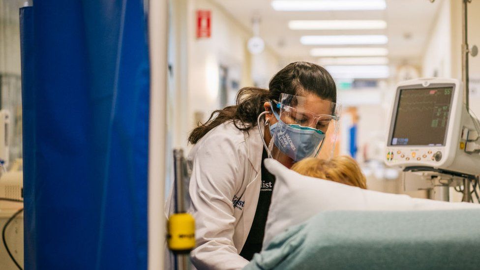
SharecloseShare pageCopy linkAbout sharingimage sourceGetty ImagesThe European Union recommended a pause on all non-essential travel from the US as Covid-19 cases surge.The daily average for hospital admissions has risen past 100,000 for the first time since last winter.The recent wave, driven by the Delta variant, is most severe in the US South but cases are rising nationwide.Monday’s guidance from the 27-nation bloc reverses advice from June that lifted restrictions on American travellers ahead of tourism season.The recommendation is nonbinding, meaning individual countries will be allowed to decide if they still wish to allow US visitors with proof of vaccination, negative tests, or quarantine.Though the EU first lifted travel restrictions on Americans in June, the US has kept their ban on European non-essential travel in place since March 2020. Earlier this month, European Commission President Ursula von der Leyen said the lack of reciprocity would not be allowed to “drag on for weeks”. Israel, Kosovo, Lebanon, Montenegro, and North Macedonia have also been removed from the EU’s safe travel list.Hospital admissions in the US for Covid-19 patients have reached levels not seen since January, when the country reached its all-time high with more than 142,000 coronavirus patients in hospital beds. Florida has more than 16,000 Covid-19 patients in hospital – the most of any state – followed by Texas and California, according to data from the US Department of Health and Human Services. The latest surge is straining hospitals and health care workers. Roughly one in five intensive care units have reached at least 95% capacity. Death rates have risen too – reaching an average of more than 1,000 per day. Just over half of all Americans are fully vaccinated against Covid-19. With the full approval of the Pfizer vaccine by the US Food and Drug Administration last week, the Biden administration has doubled down on its efforts to increase vaccination rates. Unvaccinated people are about 29 times more likely to be admitted to hospital with Covid-19 than those who are fully vaccinated, according to a study released by the US Centres of Disease Control and Prevention (CDC) last week.

Read more →

At least 80% of opioid overdoses aren’t fatal, but how do they affect the brain?

An opioid overdose isn’t a death sentence. In fact, estimates indicate that in the United States, only 4% to 18% of opioid overdoses that are treated in a hospital or pre-hospital setting — such as an ambulance or someone’s home — actually kill the patient. But there’s a lot that scientists still don’t know about what nonfatal overdoses do to the brain.
To better understand the topic, Erin Winstanley and James Mahoney — researchers with the West Virginia University School of Medicine and Rockefeller Neuroscience Institute — reviewed 79 studies of neurocognitive impairments and brain abnormalities associated with nonfatal opioid overdoses in humans. The studies all had limitations, a sign that more precise research is needed in this area. Yet despite the studies’ shortcomings, their findings still suggested that brain abnormalities and cognitive impairments are linked to overdose.
“I think there’s probably a host of morbidities associated with experiencing a nonfatal opioid-related overdose that’s largely neglected in the published literature and — to a certain extent — from a public health standpoint,” said Winstanley, an associate professor in the Department of Behavioral Medicine and Psychiatry and the Department of Neuroscience. “We should first focus on saving lives. That’s definitely the appropriate step. But we probably should start paying attention to some of these other issues, too. Given the proportion of people who have experienced a nonfatal overdose, the number of deaths is just the tip of the iceberg.”
Their findings appear in Drug and Alcohol Dependence.
Winstanley, Mahoney and their colleagues — Felipe Castillo and Sandra Comer of Columbia University — performed a systematic review of journal articles published between 1973 and 2020.
Overall, the studies lacked the level of detail and consistency that would have made many direct comparisons and inferences possible.

Read more →

Hobit turns immune cells into killers

When pathogens invade the human body, a rapid response is required. At the forefront of the immune response are special immune cells. They reside in various tissues such as the lungs, liver, skin and intestines, where they take up the fight against invaders at an early stage. Their name: innate lymphoid cells, or ILCs for short.
A special property of these cells: They do not have to be alerted first in lymphoid organs, like many other immune cells, in order to then migrate to their place of action. Instead, they settle in the tissues and organs shortly after birth and remain there permanently.
Single cell mRNA atlas of ILC1
ILCs can arise in tissues from immature precursor cells and mature into operational immune cells. This was recently shown by scientists of the Max Planck Research Group at the Institute of Systems Immunology of Julius-Maximilians-Universität (JMU) Würzburg in Bavaria, Germany. Until now, it was unclear how this maturation proceeds in detail.
“We wanted to understand how immature ILCs become effector cells that can, for example, kill tumour cells or fight infections with the help of cytokines,” explains Professor Georg Gasteiger, chair and head of the Max Planck Research Group at the JMU Institute of Systems Immunology.
To do this, the Würzburg researchers studied the group of ILC1s that play a role in viral infections and in tumour defence. They have recorded all mRNA molecules of individual ILC1s in the liver and created a virtual cell atlas from these analyses.

Read more →

Research finds ‘very low rates’ of dental fluoride varnish treatment for young children

Fewer than 5% of well-child visits for privately insured young children included a recommended dental fluoride varnish application, despite mandatory insurance coverage for this service, according to a University of Massachusetts Amherst study.
Fluoride varnish helps reduce tooth decay, which affects nearly 25% of 2- to 5-year-olds and more than half of 6- to 8-year-olds in the U.S., according to earlier studies.
The new research, published Aug. 30 in JAMA Network Open, was the first to assess delivery of this evidence-based service recommended by the U.S. Preventive Task Force and the American Academy of Pediatrics for privately insured children. Previous research showed that fewer than 8% of 1- to 5-year-olds covered by Medicaid receive fluoride varnish in medical settings.
While Medicaid in most states has paid for fluoride varnish applications for at least a decade, private insurance coverage was mandated in 2015 under the Affordable Care Act with no cost-share for families, explains lead author Kimberley Geissler, associate professor of health policy and management in the UMass Amherst School of Public Health and Health Sciences.
“Most kids are not getting fluoride varnish applications in the medical office,” Geissler says. “I wasn’t surprised due to the low rates we’ve seen in Medicaid-covered children, but I was disappointed that it wasn’t higher. Medical providers are not required to do this; it’s like a mammogram. It’s recommended and it’s good and they should do it, and the questions we were looking at were: do they do it and how often?”
Co-author and pediatrician Dr. Sarah Goff, associate professor of health policy and management at UMass Amherst, saysknowing that very low rates of fluoride varnish application are found in both Medicaid and commercially insured populations is important because it suggests that broader, system-level barriers exist. “As a health services researcher and primary care pediatrician, I am excited about our next steps of learning what sorts of things are presenting barriers to applying fluoride varnish and developing strategies for overcoming these barriers so all eligible children receive this important preventive intervention,” Goff says.
The JAMA study is part of a larger project in Massachusetts that will delve into more complex questions, such as whymedical providers aren’t applying fluoride varnish during well-child visits. The preventive treatment is especially critical in light of the statistic that fewer than one in three children under age 5 have an annual dentist visit, where this service also could be provided.
The research team, including senior author Ashley Kranz of the RAND Corporation, examined data from 2016-2018 for privately insured young children in Connecticut, Maine, New Hampshire and Rhode Island.
The sample included 328,661 well-child visits in the four states. Fluoride varnish application was more common among visits for younger children. A 2-year-old was nearly 8 percentage points more likely to receive fluoride varnish than a 5-year-old, an analysis of the data showed.
Fluoride varnish applications were most common in Rhode Island, with a regression-adjusted probability of 8.7%. New Hampshire had the lowest rate, with a regression-adjusted probability of 2.2%.
Geissler points to one “hopeful takeaway” from the study: the regression-adjusted probability of fluoride varnish application increased from 3.6% in 2016 to 5.8% in 2018. “That’s still really low,” she says, “but it did go up over time.”
The study concludes, “Although increases over time were encouraging, very low rates of fluoride varnish in medical settings suggest substantial expansion of this service in medical settings is critical for improving children’s oral health and overall well-being.”

Read more →

AI helps to spot single diseased cells

The Human Cell Atlas is the world’s largest, growing single-cell reference atlas. It contains references of millions of cells across tissues, organs and developmental stages. These references help physicians to understand the influences of aging, environment and disease on a cell — and ultimately diagnose and treat patients better. Yet, reference atlases do not come without challenges. Single-cell datasets may contain measurement errors (batch effect), the global availability of computational resources is limited and the sharing of raw data is often legally restricted.
Researchers from Helmholtz Zentrum München and the Technical University of Munich (TUM) developed a novel algorithm called “scArches,” short for single-cell architecture surgery. The biggest advantage: “Instead of sharing raw data between clinics or research centers, the algorithm uses transfer learning to compare new datasets from single-cell genomics with existing references and thus preserves privacy and anonymity. This also makes annotating and interpreting of new data sets very easy and democratizes the usage of single-cell reference atlases dramatically,” says Mohammad Lotfollahi, the leading scientist of the algorithm.
Example COVID-19
The researchers applied scArches to study COVID-19 in several lung bronchial samples. They compared the cells of COVID-19 patients to healthy references using single-cell transcriptomics. The algorithm was able to separate diseased cells from the references and thus enabled the user to pinpoint the cells in need for treatment, for both mild and severe COVID-19 cases. Biological variation between patients did not affect the quality of the mapping process.
Fabian Theis: “Our vision is that in the future we will use cell references as easily as we nowadays do for genome references. In other word, if you want to bake a cake, you usually do not want to try coming up with your own recipe — instead you just look one up in a cookbook. With scArches, we formalize and simplify this lookup process.”
Story Source:
Materials provided by Helmholtz Zentrum München – German Research Center for Environmental Health. Note: Content may be edited for style and length.

Read more →

Low-sodium salt prevents stroke

Replacing salt with a low-sodium alternative lowers the risk of stroke in people with high blood pressure or prior stroke, according to late breaking research presented in a Hot Line session today at ESC Congress 20211 and published in the New England Journal of Medicine2.
Both elevated sodium intake and low potassium intake are associated with high blood pressure and an increased risk of cardiovascular disease and premature death.3,4 Salt substitutes, which replace part of the sodium chloride in regular salt with potassium chloride, have been shown to lower blood pressure5 but their effects on heart disease, stroke, and death had been uncertain. In addition, there had been concerns about causing hyperkalaemia in people with chronic kidney disease leading to cardiac arrhythmias and sudden death.
The Salt Substitute and Stroke Study (SSaSS) compared the effect of reduced sodium salt substitute versus regular salt on stroke, cardiovascular events, mortality and clinical hyperkalaemia.6 SSaSS was an open, cluster-randomised, trial that enrolled participants between April 2014 and January 2015. Participants were adults with either previous stroke or age 60 years and above with poorly controlled blood pressure.7
The trial was conducted in 600 villages in rural areas of five provinces in China. Two counties within each province were chosen that represented the socioeconomic development level of rural counties in that province. Approximately 35 individuals were recruited from each village — for a total of 20,995 participants. Participants were cluster-randomised by village in a 1:1 ratio to provision of salt substitute or continued use of regular salt.
Participants in intervention villages were given free salt substitute (about 75% sodium chloride and 25% potassium chloride) as a replacement for regular salt and advised to use it for all cooking, seasoning and food preservation. They were also encouraged to use the salt replacement more sparingly than they previously used salt to maximise their sodium reduction. Sufficient salt substitute was provided to cover the needs of the entire household (about 20 g per person per day). Participants in control villages continued their usual habits.
The average age of participants was 65.4 years and 49.5% were female. Some 72.6% had a history of stroke and 88.4% had a history of hypertension.

Read more →

Researchers identify new biomarkers to detect consumption of emerging illicit drug

A team of researchers from the National University of Singapore (NUS) has come up with a new solution to boost the surveillance of designer drug abuse. Led by Professor Eric Chan from the NUS Department of Pharmacy, the team has identified three new urinary biomarkers that could be used to detect consumption of ADB-BUTINACA, an emerging synthetic cannabinoid which is a type of new psychoactive substance (NPS). The innovative approach used to identify the biomarkers can be applied to other existing and new synthetic cannabinoids.
NPS are drugs designed to mimic the effects of illegal substances such as cannabis, cocaine, heroin, ‘Ice’, Ecstacy and LSD. The intention of the clandestine laboratories to introduce synthetic cannabinoids with different chemical structures is to try to circumvent legislative bans.
Over the past two years, users of NPS made up the third largest proportion of drug abusers in Singapore, while synthetic cannabinoids have dominated Singapore’s NPS market for the past four years. As most synthetic cannabinoids are extensively metabolised in the body after consumption, they become virtually undetectable in urine samples.
Commenting on the significance of the team’s research, Prof Chan said, “Prior to our study, the metabolism and urinary biomarkers of ADB-BUTINACA were unclear. Our discovery and unique methodology offer assistance to the forensic fraternity who is constantly being challenged by the emergence of novel synthetic cannabinoids, and can also bring benefits to the international public communities to tackle the increasing abuse of this synthetic cannabinoid. This will bring us closer to the goal of having a drug-free world.”
The study, which was carried out in collaboration with the Analytical Toxicology Laboratory of Singapore’s Health Sciences Authority, was first published in the journal Clinical Chemistry on 13 August 2021.
New biomarkers for accurate detection of synthetic drug abuse
ADB-BUTINACA is a new synthetic cannabinoid that was first identified in Europe in 2019, and it entered Singapore’s drug scene last year. Although three existing metabolites of ADB-BUTINACA are available as reference standards for routine forensic monitoring, they have been found to be absent or detected at lower concentrations in some urine samples of abusers. This created an impetus to identify other potential metabolites for use as urinary biomarkers for the cannabinoid’s consumption.
Instead of using the conventional and more time-consuming method of chemically synthesising metabolites of ADB-BUTINACA, Prof Chan and his team introduced an innovative method to identify the cannabinoid’s unique metabolites using the concepts of drug metabolism and pharmacokinetics.
The team synthesised key metabolites of ADB-BUTINACA using human liver enzymes in the laboratory for investigating their disposition and identifying novel biomarker metabolites in urine. From their studies, a total of 15 metabolites of ADB-BUTINACA and their respective pathways of biotransformation in the body were identified for the first time using this method.
Of the 15 new metabolites, the researchers proposed four as urinary metabolite biomarkers due to their metabolic stability, including one metabolite where its reference standard is currently available. A panel comprising either one or a combination of these four newly-established urinary biomarkers was developed for diagnosing the consumption of ADB-BUTINACA.
Moving forward, the team plans to extend their current strategy to better understand the disposition of novel metabolites of synthetic cannabinoids by kidneys and their eventual occurrence in urine.
Story Source:
Materials provided by National University of Singapore. Note: Content may be edited for style and length.

Read more →

Integrated treatment for depression and heart failure improves quality of life and mood

A telephone-delivered nursing care strategy that combined heart failure care management with depression treatment improved patients’ clinical outcomes, discovered clinicians from the University of Pittsburgh. The findings of the clinical trial, called Hopeful Heart, were published today in JAMA Internal Medicine.
The Hopeful Heart Trial is the first study to apply a ‘blended’ collaborative care approach to treating heart failure and depression, whereby investigators trained medical nurses to deliver depression and heart failure care under guidance of a study cardiologist, psychiatrist and primary care physician.
“Heart failure is one of the most common cardiovascular diseases in the United States, and it’s growing even more prevalent as the population ages,” said lead author Bruce Rollman, M.D., M.P.H., UPMC endowed chair and professor of medicine at Pitt. “I’m very excited about our results because they show that we can successfully train medical nurses to deliver effective depression care as part of heart failure care management they may already be delivering, and that this pragmatic approach can significantly improve patients’ mood and help them regain a better quality of life.”
Cardiologists rarely screen their patients for depression, even though it occurs in up to half of all heart failure patients and has been associated with reduced adherence to recommended heart failure care, higher rates of hospital readmission and increased mortality. One potential explanation is that few studies have examined the benefits of depression treatment on heart failure patients’ recovery.
To find out if effective depression treatments can be delivered as part of routine heart failure care, the researchers tested a telephone-delivered ‘blended’ model of collaborative care. Medical nurses who were trained to administer depression care had weekly care-review conference calls with a study psychiatrist and a study cardiologist, and then relayed treatment recommendations to patients and their primary care physicians. Afterward, study nurses monitored patients via regular telephone calls and made recommendations for adjustments in care depending on patients’ responses to treatment.
“Collaborative ‘blended’ care model provides extra layers of emotional and educational support for patients and their families,” said co-author Amy Anderson, M.S., clinical coordinator for the Hopeful Heart Trial at Pitt. “When we sit in on case review sessions with doctors and nurses, we end up learning a great deal about these patients’ lives; it becomes personal. So, it is always very rewarding to see these patients overcome hurdles and improve over time.”
Hopeful Heart recruited 756 participants with heart failure from eight Pittsburgh-area hospitals, including 629 patients who screened positive for depression. At 12-months follow-up, ‘blended’ care patients reported better mental health-related quality of life — including fewer limitations in social activities, improved general well-being, higher energy and less fatigue, and improved mood — compared to patients receiving usual care, and improved mood compared to those who received collaborative care for heart failure alone.
The researchers hope that this innovative and practical approach to patient care could be implemented more broadly, especially as both patients and health care workers have become more accustomed to telemedicine than ever before.
“Depression often goes unrecognized and untreated in heart failure patients, and we are encouraged that our integrated approach to addressing depression was not only effective, but that it can be easily scaled up and expanded nationally,” Rollman said. “A ‘blended’ collaborative care that is built on existing systems of care also may enable organized health care systems such as UPMC to deliver effective first-line care for depression and other mental health conditions to patients with complex medical conditions.”
Other authors of the study include Scott Rothenberger, Ph.D., Kaleab Abebe, Ph.D., Ravi Ramani, M.D., Matthew Muldoon, M.D., M.P.H., John Jakicic, Ph.D., Bea Herbeck Belnap, Dr.Bio.Hum., and Jordan Karp, M.D., all of Pitt.
This research was supported by the National Heart, Lung, and Blood Institute (grant # R01 HL114016).

Read more →

Drug delivery capsule could replace injections for protein drugs

In recent years, scientists have developed monoclonal antibodies — proteins that mimic the body’s own immune defenses — that can combat a variety of diseases, including some cancers and autoimmune disorders such as Crohn’s disease. While these drugs work well, one drawback to them is that they have to be injected.
A team of MIT engineers, in collaboration with scientists from Brigham and Women’s Hospital and Novo Nordisk, is working on an alternative delivery strategy that could make it much easier for patients to benefit from monoclonal antibodies and other drugs that usually have to be injected. They envision that patients could simply swallow a capsule that carries the drug and then injects it directly into the lining of the stomach.
“If we can make it easier for patients to take their medication, then it is more likely that they will take it, and healthcare providers will be more likely to adopt therapies that are known to be effective,” says Giovanni Traverso, the Karl van Tassel Career Development Assistant Professor of Mechanical Engineering at MIT and a gastroenterologist at Brigham and Women’s Hospital.
In a study appearing today in Nature Biotechnology, the researchers demonstrated that their capsules could be used to deliver not only monoclonal antibodies but also other large protein drugs such as insulin, in pigs.
Traverso and Ulrik Rahbek, vice president at Novo Nordisk, are the senior authors of the paper. Former MIT graduate student Alex Abramson and Novo Nordisk scientists Morten Revsgaard Frederiksen and Andreas Vegge are the lead authors.
Targeting the stomach
Most large protein drugs can’t be given orally because enzymes in the digestive tract break them down before they can be absorbed. Traverso and his colleagues have been working on many strategies to deliver such drugs orally, and in 2019, they developed a capsule that could be used to inject up to 300 micrograms of insulin.

Read more →

One in two pregnancies are affected by iron deficiency, yet many women don’t get a simple screening test to check

Half of pregnant women who had a simple blood test to check their iron stores had low iron levels, and one in four had severe iron deficiency, according to a paper published today in the journal Blood Advances. But despite how common iron deficiency is, 40% of pregnant women in this large regional study never had their iron levels checked, and women of lower socioeconomic status were less likely to get tested. Researchers said the findings underscore the need to revisit clinical guidelines to ensure that ferritin testing, the standard measure of iron deficiency, is included as a routine part of maternal care and pregnancy health screenings.
“Despite the very high prevalence of iron deficiency in pregnancy, and how easy it is to treat, we are not doing a very good job of checking for it,” said lead study author Jennifer Teichman, MD, of the University of Toronto, Canada and its affiliated hospitals including St. Michael’s Hospital/Unity Health Toronto, where the study was conducted. “It’s not top of mind, in part, because of inconsistent recommendations for ferritin testing across clinical guidelines.”
Iron requirements in pregnancy are high to support the developing fetus, the growing placenta, and the increased blood supply needed to sustain the pregnancy. This demand for iron increases over the course of pregnancy. Iron deficiency is the most common cause of anemia (low hemoglobin or red blood cell count) during pregnancy, which has been linked to poorer outcomes for both mother and baby, including a higher risk of premature delivery, low birth weight, post-partum depression, and even maternal death. Anemia early in pregnancy has also been associated with neurodevelopmental delays in the offspring, even as the child approaches school age and beyond, which points to potentially long-lasting effects. Even low levels of iron alone can cause pregnant women to experience fatigue, weakness, and brain fog, Dr. Teichman explained.
The study included 44,552 pregnant women who received prenatal testing at community laboratories in Ontario, Canada, between 2013 and 2018 to determine how often ferritin testing was offered. Researchers also sought to provide more robust data about the prevalence and severity of iron deficiency among pregnant women and to identify whether certain clinical or demographic factors played a role in the likelihood of someone receiving a ferritin test.
Altogether, about 60% of patients got a ferritin test during pregnancy; 40% did not. Most tests were ordered by general practitioners (48%) and obstetricians/gynecologists (32%). The vast majority of ferritin testing (71%) occurred at or around the time of the first prenatal visit, when the risk of iron deficiency is lowest and, often, patients’ iron levels were only checked once during their pregnancy.
“Iron deficiency becomes more common as women progress through pregnancy,” said Dr. Teichman. “If we don’t re-evaluate iron stores later in pregnancy, we miss a lot of women who are becoming iron deficient in later trimesters.”
Dr. Teichman emphasized that the women in the study received care in Canada, a publicly funded health care system, which means patients don’t incur the cost of ferritin testing. Despite this, the researchers found that women of lower socioeconomic status were less likely to be tested for iron deficiency, which further underscores differences in access to care and how clinicians may treat these patients differently.

Read more →