
Ros Atkins On… the ethics of Covid booster jabs


SharecloseShare pageCopy linkAbout sharingimage source, Getty ImagesUK researchers have developed a calculator to identify those at highest risk of severe illness from Covid despite having one or two vaccines. They say jabs provide protection against serious illness and death for most people, but for some the risks – while much lowered by jabs – remain.This includes older people, people with Down’s Syndrome, and those with weakened immune systems.The risks are highest for those who have not been fully vaccinated. Writing in the BMJ, scientists say those at risk should ensure they have both jabs. They suggest their risk calculator could be used to help spot fully vaccinated individuals who may additionally benefit from boosters or early treatment – including newer therapies such as monoclonal antibodies.Patterns of riskThe research team – which includes scientists from the universities of Edinburgh, Oxford and Nottingham – examined hospital records involving some five million people in England who had had two jabs and about two million who had had one. There were about 2,000 deaths linked to Covid and almost the same number of Covid hospital admissions – most occurred 14 or more days after the first vaccine (when substantial immunity is expected to have developed). Just 81 deaths and 71 admissions happened 14 or more days after the second vaccine dose.From this data, the groups most at risk of severe illness or death, included:People living in deprived areasPeople with kidney transplantsPeople undergoing chemotherapyCare home residentsPeople with neurological conditions such as dementiaPeople from Indian and Pakistani backgrounds Researchers say while overall the risks would be lower for people who have had two jabs, they would expect the general pattern to remain in terms of the groups identified to be at higher risk.But they say because there were relatively few Covid-related hospital admissions and deaths in people who had two jabs it was harder to build up a precise picture. And they say certain risks – such as people who do high-risk jobs – could not be taken into account. Prof Julia Hippisley-Cox, from the University of Oxford, said: “Individual risk will always depend on individual choices as well as the current prevalence of the disease. However we hope that this new tool will help shared decision making and more personalised risk assessment.”The risk calculator is available online for academic use and there are plans to make a version that will be available to the public. An earlier version of the risk calculator helped spot people who would benefit from shielding in England. Related Internet LinksThe BMJ Leading Medical Research, News, Education, Opinion.websiteThe BBC is not responsible for the content of external sites.
Read more →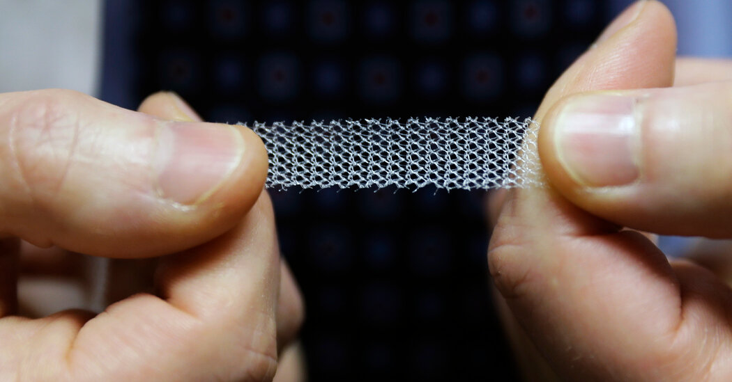
A Florida doctor and a medical consultant pleaded guilty this week to federal charges arising from a scheme to pressure hundreds of women to get their pelvic mesh implants removed. The effort was intended to increase the value of personal injury claims against the manufacturers of the medical devices.The guilty pleas — by Dr. Christopher Walker on Friday and Wesley Blake Barber on Tuesday — bring to a close a criminal case brought two years ago by federal prosecutors in Brooklyn. The two men pleaded guilty to violations of the federal Travel Act, a law that prosecutors have used to pursue charges of bribery in the health care profession.Mr. Barber, 51, could face at least four years in federal prison when he is sentenced in December by Judge Raymond J Dearie of U.S. District Court for the Eastern District of New York. Dr. Walker, also 51, who pleaded guilty to two counts, could be sentenced to at least eight years when he appears before the judge in January. Lawyers for both men are expected to ask for lesser sentences under the federal guidelines.The scheme was one of the more unsavory sides of the mass tort case against a half-dozen manufacturers of pelvic mesh including Boston Scientific and Johnson & Johnson. The case has led to more than $8 billion in settlements for roughly 100,000 women in the United States. Dozens of personal injury firms also collected billions in fees in the process.Prosecutors charged the two men in 2019 with participating in a network to get women from across the country who had pending personal injury claims against the device manufacturers to agree to hastily arranged surgical procedures to remove the implants. The pitch was that women who had the implants removed were getting bigger settlements than women who still had the implants inside them.However, removing the implants, a synthetic product that can bond with a woman’s internal organs, can be tricky. Some women who had mesh removed were worse off, or no better, than they were before the operation. Many of the women lured by the network had the procedures done without getting a second opinion.Mr. Barber was one of the architects of the scheme, according to prosecutors, and a firm he worked with helped arrange for the surgical procedures to be performed at outpatient medical centers in Florida — some in nondescript strip malls. The procedures were paid for with money from high-interest cash advances arranged by a group of so-called litigation finance firms. But the women had to pay the money back to those lenders after receiving the settlement proceeds.The charging document filed by prosecutors in 2019 said the women had been unaware that Dr. Walker and others who had performed the implant removal surgery had paid kickbacks and bribes to Mr. Barber’s firm and others for the surgical referrals.Brooklyn prosecutors filed the charges after an article in 2018 in The New York Times about how some women said they felt pressured into getting the mesh removed by a network of consultants, personal injury lawsuit marketers, lawyers and doctors. Most of the women said they did not fully understand what they had agreed to.The use of mesh to strengthen weakened pelvic muscles that can cause the bladder, the uterus and other organs to sag into the vaginal area long has been controversial. After years of complaints from women about bleeding, leaking bladders, searing pain and other ailments, the Food and Drug Administration stopped the sale and distribution of pelvic mesh in 2019 for treating organ prolapse. By then, the mesh had been implanted in millions of women around the globe.“Both defendants have admitted to participating in a reprehensible bribery and kickback scheme to exploit women across the country,” said Jacquelyn M. Kasulis, the acting U.S. attorney in Brooklyn.Dr. Walker agreed to forfeit about $800,000, and Mr. Barber agreed to forfeit about $1.1 million.Jodi Avergun, a lawyer for Dr. Walker, said that her client’s involvement in the scheme “was unfortunate” and that he “accepted responsibility for his actions in order to move forward and resume his career caring for patients.”The criminal case may be over, but civil litigation continues. In December, a lawsuit filed on behalf of 188 women who claimed to have been victimized by the network was filed in a Florida circuit court. The lawsuit, which seeks monetary damages, named some two dozen defendants, including Mr. Barber, Dr. Walker and other doctors.
Read more →
WASHINGTON — A scientific advisory committee to the Food and Drug Administration on Friday overwhelmingly recommended against approving a booster shot of the Pfizer-BioNTech coronavirus vaccine for people 16 and older.The panel agreed to discuss later Friday afternoon whether Pfizer vaccine recipients who are 65 or older, or at higher risk because of their jobs or underlying health conditions, should be given a third shot.The committee voted 16 to 2 against a broader recommendation after an intense daylong public discussion on whether booster shots are necessary and if so, for whom. The Biden administration had been hoping the F.D.A. and the Centers for Disease Control and Prevention would approve a third shot of the Pfizer vaccine in time to begin rolling out boosters for Pfizer recipients next week.Although the F.D.A. is not obliged to follow its advisory committee’s recommendations, it typically does. The agency will likely make a decision by early next week.The vote came after a sharp debate in which many of the panel’s independent experts, including infectious disease doctors and statisticians, challenged whether the data justified extra shots for so much of the population when the vaccines appear to still offer robust protection against severe Covid-19 disease and hospitalization, at least in the United States.“It’s unclear that everyone needs to be boosted, other than a subset of the population that clearly would be at high risk for serious disease,” said Dr. Michael G. Kurilla, a committee member and official at the National Institutes of Health.The negative vote was the latest in a series of setbacks for President Biden’s booster plan since he first announced it a month ago. Mr. Biden said at the time that he wanted most adults who had gotten a second Pfizer or Moderna vaccine at least eight months ago to start receiving booster shots the week of Sept. 20.But two weeks after his announcement, leaders of the F.D.A. and the C.D.C. told the White House that it would be impossible to authorize booster shots for recipients of the Moderna vaccine that soon. It is now unclear whether extra injections will be offered to Pfizer recipients, and if so, to how many.In a remarkable public display of internal dissension, two F.D.A. scientists co-authored a medical journal article earlier this week arguing that there was no credible evidence yet in support of booster shots for the general population. Those officials, who are leaving the agency this fall, joined outside experts and other federal health officials who cast doubt at the meeting on whether Pfizer’s request should be approved.On the other hand, Dr. Peter Marks, their superior and the official who oversees the F.D.A.’s vaccine division, noted that many well-known vaccines require booster shots and urged the committee to consider the importance of not just of preventing severe disease but of curbing the spread of infection.After the F.D.A. rules on Pfizer’s request, an advisory committee to the C.D.C. will meet to recommend how exactly the extra doses should be used. Earlier public discussions suggest the C.D.C. committee may be leaning toward tailoring booster shots toward the elderly and others particularly vulnerable to worse outcomes from Covid-19, instead of to all those who received their second injection eight months earlier.The F.D.A. committee’s vote followed hours of presentations by officials from Pfizer, the C.D.C., the Israeli government and independent experts on the complex array of data they have collected up until now about the waning effectiveness of Pfizer and other vaccines over time.Dr. Sara Oliver of the C.D.C. presented data showing that vaccines continue to strongly protect against severe forms of Covid-19 in the United States, even in people 75 and older.Jonathan Sterne, a professor of medical statistics and epidemiology in the United Kingdom, said he had analyzed 76 different studies on the vaccines’ real world effectiveness and found that multiple factors can skew the results, including how many unvaccinated people in a study have natural immunity from prior Covid-19 disease. He also warned against drawing conclusions from short-term results from booster shots; data from Israel, for example, only included a follow-up period of several weeks for older adults.Israeli experts made a different argument, telling the committee that they believed third Pfizer shots helped dampen a fourth wave of transmission as the Delta variant swept the nation this summer. The Israeli government, which has relied almost entirely on the Pfizer vaccine, began offering booster shots in late July, starting with the elderly..css-1xzcza9{list-style-type:disc;padding-inline-start:1em;}.css-3btd0c{font-family:nyt-franklin,helvetica,arial,sans-serif;font-size:1rem;line-height:1.375rem;color:#333;margin-bottom:0.78125rem;}@media (min-width:740px){.css-3btd0c{font-size:1.0625rem;line-height:1.5rem;margin-bottom:0.9375rem;}}.css-3btd0c strong{font-weight:600;}.css-3btd0c em{font-style:italic;}.css-w739ur{margin:0 auto 5px;font-family:nyt-franklin,helvetica,arial,sans-serif;font-weight:700;font-size:1.125rem;line-height:1.3125rem;color:#121212;}#NYT_BELOW_MAIN_CONTENT_REGION .css-w739ur{font-family:nyt-cheltenham,georgia,’times new roman’,times,serif;font-weight:700;font-size:1.375rem;line-height:1.625rem;}@media (min-width:740px){#NYT_BELOW_MAIN_CONTENT_REGION .css-w739ur{font-size:1.6875rem;line-height:1.875rem;}}@media (min-width:740px){.css-w739ur{font-size:1.25rem;line-height:1.4375rem;}}.css-9s9ecg{margin-bottom:15px;}.css-16ed7iq{width:100%;display:-webkit-box;display:-webkit-flex;display:-ms-flexbox;display:flex;-webkit-align-items:center;-webkit-box-align:center;-ms-flex-align:center;align-items:center;-webkit-box-pack:center;-webkit-justify-content:center;-ms-flex-pack:center;justify-content:center;padding:10px 0;background-color:white;}.css-pmm6ed{display:-webkit-box;display:-webkit-flex;display:-ms-flexbox;display:flex;-webkit-align-items:center;-webkit-box-align:center;-ms-flex-align:center;align-items:center;}.css-pmm6ed > :not(:first-child){margin-left:5px;}.css-5gimkt{font-family:nyt-franklin,helvetica,arial,sans-serif;font-size:0.8125rem;font-weight:700;-webkit-letter-spacing:0.03em;-moz-letter-spacing:0.03em;-ms-letter-spacing:0.03em;letter-spacing:0.03em;text-transform:uppercase;color:#333;}.css-5gimkt:after{content:’Collapse’;}.css-rdoyk0{-webkit-transition:all 0.5s ease;transition:all 0.5s ease;-webkit-transform:rotate(180deg);-ms-transform:rotate(180deg);transform:rotate(180deg);}.css-eb027h{max-height:5000px;-webkit-transition:max-height 0.5s ease;transition:max-height 0.5s ease;}.css-6mllg9{-webkit-transition:all 0.5s ease;transition:all 0.5s ease;position:relative;opacity:0;}.css-6mllg9:before{content:”;background-image:linear-gradient(180deg,transparent,#ffffff);background-image:-webkit-linear-gradient(270deg,rgba(255,255,255,0),#ffffff);height:80px;width:100%;position:absolute;bottom:0px;pointer-events:none;}.css-uf1ume{display:-webkit-box;display:-webkit-flex;display:-ms-flexbox;display:flex;-webkit-box-pack:justify;-webkit-justify-content:space-between;-ms-flex-pack:justify;justify-content:space-between;}.css-wxi1cx{display:-webkit-box;display:-webkit-flex;display:-ms-flexbox;display:flex;-webkit-flex-direction:column;-ms-flex-direction:column;flex-direction:column;-webkit-align-self:flex-end;-ms-flex-item-align:end;align-self:flex-end;}.css-12vbvwq{background-color:white;border:1px solid #e2e2e2;width:calc(100% – 40px);max-width:600px;margin:1.5rem auto 1.9rem;padding:15px;box-sizing:border-box;}@media (min-width:740px){.css-12vbvwq{padding:20px;width:100%;}}.css-12vbvwq:focus{outline:1px solid #e2e2e2;}#NYT_BELOW_MAIN_CONTENT_REGION .css-12vbvwq{border:none;padding:10px 0 0;border-top:2px solid #121212;}.css-12vbvwq[data-truncated] .css-rdoyk0{-webkit-transform:rotate(0deg);-ms-transform:rotate(0deg);transform:rotate(0deg);}.css-12vbvwq[data-truncated] .css-eb027h{max-height:300px;overflow:hidden;-webkit-transition:none;transition:none;}.css-12vbvwq[data-truncated] .css-5gimkt:after{content:’See more’;}.css-12vbvwq[data-truncated] .css-6mllg9{opacity:1;}.css-qjk116{margin:0 auto;overflow:hidden;}.css-qjk116 strong{font-weight:700;}.css-qjk116 em{font-style:italic;}.css-qjk116 a{color:#326891;-webkit-text-decoration:underline;text-decoration:underline;text-underline-offset:1px;-webkit-text-decoration-thickness:1px;text-decoration-thickness:1px;-webkit-text-decoration-color:#326891;text-decoration-color:#326891;}.css-qjk116 a:visited{color:#326891;-webkit-text-decoration-color:#326891;text-decoration-color:#326891;}.css-qjk116 a:hover{-webkit-text-decoration:none;text-decoration:none;}Dr. Sharon Alroy-Preis, Israel’s head of public health services said the summer’s rise in the number of hospitalized patients who had been fully vaccinated with Pfizer’s vaccine was “scary.” She said 60 percent of severely or critically ill patients and 45 percent of those who died during what she called the fourth surge had received two injections of Pfizer’s vaccine.After offering boosters to the general population, she said, Israel is now averaging about half as many severe or critically ill patients as anticipated. She said boosters not only helped curb the spread of infection, but “actually saved lives.”Another Israeli scientist walked the panel through a new study of health records of more than 1.1. million people over age 60. It found that at least 12 days after the booster, the rates of severe disease were nearly twenty-fold lower among those who received a third Pfizer shot compared to those who did not.Dr. William C. Gruber, a senior Pfizer vice president in charge of vaccine development, suggested that if the United States does not follow Israel’s lead, it could potentially face more than five million more infections a year among people who received their second dose 10 months earlier, compared to those who got the second shot five months earlier.“Israel could portend the U.S. Covid-19 future, and soon,” he said.He said Pfizer’s data shows a third shot elicits a robust antibody immune response that equals or greatly exceeds the response after the second dose. Data also show, he argued, that breakthrough infections among vaccinated Americans are linked more to the ebbing power of the vaccine over time instead than to the Delta variant.But committee members, including some government officials, appeared deeply skeptical of the Pfizer’s data and Israel’s analyses. Dr. Philip Krause, one of the F.D.A. vaccine experts who authored the medical journal review, criticized Pfizer’s presentation of data that had not been peer-reviewed or evaluated by the F.D.A., arguing that possible problems in the modeling within could understate the vaccine’s efficacy.Dr. Oliver, the C.D.C. official, questioned attempts to draw a parallel between the United States and Israel, noting that Israel has only nine million residents and is less diverse than the United States. Notably, she also said that Israel defines a severe case of Covid-19 more broadly than the United States does, which might help explain why Israel reports more serious breakthrough infections among its vaccinated.Another C.D.C. official, Dr. Amanda Cohn, asked Israeli officials why the spread of the virus there had recently intensified, despite a broad rollout of boosters. Dr. Alroy-Preis said that the Jewish holidays, together with the start of the school year, had contributed to what she suggested would be a temporary surge in cases.Committee members also said they were concerned about a paucity of safety data in younger recipients of a booster dose, since studies have shown a higher risk of the heart condition myocarditis in young men who received Pfizer-BioNTech’s vaccine. Several of them asked whether it would be better to wait for a booster vaccine designed specifically to fend off the Delta variant of the virus.Apoorva Mandavilli and Sheryl Gay Stolberg contributed reporting.
Read more →
Allergic reactions to the new mRNA-based COVID-19 vaccines are rare, typically mild and treatable, and they should not deter people from becoming vaccinated, according to research from the Stanford University School of Medicine.
The findings will be published online Sept. 17 in JAMA Network Open.
“We wanted to understand the spectrum of allergies to the new vaccines and understand what was causing them,” said the study’s senior author, Kari Nadeau, MD, PhD, the Naddisy Foundation Professor in Pediatric Food Allergy, Immunology, and Asthma.
The study analyzed 22 potential allergic reactions to the first 39,000 doses of Pfizer and Moderna COVID-19 vaccines given to health care providers at Stanford soon after the vaccines received emergency use authorization from the Food and Drug Administration.
Most of those in the study who developed reactions were allergic to an ingredient that helps stabilize the COVID-19 vaccines; they did not show allergies to the vaccine components that provide immunity to the SARS-CoV-2 virus. Furthermore, these allergic reactions occurred via an indirect activation of allergy pathways, which makes them easier to mitigate than many allergic responses.
“It’s nice to know these reactions are manageable,” said Nadeau, who directs the Sean N. Parker Center for Allergy and Asthma Research at Stanford. “Having an allergic reaction to these new vaccines is uncommon, and if it does happen, there’s a way to manage it.”
The study’s lead author is former postdoctoral scholar Christopher Warren, PhD, now an assistant professor at Northwestern University Feinberg School of Medicine.
WEHI researchers have made a surprise discovery about how immune ‘sentinel’ cells are maintained, which could have implications for drugs in development for treating cancer.
The researchers studied the impact of deleting specific proteins in immune cells that were responsible for controlling the ability of cells to silence or switch off genes.
They were surprised to find that one population of ‘sentinel’ immune cells was affected by deletion of a component of the machinery, causing the cells to disappear from skin and lungs completely. This suggests that drugs which inhibit this component to treat diseases, such as cancer, could have unintended consequences for the immune system.
The research was led by Dr Yifan Zhan, Dr Yuxia Zhang, Mr Shengbo Zhang, Dr Michael Chopin, Professor Stephen Nutt and colleagues, and was published in Science Immunology.
A complex issue
The research team studied the role of the polycomb repressive complex 2 (PRC2) in frontline responder immune cells.
Systemic lupus erythematosus (SLE) is an autoimmune disease in which the immune system attacks the body’s own tissues, causing widespread inflammation and damage in the affected organs. It can affect the joints, skin, brain, lungs, kidneys, and blood vessels. Asian SLE patients suffer from more severe disease and damage compared to patients in western countries.
In Singapore, the prevalence of childhood SLE has been reported to be 14.2 per 100,000 children, while that of adult SLE is 40 per 100,000 adult population. Cognitive dysfunction is common in patients with SLE. Earlier studies have shown that 25% of SLE patients were found to have cognitive dysfunction, compared to 7.3% in age- and gender-matched healthy individuals. Common cognitive dysfunction demonstrated by SLE patients includes impairment of simple reaction time, sustained and selected attention, memory search, working memory, and short-term memory for learnt associations, which result in reduced health-related quality of life and a negative impact on vocational capability.
The neural mechanism leading to cognitive dysfunction in patients with SLE remains unknown. Researchers at the NUS Yong Loo Lin School of Medicine attempted to unravel such mechanisms by adopting non-invasive diffusion magnetic resonance imaging (MRI) to study the brains of SLE patients, particularly the white matter, coupled with computerised neuropsychological assessment. White matter lies beneath the grey matter cortex in the human brain and comprises millions of bundles of nerve fibres that transmit signals to different brain regions.
Led jointly by Associate Professor Juan Helen Zhou, from the Centre for Sleep and Cognition and Deputy Director for the Centre for Translational Magnetic Resonance Research at NUS Medicine, and Associate Professor Anselm Mak, a clinician scientist with the Division of Rheumatology at the Department of Medicine, the group investigated brain white matter free water changes in SLE. White matter free water refers to water molecules surrounding white matter in the brain that are able to diffuse unhindered
In the paper published in Rheumatology, a Tier 1 journal in the field of Rheumatology with the Oxford University Press, they compared the free-water signals in SLE patients without clinically overt neuropsychiatric manifestations with a group of matched healthy participants using the novel free water diffusion MRI technique. They discovered that patients with SLE had significantly higher white matter free water than their healthy counterparts, suggesting possible microvascular degradation and/or inflammation. Such increases in free water were significantly related to cognitive dysfunction, especially sustained attention, as well as cumulative dosage of medical steroids.
“The clinical implications of the study would indicate to physicians that steroids should be judiciously prescribed, aiming for the lowest possible dose for the shortest possible duration. While systemic use of steroids will continue to be the mainstay of treatment for moderate to severe SLE-related inflammation, there are other therapeutic options that could produce similar results: these should be considered first in order to reduce steroid use or shorten the necessary course,” said Assoc Prof Anselm Mak.
He also added that there are other SLE manifestations that could be symptomatically treated without the prescription of steroids. For example, non-steroidal anti-inflammatory drugs could be used for milder inflammation, topical treatment for hair loss and topical analgesics for oral ulcers.
SLE patients on long-term steroid treatments should also be regularly assessed and monitored for cognitive dysfunction. The researchers noted that the use of the Automated Neuropsychiatric Assessment Matrix (ANAM), which has been validated for use in adult and paediatric SLE patients, is an efficient and effective tool for these regular evaluations.
Further research will have to be conducted to find out if a reduction of steroid dosage would be related to reduction of white matter free water that would lead to an improvement to cognitive function in SLE patients. The team is also keen to further investigate the mechanism of the increase in white matter free water underlying cognitive decline using multimodal imaging, behavioral, and blood assays in SLE patients longitudinally.
Ancient DNA extracted from human bones has rewritten early Japanese history by underlining that modern day populations in Japan have a tripartite genetic origin — a finding that refines previously accepted views of a dual genomic ancestry.
Twelve newly sequenced ancient Japanese genomes show that modern day populations do indeed show the genetic signatures of early indigenous Jomon hunter-gatherer-fishers and immigrant Yayoi farmers — but also add a third genetic component that is linked to the Kofun peoples, whose culture spread in Japan between the 3rd and 7th centuries.
Rapid cultural transformations
The Japanese archipelago has been occupied by humans for at least 38,000 years but Japan underwent rapid transformations only in the last 3,000 years, first from foraging to wet-rice farming, and then to a technologically advanced imperial state.
The previous, long-standing hypothesis suggested that mainland Japanese populations derive dual-ancestry from the indigenous Jomon hunter-gatherer-fishers, who inhabited the Japanese archipelago from around 16,000 to 3,000 years ago, and later Yayoi farmers, who migrated from the Asian continent and lived in Japan from around 900 BC to 300 AD.
But the 12 newly sequenced ancient Japanese genomes — which came from the bones of people living in pre- and post-farming periods — also identify a later influx of East Asian ancestry during the imperial Kofun period, which lasted from around 300 to 700 AD and which saw the emergence of political centralisation in Japan.
A paper published in the journal Science Immunology explores the idea that studying bats’ responses to SARS-CoV-2 may provide key insights into how and when to best use existing therapies for COVID-19, and to develop new treatments.
The review, led by Professor Marcel Nold and Associate Professor Claudia Nold, from Monash University’s Department of Paediatrics and Hudson Institute of Medical Research, written in collaboration with colleagues in Australia and China, is a major review of how the virus that has caused the current pandemic wreaks havoc on the human immune system.
Since first identified in December 2019, SARS-CoV-2 has mutated, and the variant strains Alpha, Beta and Delta are more infectious than the original strain. Specifically, the Delta strain is 60-79 per cent more transmissible again than the Alpha mutant, and presumably more deadly, according to Professor Nold.
He says there remains an urgent “need for effective therapies, at least in part because of the emergence of mutations.”
The authors warn that “preventing SARS-CoV-2 infection, or enabling patients to eradicate it, represents the ultimate goal in combating COVID-19: but it is uncertain when either will become reliably possible.”
“Therefore, efforts at identifying safe and effective therapies to prevent COVID from progressing to the moderate and severe disease stages, are critical in the fight against the disease,” Professor Nold said.
Researchers at Washington University School of Medicine in St. Louis have developed an approach to estimating when a person who is likely to develop Alzheimer’s disease, but has no cognitive symptoms, will start showing signs of Alzheimer’s dementia.
The algorithm, available online in the journal Neurology, uses data from a kind of brain scan known as amyloid positron emission tomography (PET) to gauge brain levels of the key Alzheimer’s protein amyloid beta.
In those who eventually develop Alzheimer’s dementia, amyloid silently builds up in the brain for up to two decades before the first signs of confusion and forgetfulness appear. Amyloid PET scans already are used widely in Alzheimer’s research, and this algorithm represents a new way of analyzing such scans to approximate when symptoms will arise. Using a person’s age and data from a single amyloid PET scan, the algorithm yields an estimate of how far a person has progressed toward dementia — and how much time is left before cognitive impairment sets in.
“I perform amyloid PET scans for research studies, and when I tell cognitively normal individuals about positive results, the first question is always, ‘How long do I have until I get dementia?’,” said senior author Suzanne Schindler, MD, PhD, an assistant professor of neurology. “Until now, the answer I’d have to give was something like, ‘You have an increased risk of developing dementia in the next five years.’ But what does that mean? Individuals want to know when they are likely to develop symptoms, not just whether they are at higher risk.”
Schindler and colleagues analyzed amyloid PET scans from 236 people participating in Alzheimer’s research studies through Washington University’s Charles F. and Joanne Knight Alzheimer Disease Research Center. The participants were an average of 67 years old at the beginning of the study. All participants underwent at least two brain scans an average of 4½ years apart. The researchers applied a widely used metric known as the standard uptake value ratio (SUVR) to the scans to estimate the amount of amyloid in each participant’s brain at each time point.
The researchers also accessed over 1,300 clinical assessments on 180 of the participants. The assessments typically were performed every one to three years. Most participants were cognitively normal at the start of data collection, so the repeated assessments allowed the researchers to pinpoint when each participant’s cognitive skills began to slip.