Lung autopsies of COVID-19 patients reveal treatment clues

Lung autopsy and plasma samples from people who died of COVID-19 have provided a clearer picture of how the SARS-CoV-2 virus spreads and damages lung tissue. Scientists at the National Institutes of Health and their collaborators say the information, published in Science Translational Medicine, could help predict severe and prolonged COVID-19 cases, particularly among high-risk people, and inform effective treatments.
Although the study was small — lung samples from 18 cases and plasma samples from six of those cases — the scientists say their data revealed trends that could help develop new COVID-19 therapeutics and fine-tune when to use existing therapeutics at different stages of disease progression. The findings include details about how SARS-CoV-2, the virus that causes COVID-19, spreads in the lungs, manipulates the immune system, causes widespread thrombosis that does not resolve, and targets signaling pathways that promote lung failure, fibrosis and impair tissue repair. The researchers say the data are particularly relevant to caring for COVID-19 patients who are elderly, obese, or have diabetes — all considered high-risk populations for severe cases. Study samples were from patients who had at least one high-risk condition.
The study included patients who died between March and July 2020, with time of death ranging from three to 47 days after symptoms began. This varied timeframe allowed the scientists to compare short, intermediate, and long-term cases. Every case showed findings consistent with diffuse alveolar damage, which prevents proper oxygen flow to the blood and eventually makes lungs thickened and stiff.
They also found that SARS-CoV-2 directly infected basal epithelial cells within the lungs, impeding their essential function of repairing damaged airways and lungs and generating healthy tissue. The process is different from the way influenza viruses attack cells in the lungs. This provides scientists with additional information to use when evaluating or developing antiviral therapeutics.
Researchers at NIH’s National Institute of Allergy and Infectious Diseases led the project in collaboration with the National Institute of Biomedical Imaging and Bioengineering and the U.S. Food and Drug Administration. Other collaborators included the Institute for Systems Biology in Seattle; University of Illinois, Champaign; Saint John’s Cancer Institute in Santa Monica, California.; the USC Keck School of Medicine in Los Angeles; University of Washington Harborview Medical Center, Seattle; University of Vermont Medical Center, Burlington; and Memorial Sloan Kettering Cancer Center in New York City.
Story Source:
Materials provided by NIH/National Institute of Allergy and Infectious Diseases. Note: Content may be edited for style and length.

Read more →

Chemistry breakthrough leads way to more sustainable pharmaceuticals

Chemistry researchers at the University of Bath have developed a new method using blue light to create pharmaceuticals in a more sustainable way, significantly reducing the amount of energy needed and the chemical waste created in the manufacture process.
Synthesising small-molecule drugs normally requires several steps, each one creating waste products and solvent waste — these are often toxic and difficult to dispose of safely.
Currently, it is estimated that for every kilogram of drug made, around 100 kg of waste is produced, making it a hugely inefficient process.
The team at Bath, led by Dr Alex Cresswell, a Royal Society University Research Fellow in the University’s Department of Chemistry, has developed a new way of synthesising nitrogen-containing chemicals called primary amines, which are used in more than half of all pharmaceuticals.
The method uses a catalyst, activated by blue light, to speed up the reaction, and uses fewer steps, less energy and dramatically cuts down the waste created by drug development.
The team has tested the method by synthesising a drug used for multiple sclerosis (MS), Fingolimod (brand name Gilenya), which is made by Novartis and had worldwide sales of $3 billion in 2020.

Read more →

Researchers reveal structure of itch receptors on cells

Ever wonder what’s going on when you get itchy skin, whether from a rash or medication or some other bodily reaction? And why do some strong anti-itching medications make us nauseous, dry-mouthed zombies? Scientists at the UNC School of Medicine and the University of California at San Francisco conducted research showing in precise detail how chemicals bind to mast cells to cause itch, and the scientists figured out the detailed structure of receptor proteins on the surface of these cells when a compound is bound to those proteins.
This work, published in Nature, was led by the labs of Bryan L. Roth, MD, PhD and Jonathan Fay, PhD at UNC-Chapel Hill, and Brian Shoichet, PhD, at UC San Francisco, co-senior authors who have collaborated on previous studies of important cell receptors — protein complexes that chemicals (including drugs) bind to cause or stop a reaction inside cells.
“Our work provides a template for the design of new anti-itch medications,” said Roth, the Michael Hooker Distinguished Professor of Pharmacology. “Also, our research team did a truly remarkable job showing precisely how chemically distinct compounds induce itching through one of two distinct receptors known to be involved in itching.”
First author Can Cao, PhD, a postdoctoral research in the Roth lab, and co-senior author Jonathan Fry, PhD, now an assistant professor in the UNC Department of Biochemistry and Biophysics, led the experiments during the COVID pandemic.
On the surface of cells sit receptor proteins you can think of as complex locks. When a chemical key enters the lock, not only does the cell “open,” but the chemical causes a chain reaction of signals inside cells. Many chemicals do this, from naturally occurring dopamine in the brain to caffeine and cocaine.
When it comes to itch, Roth’s lab identified two receptors called MRGPRX2 on the surface of mast cells and MRGPRX4 on itch-sensing neurons that live in connective tissue and play roles in allergies, immune tolerance, wound healing and other factors in health and disease.
Several drugs unintentionally flood these receptors to trigger the release of histamines, causing the side effect of itching. Drugs such as nateglinide for diabetes, as well as morphine, codeine, and the cough suppressant dextromethorphan are known to cause this reaction. Antihistamines are designed to tamp down the itch response, but they and other anti-itching medications do so clumsily, tripping other cell signaling pathways to cause side effects such as drowsiness, blurred vision, dry mouth, nausea, etc.
The researchers used the experimental technique electron microscopy to create high-resolution maps of these complex receptor proteins when bound to a compound that causes the release of histamines to cause itchiness. They also clarified how drugs bind to MRGPRX4 to cause itch related to various drugs and liver diseases. The researchers used the CryoEM Core Facility at UNC-Chapel Hill to determine the receptor structures.
“Knowing precisely how all this plays out at the molecular level will help us and others create better ways to control the role of these two receptors in itchiness and other conditions,” Roth said.
MRGPRX2 and MRGPRX4 have also been implicated in inflammation arising from the nervous system, eczema, ulcerative colitis, and pain.
“The relatively potent agonists and antagonists described in our Nature paper provide chemical probes we can use to explore the biology of these receptors,” Roth said, “And the structures we revealed so far should accelerate the search for specific medications targeting MRGPRs.”
The National Institutes of Health funded this research.

Read more →

When the gut's internal ecosystem goes awry, could an ancient if gross-sounding treatment make it right?

Dr. Cathy Williams knew something wasn’t right. The veterinarian had felt off for weeks after her 2014 trip to Madagascar.
At first she just felt bloated and uncomfortable and wasn’t interested in eating much. But eventually she developed a fever and chills that sent her to the emergency room.
When tested, doctors found that what she had wasn’t just a stomach bug. She was suffering from an infection of Clostridium difficile, a germ that causes severe diarrhea and abdominal pain and can quickly become life-threatening if not treated promptly.
“It was horrible,” Williams said.
The condition is often triggered when antibiotics disrupt the normal balance of bacteria that inhabit the gut, allowing “bad” bacteria such as C. difficile to multiply unchecked and wreak havoc on the intestines.
To get her infection under control, Williams asked her doctors if they could try an approach she and other veterinarians had used for decades to treat lemurs with digestive problems at the Duke Lemur Center. The procedure, known as a fecal microbiota transplant, involves taking stool from a healthy donor and administering it to the patient to add back “good” microbes and reset the gut.

Read more →

Advanced microscopes help scientists understand how cells break down proteins

Proteins are the building blocks of all living things. A vast amount research takes place on how these proteins are made and what they do, from enzymes that carry out chemical reactions to messengers that transmit signals between cells. In 2004, Aaron Ciechanover, Avram Hershko, and Irwin Rose won the Nobel Prize in Chemistry for a different but just as important process of protein machinery: how organisms break down proteins when they are finished doing their job.
Protein degradation is a carefully orchestrated process. Proteins are marked for disposal with a molecular label called ubiquitin, and then fed into proteasomes, a kind of cellular paper shredder that chops up the proteins into small pieces. This process of ubiquitination, or labeling proteins with ubiquitin, is involved in a wide range of cellular processes, including cell division, DNA repair, and immune responses.
In a new study published in Nature on November 17, 2021, researchers from the University of Chicago used advanced electron microscopes to delve deeper into the process of protein degradation. They described the structure of a key enzyme that helps mediate ubiquitination in yeast, part of a cellular process called the N-degron pathway that may be responsible for determining the rate of degradation for up to 80% of equivalent proteins in humans. Malfunctions in this pathway can lead to accumulation of damaged or misfolded proteins, which underlies the aging process, neurodegeneration, and some rare autosomal recessive disorders, so understanding it better provides an opportunity to develop treatments.
Minglei Zhao, PhD, Assistant Professor of Biochemistry and Molecular Biology, and his colleagues studied an E3 ligase — a type of enzyme that helps join larger molecules together — called Ubr1. In baker’s yeast, Ubr1 helps initiate the ubiquitination process as it attaches ubiquitin to proteins and elongates it into a chain of molecules known as a polymer. Polymers, which are more commonly known as the building blocks of synthetic materials like plastics, also occur naturally when large molecules (in this case ubiquitin) are connected in repeating subunits.
“Until this study, we didn’t know that much about how ubiquitin polymers are structurally formed,” Zhao said. “Now we are starting to get an idea of how it’s first installed onto the protein substrate, and then how the polymers are formed in a linkage-specific manner. This is a milestone in terms of understanding polyubiquitination at a near atomic level.”
In this study, Zhao and his team used some chemical biology techniques to mimic the initial steps of the process for attaching ubiquitin to proteins. Then, they employed another Nobel Prize-winning innovation called cryo-electron microscopy (cryo-EM) to capture the process. Cryo-EM involves flash-freezing solutions of proteins and then using a powerful electron microscope to produce images of individual molecules or subcellular structures. About 10 years ago, breakthroughs in hardware and software produced microscopes and detectors that could capture molecular images at much higher resolution. In 2017, Jacques Dubochet, Joachim Frank, and Richard Henderson won the Nobel Prize in Chemistry for developing cryo-EM techniques, which allow researchers to create a snapshot that literally freezes “live” action of a biological process.
Zhao’s team took advantage of a $10 million investment by the Biological Sciences Division in the Advanced Electron Microscope Facility to use cryo-EM to study ubiquitination in more detail. They were able to describe the structure of several intermediate enzyme complexes involved in the pathway, which will help researchers looking for ways to target proteins with drugs or intervene in a malfunctioning protein degradation process.
“Cryo-EM is exciting because after the data processing is done, a new structure pops out that you’ve never seen before,” Zhao said. “Now we can use what we’ve learned and repurpose the enzymes by introducing small molecules or mixture of peptides to degrade the proteins we want.”
Story Source:
Materials provided by University of Chicago. Original written by Matt Wood. Note: Content may be edited for style and length.

Read more →

How herpes checks into the nervous system for life

Herpes type 1 is sealed with a kiss for a lifetime. More than half of U.S. adults are carriers of HSV1 (herpes simplex virus type 1) which hibernates in the peripheral nervous system and can never be eradicated.
A new Northwestern Medicine study has uncovered the virus’s sneaky strategy for infecting the nervous system, opening a path to long-needed vaccine development for both HSV1 and its close sibling HSV2.
Some carriers will never even experience so much as a cold sore from HSV1. But for others, it can cause blindness or life-threatening encephalitis. There is increasing evidence it contributes to dementia.
And HSV2, which is more commonly transmitted via sexual contact, can be passed from a mother to newborn during the birthing process asneonatal herpes, appearing as lesions all over the body of the infant. Most babies recover, but in the worst cases, it can cause brain damage or disseminate through all the organs and be lethal.
“We desperately need a vaccine to prevent herpes from invading the nervous system,” said Greg Smith, professor of microbiology and immunology at Northwestern University Feinberg School of Medicine.
The new Northwestern Medicine study from Smith’s lab has uncovered a route to that. The study discovered how herpes kidnaps a protein from epithelial cells and turns it into a defector to help it travel into the peripheral nervous system. They have termed the process “assimilation.” It’s a discovery that may have wide-ranging implications for many viruses, including HIV and SARS-CoV-2, Smith said.

Read more →

Researchers identify technique to detect false positive COVID-19 results

Researchers from the University of Missouri School of Medicine have developed and tested a process to identify potential false-positive COVID-19 results. The method, used at MU Health Care, could help other laboratories prevent unnecessary quarantining and repeated testing of people who are not actually infected.
COVID-19 testing is an important tool for managing the virus during the pandemic, and reverse transcriptase polymerase chain reaction (RT-PCR) testing is the most widely used method. But while this type of test is considered reliable, it is associated with a small number of false positive results, most easily recognized in asymptomatic, nonexposed patients.
“False positive diagnoses have important implications for patient management,” said Lester Layfield, MD, professor of pathology and anatomical sciences and director of the Molecular Diagnostics Laboratory. “False positives may lead to inappropriate quarantine, delay of other necessary medical treatment or transfer to a COVID-19 ward.”
To help ensure the accuracy of positive tests, Layfield developed a protocol for repeat testing of all positive results involving asymptomatic and unexposed patients, and in all cases in which a specimen with a positive result was located in a testing well next to another specimen with a high virus load.
Layfield and his team of researchers implemented the quality control protocol in September 2020. Over an eight-week period, 24,717 RT-PCR tests were performed. Of those, 6,251 came from asymptomatic patients. In that group, 288 specimens initially returned a positive result. A second test revealed 20 of these to be false positives.
“Retesting of positive results from asymptomatic individuals revealed some technologist errors but also contamination from positive specimens in adjacent specimen wells,” Layfield said. “This study should alert the laboratory testing community of the possibility of false positive COVID-19 tests.”
In addition to Layfield, the study authors include Douglas C Miller, MD, PhD, chair of pathology and anatomical sciences; resident physician Kelly Bowers, DO; and Simone Camp. The authors declare they have no conflict of interest to report in relation to this study.
Their study, “SARS-CoV-2 detection by reverse transcriptase polymerase chain reaction testing: Analysis of false positive results and recommendations for quality control measures,” was published in the Journal Pathology — Research and Practice.
Story Source:
Materials provided by University of Missouri-Columbia. Note: Content may be edited for style and length.

Read more →

Biogen’s Alzheimer’s drug is unlikely to win E.U. approval.

The drug maker Biogen said on Wednesday that a panel of drug reviewers in the European Union had indicated that its new Alzheimer’s drug was unlikely to be approved there, the latest setback for a medication that has been mired in controversy since it was in approved in the United States in June.Biogen said a committee of experts that advises the European Medicines Agency had issued a “negative trend vote” — a preliminary signal that typically precedes a recommendation that the drug not be approved — on the company’s application for the drug, Aduhelm, this month. The panel will formalize its recommendation at a meeting next month.The company’s interim research chief, Dr. Priya Singhal, said Biogen was “disappointed” with the panel’s vote. Biogen said in a statement it would continue to work with European Union regulators “as it considers next steps” to try to get the drug approved in Europe.In the United States, the Food and Drug Administration approved the drug despite conflicting clinical trial results and the objections of its own independent advisers and many Alzheimer’s experts, who believed there was not enough evidence to show that Aduhelm is effective.In one study that yielded a positive result, a high dose of the drug only modestly slowed decline. Typically mild but potentially serious side effects like brain swelling or bleeding occurred in 40 percent of clinical trial participants.Biogen introduced the drug with a $56,000 annual price tag, on average, fueling expectations that it would strain government budgets within a few years. But the drug has had a stunningly slow start in its first few months of commercial availability. The company reported that the drug brought in just $1.9 million in revenue from the time it became available in the United States in June to the end of September.In the United States, the federal agency that administers Medicare is reviewing whether to standardize coverage of the drug nationwide, a step that could restrict which patients receive it. A draft decision is expected in January, with a final decision by April.The company announced on Monday that its research chief who had championed the internal effort to develop Aduhelm, Al Sandrock, would retire from the company at the end of the year.

Read more →

Covid : Children told to delay jab for 12 weeks after infection

SharecloseShare pageCopy linkAbout sharingImage source, Getty ImagesHealthy children aged 12 to 17 are being advised to wait 12 weeks after a coronavirus infection before having a Covid jab in the UK.Previously the advice was to leave a four-week gap.The UK Health Security Agency said the change was a precaution against the small risk of heart inflammation.UK vaccine advisers recommended a 12-week gap between doses when they gave a green light on Monday for 16 and 17-year-olds to get a second jab.Evidence is emerging that this length of gap may reduce the already low risk of heart inflammation after a vaccine in children whose risk from the virus is also very low.UKHSA stressed the risk of the heart problem was extremely low – latest data suggests nine cases of myocarditis can be expected to be seen in children for every one million doses given.Cases to date have been mild and recovered quickly with treatment.Which children are being vaccinated and why?Is my Covid vaccine wearing off?When can teenagers get a second dose?But UKHSA said it was taking a cautious approach by extending the recommended gap between infection and vaccination to bring it in line with the gap between doses.The longer gap does not apply to children who are deemed at higher risk – this includes those with health conditions and those who live with vulnerable adults.Healthy children aged 12 to 15 are only being recommended to get one dose at the moment, whereas 16 and 17-year-olds are now able to get a second.So far, over half of 16 and 17-year-olds have come forward for a first dose and nearly a third of 12 to 15-year-olds.While the advice will slow down the rollout of the vaccine programme, UKHSA said it should not affect spread of the virus given the protection offered by natural infection.It said natural infection provided good protection against re-infection for three to six months.Around 30% of children are thought to have been infected by Covid in recent months, according to official estimates based on antibody tests.Dr Mary Ramsay, head of immunisations at UKHSA, added: “Young people and parents should be reassured that myocarditis is extremely rare, at whatever point they take up the vaccine, and this change has been made based on the utmost precaution.”Coronavirus vaccines – NHSThe BBC is not responsible for the content of external sites.

Read more →

The DNA of Roma People Has Long Been Misused, Scientists Reveal

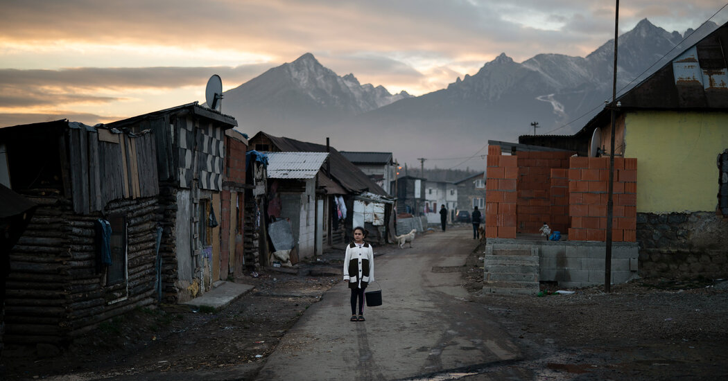
An op-ed in Nature calls for higher ethical standards in the usage and analysis of genetic information from the Romani, a marginalized group in Europe.For decades, geneticists have collected the blood of thousands of Roma people, a marginalized group living in Europe, and deposited their DNA in public databases. The ostensible purpose of some of these studies was to learn more about the history and genetics of the Roma people.Now, a group of scientists has argued this research, which has made the Roma the most intensely studied population in Europe over the past 30 years in forensic genetic journals, is rife with ethical issues and may harm the Romani people.For five years, a team of researchers in Germany and the United Kingdom pored over more than 450 papers that used the DNA of Roma people to understand how geneticists and other scholars obtained, interpreted and shared that genetic information. Their analysis, published Wednesday in an op-ed in the journal Nature, revealed many instances of clear misuse or questionable ethics.In 1981, when scientists in Hungary sampled the blood of Roma people incarcerated in Hungarian prisons, they classified prisoners as Romani based solely on their appearance, which the authors of the new paper argue is unscientific. In 1993, another group sampling Romani DNA concluded that there were three distinct ethnic groups in the country, drawing a line between “the genuine Hungarian ethnical groups” and “Jews” and “Gypsies” — a research premise the authors of the new paper argue was racist. In the 2000s, papers on the genetics of Roma people still referred to the group with the outdated term “Gypsy,” which is considered a slur, or with pejorative terms such as “inbred” or “consanguineous.”“This is an important contribution to the ongoing conversation about ethical issues in genetic research,” said Deborah Bolnick, an anthropological geneticist at the University of Connecticut who was not involved with research. Much of this conversation has taken place in North America and Australia, not Europe, she added.“The unethical practices described here are unfortunately very familiar and not a surprise,” Dr. Bolnick added.“It’s just horrifying,” said Ethel Brooks, a Romani scholar and chair of the department of women’s, gender and sexuality studies at Rutgers University in New Jersey. “But of course, it’s all things we’ve known and suspected.”The analysis spanned papers published between 1921 and 2021, most of which were published in the last 30 years. The earlier papers included “so many shocking surprises,” said Veronika Lipphardt, a science historian at the University of Freiburg, Germany, such as the samples taken from incarcerated Roma people and many instances of racist language.“Many didn’t believe us,” Dr. Lipphardt said, “because it was simply so hard to believe” that such practices were “ongoing.”In Europe, the Roma people have been oppressed for hundreds of years and still experience significant discrimination. During the Holocaust, Nazis collected blood samples from Roma people imprisoned in Auschwitz and murdered hundreds of thousands of Roma and Sinti people. In 2015, the Slovakian government defended its practice of segregating Roma children in schools, falsely citing “mild mental disabilities” tied to “high levels of inbreeding” in Romani communities.“The slip from genetics to eugenics is one that can happen quite easily,” said Dr. Brooks.Mihai Surdu, a visiting sociologist at the University of Freiburg and an author on the paper, conceptualized the project when he was writing a book on the Roma people. While searching for publications with the words “Roma” or “Gypsies” in the titles, Dr. Surdu found what seemed like an outsized number of studies on Roma DNA — nearly 20 papers.When Dr. Surdu wrote to Dr. Lipphardt in 2012 about this phenomenon, he was unsure if it was a fluke. But over the course of their study, the researchers uncovered more than 450 genetic papers with Roma subjects.With funding from the German Research Foundation, the two researchers expanded the team to include scholars from diverse disciplines, and also consulted with Anja Reuss, a spokesman for the Central Council of German Sinti and Roma, an advocacy group based in Heidelberg.They found that many studies did not adequately seek consent from the people they sampled, if they secured consent at all. Some studies cited oral consent, but “no one knows what the consent really was,” said Peter Pfaffelhuber, a mathematician at the University of Freiburg and an author on the paper.“In a way, our consent is never deemed necessary because we are not deemed able to give our consent,” Dr. Brooks said.A Roma settlement at the Lunik IX quarter of Kosice, Slovakia.Peter Lazar/Associated PressIn 2010, the main journal in the forensic genetics community, Forensic Science International: Genetics, adopted ethical requirements including informed consent. But although some papers published more recently state they were conducted with the written consent of all participants, they include DNA from earlier papers that were collected with murky procedures. “You cannot assume that consent from 30 years ago is still valid, that it can be extended forever for all possible uses,” Dr. Lipphardt said.One 2015 study pointing to Indian origins of the Roma people uploaded their amassed DNA data set to two public databases that law enforcement agencies across the world use for genetic references to solve crimes, a purpose to which the original participants likely did not consent.Even though much of this DNA was collected decades ago, its presence in public databases poses a present danger to modern communities. The 2015 study uploaded Roma DNA to the Y-STR Haplotype Reference Database, or YHRD, a searchable worldwide collection of anonymous Y-chromosome profiles that has become a crucial and contested tool helping police solve crimes. In YHRD, the national database for Bulgaria lists 52.7 percent of its data sets as “Romani” even though Roma people only make up 4.9 percent of the country’s population. If a minority population is disproportionately represented in a DNA database, this could create bias against “suspect populations,” some scholars argue. Some of these profiles came from population studies where the researchers thanked police forces for collecting the DNA.Marginalized groups like the Roma people are subject to increased surveillance and policing because of personal, institutional and cultural bias, said Matthias Wienroth, a social scientist and ethicist at Northumbria University in the United Kingdom and an author on the paper. “The continued use of genetic samples and data from marginalized communities further marginalizes these communities.”Part of the allure of Romani DNA to geneticists is the assumption that the group has been genetically isolated for hundreds of years. But the authors argue that many researchers rely on biased samples from isolated populations while intentionally excluding data from Romani people with mixed ancestry.“It was probably the most easy to get the blood samples from these places,” said Gudrun Rappold, a human geneticist at the University of Heidelberg and an author on the paper. “But then to draw conclusions with regard to these millions and millions of Roma people? This is just leading to the wrong conclusion.”Dr. Surdu added, “They’ve maintained this narrative contrary to evidence.”These highly sampled, isolated data sets, which often name specific villages, could also endanger the anonymity of individuals, especially those with rare genetic diseases, the authors argue.To ensure that Romani DNA is used ethically in the future, the researchers proposed four concrete changes. They looked to existing models for ethical DNA use for guidance, such as the Indigenous-led SING Consortium and the ethics code drafted by the San people of South Africa governing the use of their own genome, Dr. Lipphardt said.The authors recommend forming an international oversight board to investigate the DNA information from oppressed groups that is currently held in public databases, to benefit the Roma and other communities. They also call for more training on the ethics of collecting genetic data from marginalized communities, so that researchers can understand the societal implications of their work.The authors also ask journals to investigate or retract ethically fraught studies that include Romani DNA, citing Springer Nature’s recent retraction of six papers using DNA from Chinese minority ethnic groups.Finally, the researchers call for more conversations between scientists and participants, so that Roma people can learn about the benefits and risks of donating DNA.Most genetic studies of Roma DNA either seek to identify the origins of the Roma people in India or pinpoint their unique genetic mutations. But few studies aim to benefit the health and welfare of the Romani community, many of whom live in segregated settlements with less access to resources like housing and education. Dr. Lipphardt cautioned that even if genetic studies on Roma DNA led to treatments for rare diseases, there was no guarantee that those therapies would be made easily accessible to Roma people.The authors suggest scientists collaborate with and train Roma people to pursue research questions relevant to their communities. Only one paper of the 450 they examined mentioned community involvement, including training Roma doctors, nurses and midwives and conducting educational health screenings.But Dr. Surdu viewed this involvement as insufficient since the researchers did not let Romani concerns guide the research or engage the larger community, but only recruited Roma mediators to carry out a planned study. He added that he sees this access to health care and social services as a basic human right. “Informed consent for samples collected for genetic research should be fully voluntary,” Dr. Surdu said.These entrenched barriers to education are part of the reason there are fewer Romani scholars, Dr. Brooks noted. She said she felt excited about the prospect of Romani people having oversight of their DNA, both in the context of outside research and their own families.“To really open up space for these kinds of discussions within marginalized communities?” Dr. Brooks said. “It would be a scientific revolution.”

Read more →