Modern humans developed a more effective protection against oxidative stress

Very few proteins in the body have a change that makes them unique compared to the corresponding proteins in Neanderthals and apes. Researchers at the Max Planck Institute for Evolutionary Anthropology in Germany and Karolinska Institutet in Sweden have now studied one such protein, glutathione reductase, which protects against oxidative stress. They show that the risk for inflammatory bowel disease and vascular disease is increased several times in people carrying the Neanderthal variant.
What makes modern humans unique is a question that has eluded researchers for a long time. One way to approach this question is to study the proteins, or building blocks, in the body that have changes that are carried by almost all living people today and occurred after we separated from the ancestors we shared with Neanderthals about 500,000 years ago. There are around 100 proteins that have such a unique change. One of these proteins is glutathione reductase which is part of the body’s defense against oxidative stress.
The study, which is published in the journal Science Advances, examines the change in glutathione reductase in detail and was led by Hugo Zeberg at Karolinska Institutet and the Max Planck Institute for Evolutionary Anthropologyand Svante Pääbo at the Max Planck Institute. They show that the Neanderthal protein created more reactive oxygen radicals which are the cause of oxidative stress. It is the third protein change unique to present-day humans that has been studied so far.
The study also shows that the Neanderthal protein has passed over to present-day humans in low frequency when our ancestors mixed with them about 60,000 years ago. Today, it occurs mainly on the Indian subcontinent at an estimated frequency of 1 to 2 per cent of the population. The researchers found that people who carry the Neanderthal protein have a higher risk of developing vascular disease and inflammatory bowel disease, both diseases that are linked to oxidative stress.
“The risk increases we see are large; several times increased risk of inflammatory bowel disease and vascular disease,” says Hugo Zeberg.
The researchers can only speculate about why this particular change came to be one of the unique changes that almost all modern humans carry.
“Stopping oxidative stress is a bit like preventing something from rusting. Perhaps the fact that we are living longer has driven these changes,” says Svante Pääbo.
The research was supported by NOMIS Foundation, the Max Planck Society, the Jeansson Foundations and the Magnus Bergvall Foundation. The authors declare that they have no competing interests.
Story Source:
Materials provided by Karolinska Institutet. Note: Content may be edited for style and length.

Read more →

Timing of brain injury in pregnancy, birth may impact motor and language outcomes

A new UCSF study that mapped the neural connections of newborns with two different kinds of brain injuries found the maps looked very different — and were linked to significantly different developmental outcomes years later.
The study, published today in PLOS ONE and led by UCSF pediatrics, neurology and radiology researchers, used diffusion MRI to visualize the brain wiring of two sets of newborns: one set with congenital heart defects (CHD) and the other with hypoxic-ischemic encephalopathy (HIE) — otherwise known as birth asphyxia.
HIE babies suffer brain injury and oxygen deprivation within days to hours of being born, while CHD babies are steadily deprived of oxygen for longer — often months — in utero. Both groups are known to be at high risk for neurodevelopmental disabilities as they grow older, in areas ranging from motor skills to attention to behavioral issues.
“You have two sets of kids who, before or during birth, have a brain injury and then end up having some delayed or altered development and problems at school age,” said Patrick McQuillen, MD, UCSF professor of pediatrics and neurology, and the study’s corresponding author. “We wondered if the newborn brain, when faced with something challenging at different times, responds in the same way. What we found was the brains of these two sets of babies looked very, very different.”
Brain differences linked to outcomes
The researchers found that the distinct differences in brain wiring between the groups correlated with motor and language outcomes later. Specifically, they found the CHD newborns had worse language function at 12 to 18 months and worse cognitive, language and motor function at 30 months than the infants born with HIE, whose outcomes at both time points were in the normal range.
While about 20 percent of CHD babies scored below the normal language range at 12 to 18 months, the number grew to 50 percent below normal by 30 months. In addition, 37 percent of CHD children scored below normal in the cognitive domain and 25 percent scored below normal in the motor domain at 30 months. Language delays appeared to be driven by expressive, not receptive, language deficiencies, the study authors noted.
The main difference between the CHD and HIE brains that showed up in imaging was in an area called “global efficiency,” which measures how easy it is for a connection to be made from one area of the brain to the other. An efficient brain resembles a traffic system with an ideal balance of highways and local roads that take a driver where she needs to go quickly, McQuillen explained.
The researchers will continue to follow the babies in the study and were recently awarded a grant from The Children’s Heart Foundation to conduct additional brain imaging and testing of the subjects at school age. Their hope is that by understanding how the brain connections work and match to developmental outcomes, researchers will be able to link children with brain injuries to early intervention more quickly. Eventually, children may even have treatments tailored to their type of brain injury.
“We’re not quite there yet,” McQuillen said. “We are still in the stage of describing what’s different, and using those patterns to make predictions about outcomes. But I am hopeful about where this is headed.”
Additional authors on the study include Shabnam Peyvandi, MD, MAS, and Stephany Cox, PhD, from the UCSF Department of Pediatrics; Dawn Gano, MD, MAS, from the UCSF Departments of Pediatrics and Neurology; Duan Xu, PhD, and Olga Tymofiyeva, PhD, from the UCSF Department of Radiology. Alice Ramirez, MD, was a UCSF critical care fellow at the time this research was conducted.
This study was supported by NIH grants P01NS082330 and K23NS099422.

Read more →

Division of labor among genetic switches

Two X chromosomes are actually one too many. Female mammalian cells hence switch off one of them — but only when the cells start to specialize into tissues. A Berlin research team has now discovered how cells “count” their chromosomes and at the same time sense which stage of development they are in.
The cells of female mammals have a dosage problem, because they have twice as many X chromosomes as are needed in the body. Consequently, one of them is randomly selected and switched off already during early embryonic development. The Xist gene awakens and produces hundreds of RNA molecules, encasing one X chromosome and making it shrink into a small lump.
But how does the cell know to turn off one chromosome at a given time — but only if there are two of them? A research team led by Lise Meitner Group Leader Edda Schulz at the Max Planck Institute for Molecular Genetics (MPIMG) found the answer to this decades-old puzzle in mouse stem cells and published their results in the journal Molecular Cell.
A new genetic circuit
The Berlin scientists identified a genetic circuit that receives information about the developmental stage of the cell and passes it on to the Xist gene. “We found the regulatory region that senses whether the cell has left its stem cell state,” says Edda Schulz.
The newly discovered gene switch, dubbed “Xert,” is a member of the “enhancer” family of regulatory sequences. It is not sufficient to trigger the deactivation program on its own. Xist will only respond to the developmental cues if it is freely accessible and not blocked by other factors, which is the case when two X chromosomes are present in the cell. Only when both conditions are met, Xist can silence the “surplus” X chromosome.

Read more →

Immuno-CRISPR assay could help diagnose kidney transplant rejection early on

When a patient receives a kidney transplant, doctors carefully monitor them for signs of rejection in several ways, including biopsy. However, this procedure is invasive and can only detect issues at a late stage. Now, researchers reporting in ACS’ Analytical Chemistry have developed a CRISPR-based assay that can sensitively and non-invasively detect a biomarker of acute kidney rejection in urine. This could someday help diagnose rejection earlier and without a biopsy.
Kidney transplant recipients must take immunosuppressant drugs for the rest of their lives to help keep their immune systems from attacking the foreign organ. However, kidney rejection can still occur, particularly in the first few months after transplantation, which is known as acute rejection. Signs include increased serum creatinine levels and symptoms such as kidney pain and fever. Currently, the only way to definitively diagnose it is through biopsy, but this procedure can only detect problems at a relatively late stage. Being able to sensitively and non-invasively diagnose kidney rejection at an early stage would allow doctors to begin anti-rejection medication sooner. Researchers previously found that high levels of a cytokine protein called CXCL9 in the urine of kidney transplant patients was an early warning sign of rejection. But the current method for measuring CXCL9 (an enzyme-linked immunosorbent assay, or ELISA) doesn’t work very well in urine, limiting its sensitivity. So, Jonathan Dordick and colleagues wanted to develop a more sensitive technique for non-invasively diagnosing acute kidney rejection from urine.
The researchers based their detection method on CRISPR/Cas12a gene editing technology. In the presence of the CXCL9 protein, the CRISPR/Cas12a enzyme cuts a probe to produce a fluorescent signal. The researchers boosted the fluorescent signal by attaching a DNA barcode that aggregates a large number of CRISPR/Cas12a molecules, and is subsequently bound to an antibody that recognizes CXCL9. Importantly, unlike other CRISPR-based detection methods, PCR amplification is not required, which makes the method easier to adapt to a device that could be used in a doctor’s office or even a patient’s home. When tested on urine samples from 11 kidney transplant patients, the new system accurately measured CXCL9 levels, with values very similar to an ELISA. However, because the immuno-CRISPR system is about 7 times more sensitive than an ELISA, it might be able to detect kidney transplant rejection at a very early stage, the researchers say.
The authors acknowledge funding from the National Institutes of Health.
Story Source:
Materials provided by American Chemical Society. Note: Content may be edited for style and length.

Read more →

Living in outer space: Changes in blood flow volume may be at the heart of worsening eyesight

Colonizing Mars is no longer solely the work of science fiction but a potential future option for people who desire to live among the weightless. For headline grabbers like Jeff Bezos, NASA and Elon Musk, space colonization — or space settlement, a preferred term recommended by Bill Nye — is a big goal for the 21st century.
The long-term risks of living in space include bone loss, cosmic radiation and muscle weakness, just to name a few, so leaving gravity behind certainly has its obstacles. Some of these potential hurdles have already been studied extensively or are currently being investigated, but researchers at MUSC Health have found an important but underserved area of space to study: the brain and gravity’s effect on eyesight.
In a recent paper in JAMA Network Open, researchers look at Spaceflight-Associated Neuro-Ocular Syndrome (SANS) and compare brain scans before and after spaceflight.
The longer astronauts stay in space, the more they’ve reported blurry vision and eyesight problems when they return to earth, according to Mark Rosenberg, M.D., a neurology resident at MUSC Health and a researcher on the paper.
“It’s gotten to the point where astronauts actually carry extra pairs of glasses when they go into space,” said Rosenberg. “They know that their vision is going to be deteriorating up there, and they’ve even started calling them Space Anticipation Glasses. And, in fact, depending on how you define it, it affects about 70% of astronauts.”
With SANS, astronauts return to earth with altered visual acuity and struggle to distinguish between shapes at a distance. The globes of their eyes flatten, parts of their retinas show injury and their optic disks swell. Some astronauts recover from these changes in a few weeks, while others can take months or even years. There are also some who never fully recover.

Read more →

Single blood sample can detect women at risk of pre-eclampsia

A study of pregnant women’s blood RNA has found specific molecular profiles that identify women at risk of pre-eclampsia. These insights can identify complications before a woman experiences symptoms.
The study, published today in Nature, involved researchers from King’s and Guy’s and St Thomas’ NHS Foundation Trust in partnership with Mirvie. The study examines genetic material found in blood samples that can predict pregnancy complications such as pre-eclampsia.
Pre-eclampsia effects up to 1 in 12 pregnancies and is a significant cause of maternal morbidity. It is also a cause of a higher risk of cardiovascular disease. Most cases of pre-eclampsia are diagnosed when the mother experiences symptoms in the third trimester. This study could widen the window of detection and lead to quicker intervention.
Professor Rachel Tribe, Department of Women and Children’s Health, King’s College London, said: “I am delighted to be involved in this important collaborative effort to develop a new tool to predict pre-eclampsia.
“Using a cutting-edge sequencing approach, we were able to detect cell free RNA (cfRNA) in the blood of pregnant women. These provided a molecular signature that can be used to identify women at risk of pre-eclampsia.
She added: Excitingly, this requires only a single blood sample and has potential to identify women at risk much earlier in pregnancy so that they can be more closely monitored and treated by the clinicians involved.”
Researchers took 2500 blood samples from eight prospectively collected cohorts that included multiple ethnicities, nationalities, socioeconomic contexts and geographic locations. They then examined the anonymised cfRNA profiles — signals from the fetus and pregnant mother’s tissues — that reflect fetal development and healthy pregnancy progression. This provided a non-invasive window into maternal and fetal health.
In this study, researchers show the cfRNA signals which deviate from those of a healthy pregnancy. One single blood sample could reliably identify women at risk of developing preeclampsia months prior to the presentation of the disease. Using machine learning to analyse tens of thousands of RNA messages from the mother, baby and placenta, the Mirvie RNA platform can identify 75% of women who go on to develop preeclampsia. Researchers hope this test can be widened to investigate other pregnancy complications, such as preterm birth.
Professor Tribe added: “Because the study drew upon samples for a diverse group of women, including participants recruited across King’s Health Partners, the molecular signature is very reliable and has potential to outperform currently available tests.
‘We are now focused on ongoing clinical research to further validate these results and improve the understanding of other pregnancy complications. As a scientist, it was also extremely interesting to see that the molecular signature tells us something about mechanisms associated with health in pregnancy and complications including preeclampsia; such knowledge will aid development of treatment strategies in the future.”
The research was supported by the National Institute for Health Research (NIHR) through the NIHR Guy’s and St Thomas’ Biomedical Research Centre and an NIHR Doctoral Research Fellowship.
Story Source:
Materials provided by King’s College London. Note: Content may be edited for style and length.

Read more →

World’s fastest blade runner gets no competitive advantage from prostheses, study shows

Amputee sprinters using running prostheses, or blades, have no clear competitive advantage at the 400-meter distance compared to sprinters with biological legs and, in fact, appear to have a significant disadvantage at the start, according to University of Colorado Boulder research published today.
The long-awaited study, published in Royal Society Open Science, provides the most comprehensive set of data ever collected from elite runners with bilateral leg amputations, including the world’s fastest 400-meter sprinter, Blake Leeper. Leeper was ruled ineligible to compete in the Tokyo Olympics last year due to having an assumed advantage.
When comparing performance data from Leeper, South African “blade runner” Oscar Pistorius and up to six other bilateral amputee sprinters with those of the best non-amputee sprinters in the world across five performance metrics, the research found no advantage.
“A lot of assumptions have been made about running prostheses and performance with no data to support them,” said senior author Alena Grabowski, associate professor of integrative physiology at CU Boulder. “With this study, we show that the use of running prostheses provides no competitive advantage over 400 meters compared to biological legs.”
For the study, Leeper — who was born without legs — visited Grabowski’s Applied Biomechanics Lab for a series of tests in 2018. That summer, he had sprinted the 400-meter in 44.42 seconds, breaking the record of Pistorius, the first below-the-knee amputee to compete against able-bodied runners at the Olympic games.
Starting with Pistorius
After Pistorius’ history-making run in 2012, the International Association of Athletics Federations (now World Athletics) ruled that, going forward, athletes using “mechanical aids” must provide evidence that their blades do not give them a competitive edge.

Read more →

How LPS prevents or promotes development of asthma and allergic disease by airborne allergens

Immunologists have long been baffled by LPS, the bacterial lipopolysaccharide that helps form the cell walls of Gram-negative bacteria.
Many experiments show that exposure to LPS during exposure to environmental allergens protects against developing asthma or allergic disease. Yet other numerous experiments show that the presence of LPS during exposure to environmental allergens does the opposite — it promotes the development of asthma and other allergic disease.
Now Beatriz León, Ph.D., and fellow University of Alabama at Birmingham researchers have cut this Gordian knot in a study published in Cell Reports. They detail a series of mechanistic steps that reveal a surprising answer — the key to LPS’ promoting or preventing the allergic reaction lies in the allergen itself.
Greater understanding of the mechanisms underlying sensitization to allergic disease can offer new strategies to control allergic airway disease, especially asthma. A global epidemic of asthma, including a sharp increase in developed countries since the 1960s, affects about 300 million children and adults worldwide.
León and her UAB colleagues unraveled a complex trail for the antagonistic mechanisms of LPS to promote or prevent allergic disease through activation or suppression of T helper-2 immune cells.
Here are their findings, which also define specific roles for immune cells called classical monocytes and non-classical monocytes.

Read more →

India’s Rising Omicron Wave Brings a Grim Sense of Déjà Vu

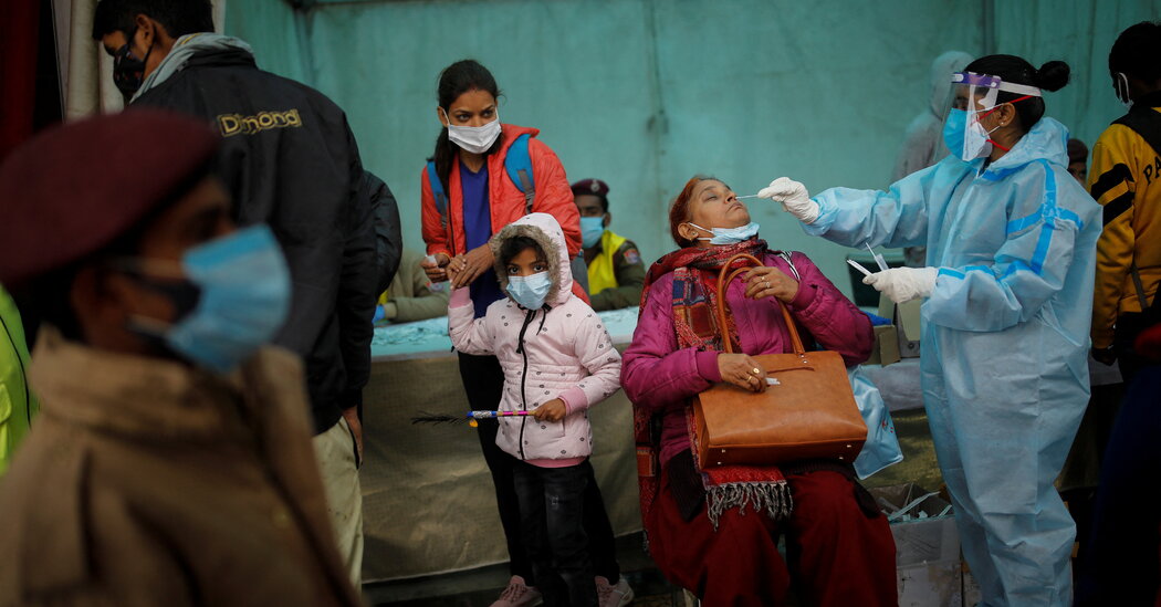
Just months after Delta fueled hospital failures and funeral pyres, India’s leaders again offer a mixed message: Their political rallies are packed even as they order curfews and work closures.NEW DELHI — When the Omicron coronavirus variant spread through India late in December, Prime Minister Narendra Modi urged the nation to be vigilant and follow medical guidelines. Arvind Kejriwal, the chief minister of the capital region of Delhi, swiftly introduced night curfews, shut down movie theaters, and slashed restaurants and public transport to half capacity.Then, both men hit the campaign trail, often appearing without masks in packed rallies of thousands.“When it is our bread and butter at stake, they force restrictions and lockdowns,” said Ajay Tiwari, a 41-year-old taxi driver in New Delhi. “There are much bigger crowds at political rallies, but they don’t impose any lockdown in those areas. It really pains us deep in the heart.”As Omicron fuels a rapid spread of new infections through India’s major urban hubs, the country’s pandemic fatigue has been intensified by a sense of déjà vu and the frustration of mixed signals.A temporary coronavirus care center in New Delhi on Wednesday. Money Sharma/Agence France-Presse — Getty ImagesIt has been just a few months since the deadly Delta variant ravaged the country, when government leaders vastly underestimated its threat and publicly flouted their own advice. The memories of overwhelmed hospitals and funeral pyres working around the clock are still all too fresh here.The metropolis of Mumbai on Wednesday reported more than 15,000 new infections in 24 hours — the highest daily caseload since the pandemic began, beating the city’s previous record of about 11,000 cases during the second wave in the spring. In New Delhi, the number of daily infections increased by nearly 100 percent overnight.The sheer size of India’s population, at 1.4 billion, has always kept experts wary about the prospects of a new coronavirus variant. In few places around world was the toll of Delta as stark as in India. The country’s official figures show about half a million pandemic deaths — a number that experts say vastly undercounts the real toll.A temporary coronavirus care facility was set up at the Chennai Trade Center in Chennai. Scientists say any optimism about Omicron is premature simply because of how many people the variant could infect.Idrees Mohammed/EPA, via ShutterstockOmicron’s high transmissibility is such that cases are multiplying at a dangerously rapid pace, and it appears to be ignoring India’s main line of defense: a vaccination drive that has covered about half of the population. Initial studies show that the Oxford-AstraZeneca vaccine, a locally manufactured version of which has been used for about 90 percent’s of India vaccinations, does not protect against Omicron infections, though it appears to help reduce the severity of the illness.Sitabhra Sinha, a professor of physics and computational biology at the Institute of Mathematical Sciences in Chennai, said his research into the reproduction rate of the virus — an indicator of how fast it is spreading that is called the “R value” — in major cities like Delhi and Mumbai shows “insanely high” numbers for cities that had built decent immunity. Both had a large number of infections in the spring, and a majority of their adult populations have been vaccinated.“Given this high R value, one is looking at incredibly large numbers unless something is done to stop the spread,” he said.A Bharatiya Janata Party (BJP) rally in Ferozepur on Wednesday. Omicron is spreading in India at a time of high public activity — busy holiday travel, and large election rallies across several states that are going to the polls in the coming months.Narinder Nanu/Agence France-Presse — Getty ImagesBut officials appear to be latching onto the optimism of the early indications from places like South Africa, where a fast spread of the variant did not cause devastating damage, rather than drawing lessons from the botched response to the Delta wave in the spring that ravaged India.Dr. Anand Krishnan, a professor of epidemiology at the All India Institute of Medical Sciences in New Delhi, said India’s messaging of the new variant as “a mild illness” has led to complacency.“The health system has stopped being complacent. But the population is complacent. People are not wearing masks or changing their behavior,” Dr. Krishnan said. “They think it is a mild illness, and whatever restrictions are being imposed are seen more as a nuisance than necessary.”Scientists say any optimism about Omicron is premature simply because of how many people the variant could infect.“Even if it is a microscopic percentage who require hospitalization,” Dr. Sinha said, “the fact is that the total population we’re talking about is huge.”A vaccination center in Bangalore. India’s vaccination drive has covered about half of the population.Manjunath Kiran/Agence France-Presse — Getty ImagesAlthough the percentage of newly infected people turning to hospitals has been increasing in recent days, data from India’s worst-hit cities — Mumbai, Delhi and Kolkata — showed that only a small number of Covid-designated beds were occupied so far. Data compiled by the Observer Research Foundation showed that about three percent of the known active cases in Delhi and about 12 percent in Mumbai have required hospitalization.Dr. J. A. Jayalal, until recently the president of The Indian Medical Association, said what worried him was not hospital beds or oxygen running out — capacity that Indian officials have been trying to expand after the deadly shortfalls during the Delta wave — but that the health system might face an acute shortfall of health workers.The Coronavirus Pandemic: Key Things to KnowCard 1 of 5The global surge.

Read more →

Antibiotic-resistant superbug evolved on hedgehogs

SharecloseShare pageCopy linkAbout sharingImage source, Pia B HansenAn antibiotic-resistant superbug – a type of MRSA – evolved naturally as a result of a battle between a fungus and bacteria on the skin of wild hedgehogs. The evidently “hedgehog-derived” bacteria developed in nature long before the antibiotics we are familiar with were discovered. An international research team found that a skin fungus common in hedgehogs naturally produces antibiotics. Bacteria on the animals’ skin developed antibiotic resistance in response.The researchers say their findings, published in the journal Nature, shows how natural biological processes – not antibiotic use – drove the emergence of this particular superbug about 200 years ago. Coronavirus: This is not the last pandemicWarning of antibiotic-resistant infection pandemicMore outbreaks if we keep exploiting wildlife – UNThe specific bacterium, called mecC-MRSA, was first found in dairy cattle and it had been assumed that the use of antibiotics on dairy farms had caused it to develop its resistance. This is, though, just one, relatively rare example of antibiotic resistance arising naturally. And the discovery “represents a tiny fraction of the risks compared to overuse of antibiotics in a human medical context”, one of the lead researchers, Prof Mark Holmes from the University of Cambridge, told BBC News. MecC-MRSA causes about one in 200 human MRSA infections. The overuse of antibiotics, both in humans and farm animals, continues to drive the emergence of other, resistant, disease-causing strains. The study itself has solved a long-standing mystery about the source of this particular type of MRSA, which veterinary scientists from the University of Cambridge discovered a decade ago. “We tried to work out how much of a problem it was – so we looked in wildlife and in farm animals and found that it was clearly very widely distributed in nature,” explained Prof Holmes. “When we looked at hedgehogs in particular, about half of the animals we sampled had this type of MRSA.” Teaming up with biologists, wildlife researchers and with hedgehog rescue centres all around Europe, the scientists then focused their investigations on hedgehogs “We wanted to know,” Prof Holmes explained, “what’s so special about a hedgehog that means there’s a lot of these resistant bacteria?”Research colleagues at the Statens Serum Institut in Copenhagen, Denmark, studied more than 1,000 samples of bacteria taken from wildlife around Europe. They built a genetic code-based timeline , which revealed that the resistant strain had emerged in European hedgehogs in the early 1800s – long before the clinical use of antibiotics. Image source, Claire L Raisen”The fungus growing on the hedgehog was releasing penicillins,” explained Prof Holmes. “The bacteria needed to be resistant because, if you want to live on the hedgehog – where there’s a fungus, you have to be resistant to the antibiotics it’s producing.”Prof Holmes added that the risk to human health from this type of MRSA was “very tiny – almost insignificant”.Hedgehog expert and co-author of the study, Dr Sophie Lund Rasmussen from the University of Oxford explained: “Hedgehogs have carried this bacterium for at least 200 years without causing any major infections in humans. “So the advice is [still] to appreciate and support the hedgehogs entering your garden and to maintain a good hand hygiene when feeding and possibly handling hedgehogs. Both for the sake of the hedgehogs and ourselves.”Hedgehog penicillinThe evolutionary “arms race” on the hedgehog’s skin is a natural example of what Alexander Fleming saw on his petri dish when he discovered penicillin in 1928 – he noticed that no bacteria could survive close to a mould – a fungus – that had contaminated the gel in his dish.Antibiotic resistance: A silent pandemicAntibiotic resistance is one of the biggest threats to global health, food security, and development todayAntibiotic resistance occurs naturally, but misuse of antibiotics in humans and animals is accelerating the processA growing number of infections – such as pneumonia, tuberculosis, gonorrhoea, and salmonellosis – are becoming harder to treat as the antibiotics used to treat them become less effective.Source: World Health OrganizationThe study is part of an ongoing effort by these scientists to understand where “reservoirs” of antibiotic resistant bacteria are, how they emerge and what practices and sources pose the greatest risk to people. “The reassuring thing is to be reasonably confident that it isn’t the overuse of antibiotics in dairy farms that leads to this type of MRSA being present in people,” said Prof Holmes. “But that does not mean that we should relax [the rules] on the use of antibiotics.”It isn’t just hedgehogs that harbour antibiotic-resistant bacteria – all wildlife carries many different types of bacteria, as well as parasites, fungi and viruses,” he added.”Wild animals, livestock and humans are all interconnected: we all share one ecosystem. It isn’t possible to understand the evolution of antibiotic resistance unless you look at the whole system.”Follow Victoria on Twitter

Read more →