To destroy cancer cells, team 'travels back in time'

When an individual suffers from cancer, the process of programmed cell death called apoptosis does not occur normally, permitting abnormal cells to thrive.
In a new study, Clay Clark, chair and professor of biology at The University of Texas at Arlington, explores the ancestry of a class of enzymes responsible for killing cells. These enzymes, known as effector caspases, provide insight into the process of cell death and how it can be manipulated in disease states.
“If drug developers want to target a cancer cell, they could activate the caspases to perform their normal function of killing cells,” Clark said. “But we first need to isolate the caspases of cancer cells and not alter their activity in normal cells. The question is, how can we target the activity of the tumor cell without disturbing its healthy neighbors.”
The study, “Evolution of the folding landscape of effector caspases,” appears in the Journal of Biological Chemistry. In it, Clark and co-author Suman Shrestha, UTA alumnus and postdoctoral fellow at Princess Margaret Cancer Center, look to the past for answers.
Cells have had effector caspases since organisms called eukaryotes emerged more than a billion years ago. Over time, the caspases evolved from one to 12 proteins that serve various functions during cell growth and division. To understand their evolution, Clark’s laboratory used a computational method called ancestral state reconstruction to recreate a 650-million-year-old protein sequence of an ascendant of caspases 3, 6 and 7.
Caspases are the central components of apoptosis. In cancerous states, cells turn off caspases and circumvent the cell death process. A goal of Clark’s research is to understand how effector caspases work in healthy conditions. Once the proteins’ normal function is understood, Clark’s team could discover methods to activate them in disease states to destroy abnormal cells while preserving healthy ones.
By examining the ancestor’s characteristics and tracing the protein’s evolution into multiple members, Clark’s team discovered commonalities and differences among caspases 3, 6 and 7 that could allow for the isolated activation of proteins in tumor cells without disrupting the activity of healthy cells.
Morteza Khaledi, dean of the College of Science, said the results of the study will advance humanity’s battle to mitigate the effects of cancer.
“By employing the innovative methods of ancestral state reconstruction, Dr. Clark’s team has discovered vital information about the essential building blocks for healthy human bodies,” Khaledi said. “The knowledge uncovered in this study provides another weapon in our fight against cancer.”
Story Source:
Materials provided by University of Texas at Arlington. Original written by Linsey Retcofsky. Note: Content may be edited for style and length.

Read more →

Chemical commonly found in consumer products may disrupt a hormone needed for healthy pregnancy

Exposure to phthalates — a group of chemicals found in everything from plastics to personal care products to electronics — may disrupt an important hormone needed to sustain a healthy pregnancy, according to a Rutgers study.
The study, which appears in Environment International, was among the first to examine the impact that phthalates, added to plastics to increase flexibility, have on the placental corticotropin releasing hormone (pCRH) that is produced by the placenta and increases throughout the course of pregnancy. The hormone plays an important role in promoting the onset of labor, but when levels are high or rise rapidly earlier in pregnancy, it may contribute to preterm birth and fetal growth problems as well as high blood pressure, diabetes, and postpartum depression.
“We are all exposed to phthalates in our environment through the products we use and the foods we eat,” says Emily S. Barrett, an associate professor at the Rutgers School of Public Health and member of the Environmental and Occupational Health Sciences Institute. “Our findings show that these chemicals may alter the production of essential placental hormones, which has important implications for the course of pregnancy as well as subsequent child health and development.”
The researchers analyzed data from 1,018 low-risk pregnant women carrying single fetuses at two time points, mid- and late pregnancy.
They found that the presence of various phthalates was associated with higher pCRH hormone levels in mid-pregnancy, but lower pCRH later in pregnancy. These levels were strongest in women who developed pregnancy complications like gestational diabetes and high blood pressure, suggesting that women who develop complications may be particularly vulnerable to this hormonal disruption. This is among the first and largest study so far to examine how these chemicals and the connection with pCRH disrupt the function of the endocrine system, which is especially delicate during pregnancy.
“Associations between phthalates and pCRH among women with pregnancy complications grew stronger across the course of pregnancy. We know very little about how women with pregnancy complications are affected by environmental exposures. This study sets the stage for future research in that area,” Barrett said.
pCRH produced by the placenta is identical in structure to corticotropin-releasing hormone (CRH) produced by the brain when responding to stress. While this study did not find that women who had experienced childhood traumas were any more vulnerable to the hormone-disrupting effects of phthalates, earlier research found that pCRH levels were higher in women who have experienced childhood trauma suggesting that traumatic events may impact pregnancy decades later.
Story Source:
Materials provided by Rutgers University. Original written by Michelle Edelstein. Note: Content may be edited for style and length.

Read more →

The unexpected benefits of fat in type 2 diabetes

With nearly 10% of the world’s population affected, type 2 diabetes is a major public health issue. An excessively sedentary lifestyle and a too-caloric diet encourage the development of this metabolic disease by altering the functioning of pancreatic cells and making blood sugar regulation less effective. However, fat, which is often cited as the ideal culprit, could be rehabilitated. Indeed, fat does not necessarily aggravate the disease and could even play a protective role: by studying insulin-producing pancreatic beta cells, scientists from the University of Geneva (UNIGE), Switzerland, have shown that these cells suffered less from excess sugar when they had previously been exposed to fat. By investigating the cellular mechanisms at work, the researchers discovered how a cycle of fat storage and mobilisation allows cells to adapt to excess sugar. These results, published in the journal Diabetologia, highlight an unexpected biological mechanism that could be used as a lever to delay the onset of type 2 diabetes.
Type 2 diabetes results from a dysfunction of pancreatic beta cells, which are responsible for insulin secretion. This impairs the regulation of blood sugar levels and can lead to serious heart, eye and kidney complications. In the 1970s, fat was singled out and the concept of lipotoxicity emerged: exposure of beta cells to fat would cause their deterioration. More recently, excess sugar has also been blamed for damaging beta cells and promoting the development of type 2 diabetes. However, while the culpability of sugar is no longer in doubt, the role of fat in beta cell dysfunction remains ambiguous. What are the cellular mechanisms involved? “To answer this key question, we studied how human and murine beta cells adapt to an excess of sugar and/or fat,” explains Pierre Maechler, a Professor in the Department of Cell Physiology and Metabolism and in the Diabetes Centre of the UNIGE Faculty of Medicine, who led this work.
When fat lends a hand to beta cells
In order to differentiate the effect of fat from that of sugar, the scientists exposed beta cells to an excess of sugar, of fat, and then to a combination of the two. The toxicity of sugar was first confirmed: beta cells exposed to high sugar levels secreted much less insulin than normal. “When cells are exposed to both too much sugar and too much fat, they store the fat in the form of droplets in anticipation of less prosperous times,” explains Lucie Oberhauser, a researcher in the Department of Cell Physiology and Metabolism at the UNIGE Faculty of Medicine, and first author of this work. “Surprisingly, we have shown that this stock of fat, instead of worsening the situation, allows insulin secretion to be restored to near-normal levels. The adaptation of beta cells to certain fats would thus contribute to maintain normal blood sugar levels.”
The essential use of fat
By further analysing the cellular changes at stake, the research team realised that fat droplets were not static reserves, but were the site of a dynamic cycle of storage and mobilisation. And thanks to these released fat molecules, beta cells adapt to the excess sugar and maintain a near-normal insulin secretion. “This release of fat is not really a problem as long as the body uses it as a source of energy,” adds Pierre Maechler. “To avoid developing diabetes, it is important to give this beneficial cycle a chance to be active, for example by maintaining regular physical activity.” Scientists are now trying to determine the mechanism by which this released fat stimulates insulin secretion, in the hope of discovering a way to delay the onset of diabetes.
Story Source:
Materials provided by Université de Genève. Note: Content may be edited for style and length.

Read more →

Images of enzyme provide insights into cause of hereditary neurological disease

WEHI researchers have produced the first molecular images of an enzyme that controls proteins to signal and communicate with each other in human cells. The discovery could help to solve the mystery cause of a rare group of hereditary neurodegenerative diseases linked to deregulation of this enzyme.
In the study, published in Molecular Cell, Dr Thomas Cotton, Dr Bernhard Lechtenberg and colleagues at WEHI solved the first three-dimensional (3D) structure of an enzyme called RNF216. The team captured molecular ‘snapshots’ of RNF216 as it assembled chains of the small protein ubiquitin, which tags the target proteins to modify their behaviour in the cell. They also showed how RNF216 dictates the type of ubiquitin chain that is formed — the first time that this has been explained.
Mutations in RNF216 have been linked to Gordon-Holmes Syndrome, a very rare neurodegenerative disorder that results in reproductive problems, movement disorders, and progressive cognitive decline and dementia.
At a glance
• Scientists from WEHI have captured the first molecular images of RNF216, an enzyme that adds a specific type of ubiquitin chain to proteins, instructing those proteins how to behave.
• They also found that RNF216 can be ‘supercharged’, greatly increasing the production of and specificity towards K63-linked ubiquitin chains, which are involved in protein-protein signalling.

Read more →

New findings may contribute to better diagnosis and treatment of liver cancer

In a new study, researchers at Karolinska Institutet have identified the presence of a specific connection between a protein and an lncRNA molecule in liver cancer. By increasing the presence of the lncRNA molecule, the fat depots of the tumor cell decrease, which causes the division of tumor cells to cease, and they eventually die. The study, published in the journal Gut, contributes to increased knowledge that can add to a better diagnosis and future cancer treatments.
Our genome gives our cells instructions that determine each cell type’s highly specialized function. The information is sent out using two different types of RNA molecules: coding RNA that converts DNA into proteins and non-coding RNA that do not produce proteins.
Because non-coding RNA molecules do not produce proteins, they have not been the main focus of research in the past, even though they amount to approximately 97 per cent of the RNA in our body. However, certain proteins, called RNA-binding proteins, have been shown to play a crucial role in cancer because of their ability to affect several different properties of RNA molecules.
“With the help of tissue material donated by patients with liver cancer, we have been able to map both the coding and non-coding part of our genome to identify which RNA-binding proteins have a high presence in liver cancer cells,” says the study’s senior author Claudia Kutter, researcher at the Department of Microbiology, Tumor and Cell Biology, Karolinska Institutet. “We found that many of these proteins interacted with a long type of non-coding RNA molecules, so-called lncRNA.”
The research team conducted a more detailed study of a specific pairing of a RNA-binding protein (CCT3) and an lncRNA molecule (LINC00326). Using advanced CRISPR technology, they were able to both reduce and increase the amount of the protein and the lncRNA to see how it affected the cancer cells. When the lncRNA was increased, the fat depots of the tumor cell decreased, the cell division ceased and many of the cancer cells died. Following the laboratory studies, the results were also verified in vivo.
Many more combinations to investigate
The researchers’ discovery provides an insight into the interaction between RNA-binding proteins and lncRNA molecules, and contributes to a better scientific understanding of their role in tumors.
“The activities of the CCT3-LINC00326 pair can already be used in liver cancer diagnosis and prognosis,” says the study’s first author Jonas Nørskov Søndergaard, researcher in Kutter’s research group. “However, the knowledge of this particular pairing is just the beginning and there are many more combinations of RNA-binding proteins and lncRNA molecules that we will further investigate. In the long run, these findings can help to contribute to new and effective treatments such as RNA-based treatments that target only the diseased cells, with the possibility of reducing side effects.”
The study was supported by grants from the Knut and Alice Wallenberg Foundation, the Ruth and Richard Julin foundation, a SFO-SciLifeLab fellowship, the Swedish Research Council, the Lillian Sagen and Curt Ericsson Research Foundation, Gösta Miltons Foundation, the Chinese Scholarship Council, KI-KID funding, SNIC projects, Nilsson-Ehle Endowments, Barts and London Charity, Cancer Research UK, AIRC Fellowship for Abroad, Tornspiran Foundation and the Swedish Society of Medicine.
Story Source:
Materials provided by Karolinska Institutet. Note: Content may be edited for style and length.

Read more →

Tomato concentrate could help reduce chronic intestinal inflammation associated with HIV

New UCLA-led research in mice suggests that adding a certain type of tomato concentrate to the diet can reduce the intestinal inflammation that is associated with HIV. Left untreated, intestinal inflammation can accelerate arterial disease, which in turn can lead to heart attack and stroke.
The findings provide clues to how the altered intestinal tract affects disease-causing inflammation in people with chronic HIV infection, suggesting that targeting the inflamed intestinal wall may be a novel way to prevent the systemic inflammation that persists even when antiviral therapy is effective in controlling a person’s HIV.
The study is published in the peer-reviewed journal PLOS Pathogens.
“Inflammation is an important process that protects the body from invading infections and toxins,” said Dr. Theodoros Kelesidis, the paper’s senior author and an associate professor of medicine in the division of infectious diseases at the David Geffen School of Medicine at UCLA. “But in individuals who are successfully treated for HIV to the point that their viral load is no longer detectable, the continuing low-grade inflammation in the cells of the intestine contributes to an increased risk of heart attack or stroke.”
People with HIV have been found to have a condition called “leaky gut,” in which products in the gut bacteria, such as lipopolysaccharides, move to other parts of the body through the bloodstream. Those products promote systemic inflammation and can accelerate coronary disease, Kelesidis said.
The researchers worked with mice that had been infected with HIV and whose immune systems had been altered to mimic those of humans. The mice were fed a diet containing the tomato concentrate Tg6F, while the rest were fed a normal diet for mice — low in fat, cholesterol and calories.

Read more →

Possibility of vaccine to prevent skin cancer

Research by the Oregon State University College of Pharmacy suggests that a vaccine stimulating production of a protein critical to the skin’s antioxidant network could help people bolster their defenses against skin cancer.
Ultraviolet radiation from the sun leads to oxidative stress, which increases the risk of skin cancers such as melanoma, explains Arup Indra, professor of pharmaceutical sciences at OSU and the study’s leader.
A messenger RNA vaccine, like the Moderna and Pfizer vaccines for COVID-19, that promoted production of the protein, TR1, in skin cells could mitigate the risk of UV-induced cancers and other skin problems, he said.
Findings of the research, in which Arup and collaborators used a mouse model to probe TR1’s role in skin cells’ health and stability, were published in the Journal of Investigative Dermatology.
Skin cancer is the most common cancer in the United States, according to the Centers for Disease Control and Prevention. Melanoma, the most lethal type of skin cancer, is a form in which malignant cells form in skin cells known as melanocytes; melanocytes produce the pigment melanin, which determines skin color.
Most cases of skin cancer, the CDC says, are linked to UV radiation exposure. People become tan from exposure to the sun or tanning beds because producing melanin is the body’s way of trying to protect the skin from burning.

Read more →

Decoding inner language to treat speech disorders

When human beings speak, different areas of their brain must be activated. However, the function of these regions can be seriously impaired after damage to the nervous system. For example, amyotrophic lateral sclerosis (or Charcot’s disease) can completely paralyze the muscles used to speak. In other cases, following a stroke for example, areas of the brain responsible for language can be affected: this is called aphasia. However, in many of those cases, the ability of patients to imagine words and sentences remains partly functional.
Decoding our internal speech is therefore of great interest to neuroscience researchers. But the task is far from easy, as Timothée Proix, scientist in the Department of Basic Neuroscience at the UNIGE Faculty of Medicine, explains: “Several studies have been conducted on the decoding of spoken language, but much less on the decoding of imagined speech. This is because, in the latter case, the associated neural signals are weak and variable compared to explicit speech. They are therefore difficult to decode by learning algorithms.” That is, through computer programmes.
A well-hidden speech
When a person speaks aloud, he or she produces sounds that are emitted at certain precise moments. Researchers can thus relate these tangible elements to the brain regions involved. In the case of imagined speech, the process is much less easy. Scientists have no obvious information on the sequencing and tempo of the words or sentences formulated internally by the individual. The areas recruited in the brain are also less numerous and less active.
In order to perceive the neural signals of this very particular type of speech, the UNIGE team used a panel of thirteen hospitalized patients, in collaboration with two American hospitals. They collected data through electrodes implanted directly into patients’ brains in order to assess their epileptic disorders. “We asked these people to say words and then to imagine them. Each time, we reviewed several frequency bands of brain activity known to be involved in language,” explains Anne-Lise Giraud, a professor in the Department of Basic Neuroscience at the UNIGE Faculty of Medicine, and newly appointed director of the Institut de l’Audition in Paris.
Tapping into the right frequency
The researchers observed several types of frequencies produced by different brain areas when these patients spoke, either orally or internally. “First of all, the oscillations called theta (4-8Hz), which correspond to the average rhythm of syllable elocution. Then the gamma frequencies (25-35Hz), observed in the areas of the brain where speech sounds are formed. Thirdly, beta waves (12-18Hz) related to the cognitively more efficient regions solicited, for example to anticipate and predict the evolution of a conversation. Finally, the high frequencies (80-150Hz) that are observed when a person speaks out” explains Pierre Mégevand, assistant professor in the Department of Clinical Neurosciences at the Faculty of Medicine of the UNIGE and associate physician at the HUG.
Thanks to these observations, the scientists were able to show that the low frequencies and the coupling between certain frequencies (beta and gamma in particular) contain essential information for the decoding of imagined speech. Their research also reveals that the temporal cortex is an important area for the eventual decoding of internal speech. Located in the left lateral part of the brain, this specific cerebral region is involved in the processing of information related to hearing and memory, but it also houses a part of Wernicke’s area, responsible for the perception of words and language symbols.
These results are a major advance in the reconstruction of speech from neural activity. “But we are still a long way from being able to decode imagined language,” concludes the research team.
Story Source:
Materials provided by Université de Genève. Note: Content may be edited for style and length.

Read more →

How Exercise May Tame Our Anxiety

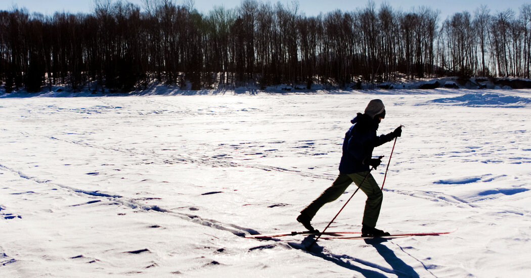
Cross-country ski racers were less likely to develop anxiety disorders. The good news is less intense aerobic activities may provide similar benefits.To better cope with all the dispiriting news this winter about rising Covid-19 cases and so much else, you might want to get out and play in the snow, according to a new report. The large-scale study of almost 200,000 cross-country skiers found that being physically active halves the risk of developing clinical anxiety over time. The study, from Sweden, focused on skiing, but the researchers said almost any kind of aerobic activity likely helps protect us against excessive worry and dread, a cheering thought as we face yet another grim pandemic season.Science already offers plenty of encouraging evidence that exercise can lift our moods. Experiments show that when people (and lab animals) start working out, they typically grow calmer, more resilient, happier and less apt to feel unduly sad, nervous or angry than before. Epidemiological studies, which often focus on the links between one type of activity or behavior and various aspects of health or longevity, likewise find that more exercise is linked with substantially lower chances of developing severe depression; conversely, being sedentary increases the risk for depression. A remarkable neurological study from 2013 even found that exercise leads to reductions in twitchy, rodent anxiety, by prompting an increase in the production of specialized neurons that release a chemical that soothes over-activity in other parts of the brain.But most of these studies were small, short term or mainly relevant to mice, leaving open many questions about what kinds of exercise might help our mental health, how long mood enhancements might potentially last, whether men and women benefit equally and whether it is possible to work out too much and perhaps increase your likelihood of feeling emotionally worse off.So, for the new study, which was published in Frontiers in Psychiatry, exercise scientists at Lund University in Sweden and other institutions decided it would be worthwhile to look into the long-term mental health of the thousands upon thousands of men and women who have raced Sweden’s famous Vasaloppet cross-country skiing event over the years.The Vasaloppet, which celebrates its centenary this winter, is the largest series of cross-country ski races in the world, with crowds of racers annually lining up in the woods of central Sweden to whoosh, glide and pant through races ranging in length from 30 kilometers, or almost 19 miles, to the showcase distance of 90K, about 56 miles. Because this kind of endurance event requires abundant health, stamina and training, researchers previously have used data about Vasaloppet racers to study how exercise influences heart health, cancer risks and longevity.“We use participation in a Vasaloppet as a proxy for a physically active and healthy lifestyle,” said Tomas Deierborg, the director of the experimental medicine department at Lund University and senior author of the new study, who has twice completed the 90K race.To start, he and his colleagues gathered finishing times and other information for 197,685 Swedish men and women who participated in one of the races between 1989 and 2010. They then crosschecked this information with data from a Swedish national registry of patients, looking for diagnoses of clinical anxiety disorder among the racers in the following 10 to 20 years. For comparison, they also checked anxiety diagnoses during the same time period for 197,684 of their randomly selected fellow citizens who had not participated in the race and were generally considered relatively inactive.The skiers, the researchers found, proved to be considerably calmer over the decades after their race than the other Swedes, with more than 50 percent less risk of developing clinical anxiety. These good spirits tended to prevail among male and female skiers of almost any age — except, interestingly, the fastest female racers. The top female finishers from each year tended to be more likely afterward to develop anxiety disorders than other racers, although their risk overall remained lower than for women of the same age in the control group.These results indicate “the link between exercise and reduced anxiety is strong,” said Dr. Lena Brundin, a lead investigator of neurodegenerative diseases at the Van Andel Research Institute in Grand Rapids, Minn., who was another author on the study.And helpfully, you probably don’t need to cross-country ski for long distances in the snowy woods of Sweden to reap the rewards, Dr. Deierborg said. Earlier studies of exercise and mood suggest that following the World Health Organization’s recommendations of about 30 minutes of brisk walking or similar activities most days “has good effects on your mental health,” he said, and these benefits appear to apply to a “broader population” than just Swedes.Still, it may be worthwhile to monitor your psychological response to intense training and competition, especially if you are a competitive woman, he said. The finding that the fastest women tended to develop anxiety more often than other racers surprised the researchers, he said, and suggests perhaps performance anxiety or other issues could be initiated or exacerbated in some people by racing.“It is not necessary to complete extreme exercise to achieve the beneficial effects on anxiety,” Dr. Brundin said.The findings have limitations, though. They cannot prove exercise causes people to enjoy better moods, only that highly active people tend to be less anxious than their more sedentary peers. The study also does not explain how skiing might reduce anxiety levels. The researchers suspect physical activity changes levels of brain chemicals related to mood, such as dopamine and serotonin, and reduces inflammation throughout the body and brain, contributing physiologically to stouter mental health. Getting outside among silent, snow-drenched pines and far from Zoom calls while training for a Vasaloppet probably does not hurt, either.Any exercise in any setting likely should help us cope better this winter, the researchers said. “A physically active lifestyle seems to have a strong effect on reducing the chances of developing an anxiety disorder,” said Dr. Deierborg, who hopes to extend those benefits to the next generation. He plans to enter and train for another Vasaloppet in a few years, he said, when his young children are old enough to join him.

Read more →

He Used to Run Several Times a Week. Now He Could Barely Stand.

Could an unusual drug habit account for such a rapid decline?The mother stood in the baggage-claim area of the Buffalo Niagara International Airport, waiting for her 37-year-old son, who had just flown in from North Carolina. The carousel was nearly empty by the time she caught sight of him. She was shocked by how sick he looked. His face was pale and thin, his hair and clothes rumpled as if he felt too awful to care. Most surprising of all: He was being rolled toward her in a wheelchair. “I had some trouble with the stairs,” he explained. He thanked the attendant and then struggled to get to his feet. He didn’t make it. Before he got more than a few inches off the seat, his arms and then his legs began to shake and wobble, and he fell heavily back into the chair. His mother collected his bag and pushed him out to where her husband was waiting in the car. On the drive home, the young man struggled to explain what was going on. He had always considered himself to be pretty strong and healthy, but these past few weeks had been rough. It started in his legs. He felt wobbly. When he walked, his hips, legs and especially his feet felt as if they might not be able to hold him up. He saw his physician assistant about it — he worried that it was caused by the cholesterol-lowering medication he had started taking — but the P.A. assured him it wasn’t. He was running a few times a week, but he had to stop because his legs were done well before the run was. And he didn’t feel as sharp as he used to be. His brain seemed foggy and slow. Then this morning he had trouble climbing the stairs to the plane. That was scary. The guy behind him helped by holding up his backpack, but his feet felt like dead weights. He had to use his arms to help get his body up high enough to take each step. Once on the plane, he supported himself on the headrests to get to his assigned seat. They offered the wheelchair when he arrived in Buffalo, and he gratefully accepted. His mother tentatively asked if he thought he should see a doctor. She knew he hated it when she tried to tell him what to do. He had flown up to see a football game with her ex-husband, his father, and a hockey game with his stepbrother. If he didn’t feel any better after that, he conceded, it would be time to see a doctor. At Highmark Stadium, the man and his father took the elevator to the third level, where their seats were located. But getting up the last rows to their usual seats was tough, even using the cane his mother had given him. He needed help to make it up the final six steps, where there was no railing to lean on. He was a lifelong Buffalo Bills fan, an allegiance passed down from his father, who always got season tickets. Even so, he found himself struggling to follow the game, though it was clear the Bills were playing poorly. When his father suggested they leave early to beat the traffic, he eagerly agreed. After leaving the game, he texted his mother. The weakness — so much worse than it was the day before — scared him. He did need to see a doctor, maybe a neurologist. Did she know how he could get an appointment soon? She did. They had a family friend, Jay Maslyn, who used to work at a small hospital outside Rochester. He would know whom to call, she assured her son.Maslyn had retired from his position as chief financial officer of the Nicholas H. Noyes Memorial Hospital the year before. Even if he were still working there, his friend’s request for a neurologist would have been a challenge. Noyes was a small community hospital and did not have a neurologist on staff. Instead, Maslyn immediately thought of an internist he’d turned to in the past. I know just the guy, he told his friend. Tell me what’s going on. Photo illustration by Ina JangUrgent Intervention Dr. Bilal Ahmed had just finished making rounds with his team at Highland Hospital in Rochester when his phone buzzed. A text from a number he didn’t recognize read: “Hi Dr. Ahmed, it’s Jay Maslyn (former Noyes CFO) I’m looking for a recommendation for a neurologist who might specialize in drug induced conditions (not for me!).” Ahmed, the chief medical officer at Noyes in addition to his responsibilities at Highland, was used to getting requests for referrals from friends and colleagues. He texted back immediately: “What are the symptoms?” Maslyn reported that the patient was a young man who was “losing mobility and needs a cane to walk.” He then revealed a crucial detail the patient’s mother had shared: that he had been a frequent user of nitrous oxide, also known as laughing gas, to get high. Commercially, the gas is used as a propellant for canned whipped cream, hence its other common name: “whippets.” Medically, nitrous oxide is used as a rapid-​acting inhaled form of anesthesia, in dental surgeries and childbirth. When Ahmed read that this previously healthy man was suddenly having trouble walking, the first thing that came to mind was Guillain-Barré syndrome (G.B.S.). This is an autoimmune disorder: Antibodies, usually triggered by an infection, mistakenly attack healthy cells, confusing them with the invaders they are there to control. In G.B.S., the targeted cells are those forming the protective shield around nerve fibers. Without this covering, the nerves are damaged and stop transmitting sensory information between the body and the brain. But as soon as Ahmed read further and saw that the man was using whippets, another much more likely diagnosis sprang to mind. Nitrous oxide is considered a relatively safe drug — unless used frequently, when it can cause weakness and paralysis that can become permanent. The drug binds to the working part of vitamin B12, inactivating this key nutrient. The lack of functional B12 causes injury and eventually destruction of the protective sheath and the nerve below, causing symptoms similar to those seen in G.B.S. Once sufficient nerve tissue is destroyed, the weakness becomes irreversible. “If [his weakness] is due to whippets, he needs to come to the ER to get IM vitamin B12,” Ahmed texted back. “Get him to an ER pronto!” Ahmed had seen this once before during his training in critical-care medicine in London. Even a brief delay can cause permanent damage. Maslyn immediately called his friend back with the urgent recommendation. A Long and Colorful HistoryDr. Matthew McIntosh was the doctor assigned to take care of the patient at Buffalo General Hospital. By the time he saw the man, blood tests to measure his B12 level had already been sent, and he’d got his first doses of the vitamin. McIntosh had heard of whippets; had heard it was a popular recreational drug. But he had never seen anyone in the hospital because of it and was completely unaware of the vitamin B12 deficiency that could come from its misuse. And so after hearing about this patient, McIntosh did what all doctors must do when confronted with something new — he went to the internet.It doesn’t take much work to uncover the long and colorful history of nitrous oxide. It was first discovered in the late 18th century, and its recreational use and euphoric properties were identified decades before its utility as an anesthetic. The link between overuse and this kind of B12 deficiency wasn’t identified until 1978. But it has been well described in the medical literature since. The blood tests revealed that although the man had normal levels of vitamin B12, it wasn’t doing its job. The nitrous oxide had made the vitamin he had in his system useless. Other blood tests and M.R.I.s of the man’s brain and spine showed no other abnormalities. The man got several doses of the vitamin over the next couple of days, and he improved rapidly. When he arrived at the hospital, he was too weak to stand. By the time he left, he could walk with the aid of a walker. After a month of vitamin replacement and intense physical therapy, the patient is now able to walk on his own. His gait is a little awkward, but he finally feels certain he will get it all back. He was using whippets on and off for a decade. The drug is not considered addictive, but he will tell you that he was fully addicted to it. He quit using the drug many times over the years but frequently relapsed when stressed or depressed. But he is confident that this time he has quit for good. There is no amount of stress that would provoke him to risk permanent disability. He was lucky this time. He is entering rehab to make sure he’ll never have to be lucky again. Lisa Sanders, M.D., is a contributing writer for the magazine. Her latest book is “Diagnosis: Solving the Most Baffling Medical Mysteries.” If you have a solved case to share, write her at Lisa.Sandersmd@gmail.com.

Read more →