Cancer treatment: A berry from Brazil helps out

Castalagin, a polyphenol from the Amazonian fruit camu-camu, increases the efficacy of immunotherapy in mice by modifying their microbiome, Canadian researchers find.
Canadian researchers have discovered that the Brazilian camu-camu berry, already recognized for its protective effects against obesity and diabetes, can also help to treat cancers.
In a study published in Cancer Discovery, the team of researcher Bertrand Routy, a professor in Université de Montréal’s Department of Medicine, shows one compound from the fruit can have a positive role to play in immunotherapy.
“With this research, conducted with our colleagues from Université Laval and McGill University, we have proved that castalagin, a polyphenol acting as a prebiotic, modifies the gut microbiome and improves immunotherapy response, even for cancers resistant to this type of treatment,” said Dr. Routy.
“Our results pave the way for clinical trials that will use castalagin as a complement to medications called immune checkpoint inhibitors in cancer patients,” added Meriem Messaoudene, a postdoctoral student in Dr. Routy’s lab and first author of the study.
In recent years, immune checkpoint inhibitors (ICI) have given patients renewed hope that their immune systems can overcome cancer resistance by revolutionizing therapies targeting melanoma and lung cancer. This type of immunotherapy activates the immune system to kill cancer cells.
A hunt for new approaches
Despite these improvements, only a minority of patients have long-lasting responses to immunotherapy akin to a cure, so researchers like Routy have been on the hunt for new therapeutic approaches. Their ultimate goal is to turn an unhealthy microbiome into a healthy one in order to strengthen the immune system.
Among the strategies Routy has come up with is one that employs prebiotics, chemical compounds that can improve the composition of the gut microbiome.
“To evaluate the beneficial effects of castalagin, we orally administered the prebiotic to mice that had received a fecal transplant from patients resistant to ICI,” he said. “We found that castalagin binds to a beneficial intestinal bacteria, Ruminococcus bromii, and promotes an anti-cancer response.”
The discovery will soon be tested in patients thanks to the launch of the first clinical trial combining the camu-camu berry and ICIs. Recruitment of 45 patients with lung cancer or melanoma will begin this month at the CHUM and the Jewish General Hospital.
Story Source:
Materials provided by University of Montreal Hospital Research Centre (CRCHUM). Note: Content may be edited for style and length.

Read more →

Genome study finds unexpected variation in a fundamental RNA gene

A genome study undertaken by Johns Hopkins Kimmel Cancer Center researchers to look for variants in a gene considered a fundamental building block for microscopic structures that synthesize proteins took a surprising twist.
Human ribosomal RNA (rRNA) genes are essential for building ribosomes, or machineries that translate proteins. The study findings, to be published in the Feb. 2 issue of the journal RNA, demonstrated that these genes — which were thought to be similar among people — instead differed significantly based upon a person’s geographic ancestry. Particularly high variations were found on a segment called 28S rRNA, an essential component of the protein-translating ribosome.
The team, directed by Marikki Laiho, M.D., Ph.D., director of molecular radiation sciences in the Department of Radiation Oncology and Molecular Radiation Sciences, veered from their regular research focus of developing new molecules that could be potentially useful in the treatment of cancer to delve into a basic biology concept they wanted to better understand.
They had developed cancer drugs that target synthesis of ribosomal rRNAs, a unique process that drives cancer development. Without these, cancer cells cannot multiply. The team wondered if the rRNA gene itself was altered in cancers, and how that could affect their targeting strategy. Despite the importance of this gene, there was no definitive reference sequence published to date.
Team members set out to take a bioinformatics approach to rRNA gene sequences, using high-performance computers at the Maryland Advanced Research Computing Center, a joint venture managed by The Johns Hopkins University and the University of Maryland. To start charting cancer alterations, they had to understand whether variants existed in the human population. The rRNA gene sequence was considered “untouchable,” or so fundamental that it seemed unlikely to have any variations.
“However, when we started that analysis, we very quickly realized that the cancer genomes were highly aberrant,” Laiho says. “In order for us to understand whether that aberration is real — meaning that it changes in a particular cancer — we needed to better understand what a normal human gene looks like.”
Next, they used whole-genome sequencing data from the 1000 Genomes Project (an international human genetics database) to analyze variants in 2,504 individuals from 26 populations. They identified 3,791 variant positions on the rRNA gene. This included 470 variant positions seen on 28S rRNA. Most of these variants were located on long protruding folds of the rRNA that vary among species. These represent positions of diversity, and are potentially under continuous evolution.

Read more →

COVID forecasting method using hospital and cellphone data proves it can reliably guide US cities through pandemic threats

Using cellphone mobility data and COVID-19 hospital admissions data, researchers at The University of Texas at Austin have reliably forecast regional hospital demands for almost two years, according to a new study published in the Proceedings of the National Academy of Sciences. The forecasting system, which municipal authorities credit with helping Austin maintain the lowest COVID-19 death rate among all large Texas cities, has been built out for use by 22 municipal areas in Texas and can be used by any city to guide COVID-19 responses as the virus continues to spread.
The scientific team — collaborating with Austin city elected leaders, public health officials and health care system executives — developed a powerful forecasting model and two public-facing dashboards that allowed city leaders to manage health care resources, ensure sufficient hospital capacity and communicate pandemic risks to the public.
When the model was developed in the first months of the pandemic, it stood out among other forecasts that were available online. For example, the UT model incorporated detailed public movement data and hospital admissions data long before the well-known Institute for Health Metrics and Evaluation (IHME) model by the University of Washington. The model also provides city-level rather than state-level forecasts that are vital for anticipating and managing COVID-19 health care surges. To do so, it incorporates detailed information about the ages and health risks of local residents.
The forecasting dashboards developed by the UT COVID-19 Modeling Consortium use intuitive graphics and spaghetti lines from hurricane forecasting to communicate the immediate and future risks of COVID-19. The daily online forecasts have been helping Austin residents and local officials make life-saving decisions since the spring of 2020. The model can be adapted to project COVID-19 health care needs in any U.S. city three weeks in advance. It uses anonymized cellphone mobility data from SafeGraph, which indicates how much time people stay at home and how frequently they visit points of interest such as bars, restaurants and schools. These data reflect how behaviors change daily in response to changing COVID-19 conditions.
“Community movement data helps us gauge changing COVID-19 transmission risks and anticipate health care surges several weeks in advance,” said Spencer Fox, corresponding author and associate director of the UT COVID-19 Modeling Consortium.
The team also measured the relationship between mobility and COVID-19 transmission and found that precautionary measures, such as face masks and social distancing, reduced the risks of transmission when people were out in public.

Read more →

New mouse model to shed light on the mystery surrounding Huntington’s disease onset and improve the targeting of potential therapies

Researchers at the Jane and Terry Semel Institute for Neuroscience and Human Behavior at UCLA and the David Geffen School of Medicine at UCLA have developed a new mouse model of Huntington’s disease that recapitulates more Huntington’s disease-like characteristics than earlier models. It is providing new clues to the mystery surrounding how genetic mutations dictate disease onset and giving researchers a powerful new tool to test new therapies engaging multiple targets to treat the devastating neurological disorder.
Huntington’s disease affects more than 30,000 people in the U.S., according to the National Institute of Neurological Disorders and Stroke, causing a variety of symptoms, such as personality changes, impaired judgment, unsteady gait and involuntary movements, and speech and swallowing impairment. Although it usually begins between ages 30 and 50, an earlier onset (under age 20) or later onset (after age 70) can occur.
Huntington’s disease is a familial neurodegenerative disorder in which a child of parents with the disease has a 50-50 chance of inheriting the causative mutated gene, named huntingtin. In people who do not have Huntington’s disease, the gene typically contains about 18 repeats of DNA letters C-A-G, but those with this disease may have 40 repeats or many more; the longest stretches found in patients so far contain more than 100 CAG repeats.
“Since Huntington’s disease is caused by a single gene mutation, conceivably it should be easier for therapeutic intervention. However, even though this mutation was found about 30 years ago and scientists around the world fight very hard to find disease-modifying treatment, so far, all the efforts are yet to be successful, especially with the halting of last year’s promising clinical trial to lower mutant huntingtin expression that was a setback to the HD community,” said Dr. X. William Yang, professor of psychiatry and biobehavioral sciences and the Terry Semel Chair in Alzheimer’s Disease Research and Treatment at the David Geffen School of Medicine at UCLA.
This study by the Yang Lab was designed to answer a genetic mystery in Huntington’s disease. Previous studies in the field had focused on the toxic protein products encoded by the CAG repeats, a string of amino acid residues (glutamine) that are toxic to neurons. However, recent human genetic studies with thousands of HD patients revealed an unexpected finding: Patients with CAA interruptions (CAA also encodes glutamine) in the CAG repeats have a later onset of the disease compared to patients without such interruptions but with the same glutamine repeat.
“In this study, we developed the first human genomic transgenic mouse model of Huntington’s disease with long — about 120 — uninterrupted CAG repeats and compared the new model to our previous HD model with frequent CAA interruptions. Together, they showed that the long CAG repeat is selectively toxic to the striatum, the brain region that controls movement and cognition and is the most affected in Huntington’s disease,” Yang said.
Yang, senior author of an article published online on Feb. 2, 2022, in the journal Neuron, added that among other findings, the study provides evidence that the new model with long CAG repeats may be toxic at the DNA, RNA and protein levels in brain regions affected by Huntington’s around the time of disease onset.
The new mouse model has a subset of Huntington’s disease-like behavioral deficits, such as motor deficits and sleep disorders, and other characteristics that are largely absent in previous mouse models carrying the human huntingtin gene, such as pathological changes in non-neuronal cells and broad dysregulation of gene expression in the HD-vulnerable brain region.
“Our new model is unique from a therapeutic perspective as it has the entire human huntingtin gene, including several DNA variants present in the patients, and it has a long and pure CAG repeat,” said first author Dr. Xiaofeng Gu, a project scientist in the Center for Neurobehavioral Genetics at the Semel Institute who was primarily responsible for engineering and characterizing the mouse model.
The new model developed in the Semel Institute at UCLA can be used to test candidate therapies to lower the human huntingtin and those targeting the toxicities originated from the pure CAG repeats in huntingtin, said Yang, adding that it also can be used to test combinatory therapies against both types of targets. Currently, the new model has already been used by two pharmaceutical companies and several academic labs to test their therapeutic interventions.
Other study authors are Jeffrey Richman, Peter Langfelder, Nan Wang, Shasha Zhang, Lucia Yang, Lalini Ramanathan, Linna Deng, Chang Sin Park, Christopher R. Choi, Jeffrey P. Cantle, Fuying Gao and Giovanni Coppola, of the Center for Neurobehavioral Genetics at the Semel Institute and the department of psychiatry and biobehavioral sciences at the Geffen School; Huei-Bin Wang and Christopher S. Colwell, of the department of psychiatry and biobehavioral sciences at the Geffen School; Monica Bañez-Coronel and Laura P.W. Ranum, of McKnight Brain Institute and Norman Fixel Institute of Neurological Diseases, University of Florida; Michelle Gray, of the Center for Neurodegeneration and Experimental Therapeutics at the University of Alabama, Birmingham; Gillian P. Bates, of the Huntington’s Disease Centre, Sobell Department of Motor Neuroscience and Movement Disorders, UCL Institute of Neurology, University College London, U.K.; and Steve Horvath, department of human genetics, David Geffen School of Medicine.

Read more →

Social isolation and loneliness increase heart disease risk in senior women

During the current pandemic, social distancing has been one tool used to reduce the spread of COVID-19. But data from a new study point to as much as a 27% increase in heart disease risk in postmenopausal women who experience both high levels of social isolation and loneliness.
The findings of the prospective study, published in the February 2, 2022 online issue of JAMA Network Open, reveal that social isolation and loneliness independently increased cardiovascular disease risk by 8% and 5% respectively. If women experienced high levels of both, their risk rose 13% to 27% compared to women who reported low levels of social isolation and low levels of loneliness.
“We are social beings. In this time of COVID-19, many people are experiencing social isolation and loneliness, which may spiral into chronic states,” said first author Natalie Golaszewski, Ph.D., a postdoctoral scholar at the Herbert Wertheim School of Public Health and Human Longevity Science at University of California San Diego. “It is important to further understand the acute and long-term effects these experiences have on cardiovascular health and overall well-being.”
Importantly, social isolation and loneliness are mildly correlated and can occur at the same time, but they are not mutually exclusive. A socially isolated person is not always lonely and conversely a person experiencing loneliness is not necessarily socially isolated.
“Social isolation is about physically being away from people, like not touching or seeing or talking to other people. Loneliness is a feeling, one that can be experienced even by people who are regularly in contact with others,” said senior author John Bellettiere, Ph.D., M.P.H., assistant professor of epidemiology at the Herbert Wertheim School of Public Health.
Social isolation and loneliness are a growing public health concern as they are associated with health conditions that increase the risk of cardiovascular disease including obesity, smoking, physical inactivity, poor diet, high blood pressure and high cholesterol.

Read more →

Different autism risk genes, same effects on brain development

Autism spectrum disorder has been associated with hundreds of different genes, but how these distinct genetic mutations converge on a similar pathology in patients has remained a mystery. Now, researchers at Harvard University and the Broad Institute of MIT and Harvard have found that three different autism risk genes actually affect similar aspects of neural formation and the same types of neurons in the developing human brain. By testing the genetic mutations in miniature 3D models of the human brain called “brain organoids,” the researchers identified similar overall defects for each risk gene, although each one acted through unique underlying molecular mechanisms.
The results, published in the journal Nature, give researchers a better understanding of autism spectrum disorder and are a first step toward finding treatments for the condition.
“Much effort in the field is dedicated to understanding whether commonalities exist among the many risk genes associated with autism. Finding such shared features may highlight common targets for broad therapeutic intervention, independent from the genetic origin of disease. Our data show that multiple disease mutations indeed converge on affecting the same cells and developmental processes, but through distinct mechanisms. These results encourage the future investigation of therapeutic approaches aimed at the modulation of shared dysfunctional brain properties,” said senior author of the study Paola Arlotta, who is the Golub Family Professor of Stem Cell and Regenerative Biology at Harvard University and an institute member in the Stanley Center for Psychiatric Research at the Broad Institute.
The Arlotta lab focuses on organoid models of the human cerebral cortex, the part of the brain responsible for cognition, perception, and language. The models start off as stem cells, then grow into a 3D tissue that contains many of the cell types of the cortex, including neurons that are able to fire and connect into circuits. “In 2019, we published a method to allow the production of organoids with the unique ability to grow reproducibly. They consistently form the same types of cells, in the same order, as the developing human cerebral cortex,” said Silvia Velasco, a senior postdoctoral fellow in the Arlotta lab and a co-lead author in the new study. “It is a dream come true to now see that organoids can be used to discover something unexpected and very new about a disease as complex as autism.”
In the new study, the researchers generated organoids with a mutation in one of three autism risk genes, which are named SUV420H1, ARID1B, and CHD8. “We decided to start with three genes that have a very broad hypothetical function. They don’t have a clear function that could easily explain what is happening in autism spectrum disorder, so we were interested in seeing if these genes were somehow doing similar things,” said Bruna Paulsen, a postdoctoral fellow in the Arlotta lab and co-lead author.
The researchers grew the organoids over the course of several months, closely modeling the progressive stages of how the human cerebral cortex forms. They then analyzed the organoids using several technologies: single-cell RNA sequencing and single-cell ATAC-sequencing to measure the changes and regulation in gene expression caused by each disease mutation; proteomics to measure responses in proteins; and calcium imaging to check whether molecular changes were reflected in abnormal activity of the neurons and their networks.

Read more →

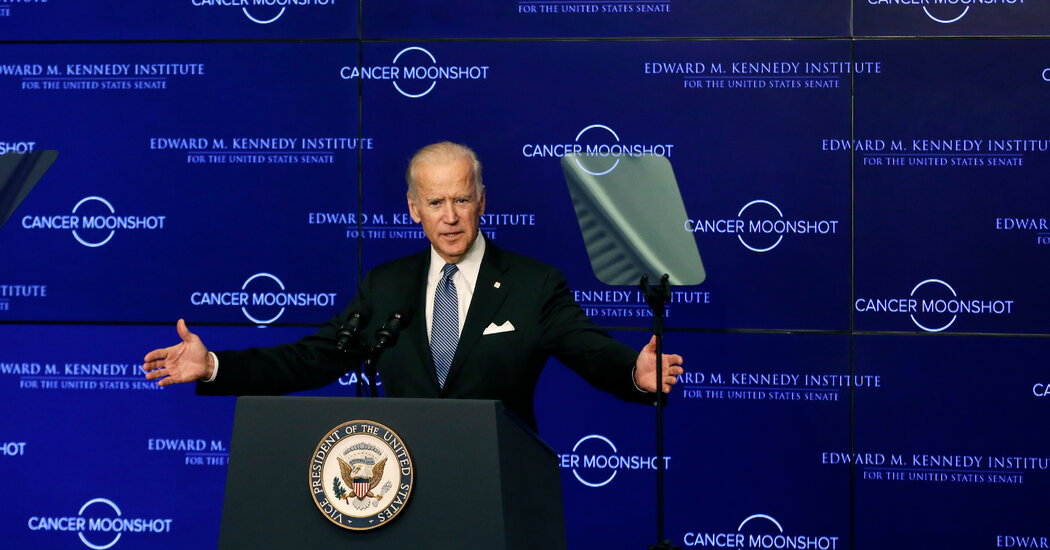
Biden to Present Plan to Cut Cancer Death Rate in Half

The president aims to revive the cancer “moonshot” program he headed as vice president.WASHINGTON — President Biden will unveil a plan on Wednesday to reduce the death rate from cancer by at least 50 percent over the next 25 years — an ambitious new goal, senior administration officials say, for the cancer “moonshot” program he initiated and presided over five years ago as vice president.Mr. Biden and his wife, Jill Biden, will also announce a campaign to urge Americans to undergo screenings that were missed during the coronavirus pandemic, according to the officials, who spoke on condition of anonymity Tuesday evening to preview the president’s announcement. Screening is important to reduce cancer deaths.The president has a deep personal interest in cancer research; in 2015, his son Beau died of glioblastoma, an aggressive brain cancer. The next year, President Barack Obama called on Mr. Biden in his State of the Union address to lead the moonshot program, with a goal of making “a decade’s worth of advances in cancer prevention, diagnosis and treatment” in five years.At the time, Congress authorized $1.8 billion over seven years; roughly $400 million of that money has yet to be allocated, and the National Cancer Institute, which oversees the initiative, says it has already spent $1 billion on more than 240 research projects. The senior officials said the White House would not be announcing any new funding commitments, but insisted that there would be “robust funding going forward.”Instead, the Bidens will set out broadly outlined goals in a showy White House ceremony to be attended by roughly 100 people, including Vice President Kamala Harris, patients, caregivers, family members, researchers and members of Congress.The White House is billing the event as a fresh push by the president to “reignite” the moonshot program and “end cancer as we know it.” Specifically, Mr. Biden will set a goal of cutting the age-adjusted death rate — a statistic that accounts for expectations that older people are more likely to grow ill and die — by more than half over the next 25 years. But there were few specifics about how that goal would be achieved.“These are audacious goals, and I have no doubt there will be mechanisms to achieve them,” said Ellen Sigal, founder of Friends of Cancer Research, which works to support cancer research and deliver new therapies to patients, who has been briefed on the plan.Mr. Biden has already named Danielle Carnival, who worked on the moonshot program during the Obama administration, to help oversee the new version of the effort. Now, the senior officials said, the president will create a “cancer cabinet” to coordinate the work of multiple government agencies.The White House says more than 9.5 million cancer screenings were missed in the United States as a result of the Covid-19 pandemic. Mr. Biden will call on the cancer institute, a branch of the National Institutes of Health, to coordinate with cancer treatment centers to offer screenings around the country, and to develop a program to fast-track the development of tests that can detect multiple types of cancer at once.Presidents since Richard M. Nixon have sought to tackle cancer, of which there are more than 100 types of disease that can vary in the way they grow, spread and respond to treatment. The cancer institute estimates that nearly 40 percent of men and women will be diagnosed with some type of cancer at some point during their lifetimes. The American Cancer Society estimates there will be 1.9 million new cases of cancer in the United States this year, and more than 609,000 cancer deaths.Most experts no longer talk of “curing” cancer; that language is far too simplistic, and the White House is not using it. But officials say it is possible to make substantial progress in the fight against cancer through early diagnosis and improved treatments.There have been great strides in cancer research, treatment and prevention in the five years since the original moonshot program was announced. Targeted therapies are helping cancer patients live longer. Doctors can now detect cancers through a simple blood draw. More refined colonoscopies are preventing more colon cancers.“The original moonshot demonstrated that it was possible to compress a decade’s worth of progress into a few short years,” Ms. Sigal said, adding, “We can’t afford to not make that opportunity a reality again.”

Read more →

3D structure of regulator protein revealed

Proteins are indispensable components in living organisms. They are not only “building material” for the body — they also make molecular communication between cells possible, they are needed for nerve impulses to occur, and they control chemical reactions. What is decisive for proteins to function is their three-dimensional structure. If this is known, conclusions can be drawn about how proteins function. A team of researchers led by Prof. Daniel Kümmel from the University of Münster and Prof. Stefan Raunser from the Max Planck Institute (MPI) of Molecular Physiology in Dortmund (Germany) has now clarified the structure of a protein complex which is an important regulator of cellular degradation processes.
The protein complex “Mon1/Ccz1” determines which intracellular vesicles deliver their content to the cellular “recycling centre,” the lysosome. To this end, it docks onto the vesicle membrane, where it introduces a label. Intracellular vesicles are membrane bubbles which transport material through the cell. In the lysosome, the content is degraded and re-used. By elucidating the structure in almost atomic resolution, the researchers were now able to clarify, among other things, how the protein complex recognises the appropriate vesicles. For example, they showed that the complex has a positively charged and relatively flat area which determines its orientation after docking onto the vesicle membrane.
“Mon1/Ccz1” belongs to a family of regulators for which no structural information exists. These complexes are involved in a range of cellular processes and are sometimes associated with the occurrence of developmental disorders such as albinism and blood clotting disorders. “Our structure now provides a basis for a better understanding of these connections,” says Daniel Kümmel.
The protein complex examined comes from the filamentous fungus Chaetomium thermophilum and is particularly stable and easy to handle under laboratory conditions. It can serve as a model for human proteins. In order to determine the protein’s structure, the researchers used high-resolution cryogenic electron microscopy. “With this method, we can study the structure of proteins at temperatures around minus 150 degrees Celsius in an almost natural state,” says Stefan Raunser.
The researchers checked their results by means of biochemical studies, for example sedimentation assays. In this case, the protein-membrane interaction is demonstrated with artificial vesicles and purified protein in vitro, i.e. outside the organism. “The structure of Mon1/Ccz1 has a unique architecture that, to our knowledge, has not been demonstrated in any other protein complex. It could serve as a blueprint for a better understanding of other related regulatory proteins. We want to continue our successful collaboration,” Daniel Kümmel and Stefan Raunser agree.
The study was published in the interdisciplinary journal Proceedings of the National Academy of Sciences. In addition to scientists from WWU Münster and MPI Dortmund, researchers from the University of Osnabrück were also involved.
Story Source:
Materials provided by University of Münster. Note: Content may be edited for style and length.

Read more →

Research advances technology of AI assistance for anesthesiologists

A new study by researchers at MIT and Massachusetts General Hospital suggests the day may be approaching when advanced artificial intelligence systems could assist anesthesiologists in the operating room.
In a special edition of Artificial Intelligence in Medicine, the team of neuroscientists, engineers and physicians demonstrated a machine learning algorithm for continuously automating dosing of the anesthetic drug propofol. Using an application of deep reinforcement learning, in which the software’s neural networks simultaneously learned how its dosing choices maintain unconsciousness and how to critique the efficacy of its own actions, the algorithm outperformed more traditional software in sophisticated, physiology-based simulations of patients. It also closely matched the performance of real anesthesiologists when showing what it would do to maintain unconsciousness given recorded data from nine real surgeries.
The algorithm’s advances increase the feasibility for computers to maintain patient unconsciousness with no more drug than is needed, thereby freeing up anesthesiologists for all the other responsibilities they have in the operating room, including making sure patients remain immobile, experience no pain, remain physiologically stable, and receive adequate oxygen said co-lead authors Gabe Schamberg and Marcus Badgeley.
“One can think of our goal as being analogous to an airplane’s auto-pilot where the captain is always in the cockpit paying attention,” said Schamberg, a former MIT postdoc who is also the study’s corresponding author. “Anesthesiologists have to simultaneously monitor numerous aspects of a patient’s physiological state, and so it makes sense to automate those aspects of patient care that we understand well.”
Senior author Emery N. Brown, a neuroscientist at The Picower Institute for Learning and Memory and Institute for Medical Engineering and Science at MIT and an anesthesiologist at MGH, said the algorithm’s potential to help optimize drug dosing could improve patient care.
“Algorithms such as this one allow anesthesiologists to maintain more careful, near continuous vigilance over the patient during general anesthesia,” said Brown, Edward Hood Taplin Professor Computational Neuroscience and Health Sciences & Technology at MIT.

Read more →

Immunological memory provides long-term protection against coronavirus

Exposure to SARS-CoV-2 by infection or vaccination generates immune cells that provide long-term immunity. These long-lived memory T cells play a key role in preventing severe cases of Covid-19. Researchers at the University of Zurich have now discovered how these memory T cells form.
Many questions about how exposure to SARS-CoV-2 by infection or immunization might result in long-term protective immunity remain unanswered. Onur Boyman, head of the Department of Immunology, and his research team at the University of Zurich and the UniversityHospital Zurich have now taken a closer look at how this long-lived protection is formed. Together with researchers from ETH Zurich, they identified specific signaling pathways that determine when immune cells develop into so-called memory T cells.
From short-lived killers to long-term memory T cells
Virus-specific antibodies produced by B cells are insufficient to effectively protect against the novel coronavirus. The cellular immune response to SARS-CoV-2 is just as important. Here, virus-specific CD8+ T cells play a crucial role, as they can identify and kill the cells that have been infected by the virus. These cytotoxic T cells eliminate viruses that are hidden inside the host cells and help prevent the spread of millions of newly formed viruses. “These T cells are usually active for only a short time and disappear quickly. When it comes to establishing long-term protective immunity, it is important to generate long-lived memory T cells that are activated very quickly upon re-exposure to the virus,” explains Onur Boyman. This latter ability is referred to as immunological memory.
Previous studies have focused on the whole CD8+ T cell population that formed in response to the virus. Boyman and his team have now succeeded in tracking individual clones of SARS-CoV-2-specific CD8+ T cells in patients with Covid-19, from the acute viral infection up to one year after recovery. The researchers were also able to identify the signaling pathways responsible for the transition of CD8+ T cells from short-lived killers to long-lived memory cells — and they found a distinct molecular signature.
Immune messengers determine the cell type
In their study, the researchers were able to demonstrate that the signature of long-lived memory CD8+ T cells was already present during acute SARS-CoV-2 infection, and these cells could thus be distinguished from their short-lived counterparts at an early stage. “The distinct signature of memory cells contained signals of immune messengers, such as interferons, which are an important part of the immune response against SARS-CoV-2 and also contribute to controlling viral infections,” says Onur Boyman.
Immune response varies from one patient to another
The study helps to unravel the complex way in which immunological memory to SARS-CoV-2 is — or is not — formed and maintained. While some infections result in robust and long-lasting T cell memory, others fail to do so. The newly identified signature makes it possible to determine which type of infection — e.g. mild or severe, systemic or limited to mucosal membranes — gives rise to sustained immunity. The immune response is also shaped by vaccines, which contain different ingredients and adjuvants. “While everyone responds differently to the virus or a vaccine, cellular immunity plays a crucial role in preventing severe cases of Covid-19 in both vaccinated and recovered people,” says Boyman.
Funding
The study was supported by the Swiss National Science Foundation (SNSF), the Clinical Research Priority Program CYTIMM-Z of the University of Zurich (UZH), an innovation grant from the UniversityHospital Zurich (USZ), the UZH Pandemic Fund, the Botnar Research Centre for Child Health (BRCCH), and the Swiss Academy of Medical Sciences (SAMS).
Story Source:
Materials provided by University of Zurich. Note: Content may be edited for style and length.

Read more →