COVID-19 increases risk of pregnancy complications, study suggests

Pregnant women with COVID-19 appear to be at greater risk for common pregnancy complications — in addition to health risks from the virus — than pregnant women without COVID-19, suggests a study funded by the National Institutes of Health.
The study, which included nearly 2,400 pregnant women infected with SARS-CoV-2, found that those with moderate to severe infection were more likely to have a cesarean delivery, to deliver preterm, to die around the time of birth, or to experience serious illness from hypertensive disorders of pregnancy, postpartum hemorrhage, or from infection other than SARS-CoV-2. They were also more likely to lose the pregnancy or to have an infant die during the newborn period. Mild or asymptomatic infection was not associated with increased pregnancy risks.
“The findings underscore the need for women of child-bearing age and pregnant individuals to be vaccinated and to take other precautions against becoming infected with SARS-CoV-2,” said Diana Bianchi, M.D., director of NIH’s Eunice Kennedy Shriver National Institute of Child Health and Human Development (NICHD), which funded the study. “This is the best way to protect pregnant women and their babies.”
The study was conducted by Torri D. Metz, M.D., of the University of Utah, Salt Lake City, and colleagues in the NICHD Maternal-Fetal Medicine Units Network. It appears in the Journal of the American Medical Association. Additional funding was provided by NIH’s National Center for Advancing Translational Sciences.
The study included more than 13,000 pregnant individuals from 17 U.S. hospitals, approximately 2,400 of whom were infected with SARS-CoV-2. Participants delivered between March 1 and December 31 2020, before SARS-CoV-2 vaccination was available. The researchers compared outcomes among those with COVID-19 to those from uninfected patients, and tabulated the study results as a primary outcome — whether the patient had died from any cause or had a serious illness or condition related to common obstetric complications. They also evaluated the results in terms of several secondary outcomes, including cesarean delivery, preterm birth, and fetal and newborn death.
Compared to uninfected patients, those with moderate to severe COVID-19 were more likely to experience the primary outcome, (26.1 vs 9.2%). They were also more likely to deliver by cesarean (45.4 vs 32.4%) or preterm (26.9 vs 14.1%) or to have a fetal or newborn death (3.5 vs 1.8%). Mild or asymptomatic COVID-19 was not associated with any of adverse outcomes.

Read more →

Genetic remodeling in tumor formation

Cancerous tumors are made up of rapidly growing, abnormally shaped, cells that can infiltrate and destroy healthy tissues, travel to other parts of the body, and form additional tumors. In part because of its rapid and invasive nature, cancer remains the second leading cause of death in the United States and a major cause of death worldwide.
Tumor formation is often driven by genes called oncogenes, which are usually involved in the normal processes of cell growth, proliferation, and death. But they can also mutate and be expressed at high levels, which can drive the multiplication of cancerous cells.
Mutated oncogenes work in various ways, including on target cells’ chromatin, compact, dense structures of DNA and selected protein molecules. Oncogenes also work with an extensive variety of protein complexes that help turn specific genes “on” or “off” by binding to certain regions of the DNA.
The most well-known oncogene is Kras. Mutant Kras is found in 20% of all human cancers, including 97% of pancreatic ductal, 45% of colorectal, and 30% of lung cancers.
Despite the attention focused on mutant Kras tumor formation, the question remains as to why Kras mutation is so essential for oncogenesis. The conventional wisdom is that Kras mutation simply promotes cell proliferation. However, as many genes can promote cell proliferation, what makes Kras so deadly and difficult to treat?
A collaborative team that includes researchers from the Terasaki Institute for Biomedical Innovation (TIBI) and Duke University led by Dr. Xiling Shen, a professor and the chief scientific officer of TIBI, has been able to shed light on these processes. In a recent study, published in Developmental Cell, they discovered that Kras mutation causes rearrangements of chromatin inside cells. This rearrangement causes tissue cells to revert to an early developmental, or “stem-like” state and erroneously start to regenerate “new tissue,” causing the onset of tumor formation.
The team revealed that Kras-induced chromatin remodeling was mediated by a protein complex called AP-1, which binds and opens chromatins to rewire the cell’s fate. The AP-1 mediated chromatin accessibility mechanism seems to be a common process in tumor initiation, including in the lung, skin, and intestine. The team demonstrated that small-molecule drugs inhibiting AP-1 deterred oncogenesis and cell proliferation, providing a promising way to treat Kras-mutant tumors. This is especially significant because, currently, the majority of Kras tumors are not treatable with drugs.
What is even more fascinating is that Kras/AP-1 induced chromatin remodeling instills plasticity, or the ability to assume different cellular characteristics in response to mutations. Hence, Kras-mutant cells from different origins such as alveolar cells in the lungs and club cells in the airway will start masking their cell identity and become alike to one another. This sheds new light on the question of how lung cancer originates.
According to Dr. Shen, “Our study demonstrates the ability of Kras to use genetic reprogramming to make cells more stem-like and plastic; it resolves the long-standing debate over why Kras is so special in tumor formation. Our elucidation of the AP-1 complex as its effector for chromatin remodeling opens new therapeutic opportunities to target Kras, a notoriously hard-to-drug target.”
“An understanding of the complex processes involved in tumorigenesis is essential for designing drug treatments and screening platforms for cancer,” said TIBI Director and CEO, Ali Khademhosseini, PhD. “Dr. Shen’s work will prove invaluable in meeting those needs and will dovetail with the drug delivery and physiological models platforms that we have here at the institute.”
Authors are Preetish Kadur Lakshminarasimha Murthy, Rui Xi, Diana Arguijo, Jeffrey I. Everitt, Dewran D. Kocak, Yoshihiko Kobayashi, Aline Bozec, Silvestre Vicent, Shengli Ding, Gregory E. Crawford, David Hsu, Purushothama Rao Tata, Timothy Reddy, and Xiling Shen.
This work was funded by NIH U01 CA217514, U01 CA214300, and R35 GM122465 to XS and DFG-SPP microbone (BO3811-1/7) grant to AB.

Read more →

Mouse experiments show how sugar molecules can be used to track stem cells

A Johns Hopkins Medicine scientist who spent 30 years figuring out how to put chemical labels into cells to track their movement in living tissues has found that certain self-renewing stem cells have built-in tracers — made out of sugars — that can do the job without added chemical “labels” when injected into mouse brains. The finding, made with stem cells widely engineered into experimental therapies for multiple sclerosis and other neurodegenerative diseases, was a welcome surprise, the investigators say.
“There is an entire scientific field dedicated to chemical and genetic cell labeling, because otherwise, we can’t see where specially and expensively engineered therapeutic cells travel and whether they get to the intended spot in a body to repair or replace diseased tissue,” says Jeff Bulte, Ph.D., professor of radiology and radiological science at the Johns Hopkins University School of Medicine and director of cell imaging for the Johns Hopkins Institute for Cell Engineering.
If confirmed with subsequent experiments, the new study should, Bulte says, streamline and advance restorative research for diseases of the brain, an organ considered the most difficult in which to track therapies because of the sensitive nature of the brain and its blood-brain barrier.
In Bulte’s experience, he says, labeling therapeutic cells that are ready for human clinical trials in any organ is a costly and difficult process, requiring extensive safety studies and keeping teams of scientists focused on finding better ways to track therapeutic cells damaged by multiple sclerosis and other neurodegenerative diseases.
For such research, scientists have long been using so-called mesenchymal stromal cells, a type of stem cell found in the bone marrow that can develop into many types of cells, and also reduce inflammation.
In the new proof-of principle study described Feb. 7 in Nature Biomedical Engineering, Bulte and his research team found that these mesenchymal stromal cells contain high levels of a sugar called mannose, which is similar to glucose and can be spotted easily and successfully with a standard imaging method based on magnetic resonance imaging (MRI).

Read more →

New benchmark could improve detection of genetic variants linked to spinal muscular atrophy, other diseases

The stretches of DNA that differ from person to person, called variants, are a major part of what makes us unique, but they can also put us at greater risk of disease. Although we can currently spell out between 80% and 90% of the millions that are in the human genome, the remaining variants may hold clues for treating an array of diseases. Today the list of variants yet to be decoded has shrunk sizably.
A team led by researchers at the National Institute of Standards and Technology (NIST), Baylor College of Medicine and DNAnexus has characterized over 20,000 variants in 273 genes of medical importance. In a study published in the journal Nature Biotechnology, the researchers applied both cutting-edge and long-standing DNA sequencing methods to decipher the genetic codes of the variants with a high degree of certainty. Using their results, they formulated benchmarks that will help labs and clinics sequence the genes more accurately, which is critical for gaining a better understanding of a host of diseases and eventually developing treatments.
“Some of these genes, which have previously been very difficult to access, are suspected to have some connection to disease. Others have very clear clinical importance,” said NIST biomedical engineer Justin Zook, a co-author of the study. “SMN1, for example, is a gene we characterized that is directly associated with spinal muscular atrophy, a rare but severe condition.”
The new benchmark is the latest produced by the Genome in a Bottle (GIAB) consortium, a NIST-hosted collaborative effort aimed at improving DNA sequencing technologies and making them practical for clinical application.
These benchmarks are highly accurate sequences of DNA that clinics and research labs can use as a kind of answer key when testing their own sequencing methods. By sequencing the same genome used to develop a benchmark and then comparing their result to the benchmark itself, they could learn how well they can detect certain variants.
Over the years, producing benchmarks for some regions of the genome has proved much more difficult than others. There are several reasons, many of which are tied to the general approach people use to sequence DNA.

Read more →

Chimps Catch Insects to Put on Wounds. Is It Folk Medicine?

They don’t eat the bugs, and they’re definitely applying them to wounds, so some scientists think the primates may be treating one another’s injuries.A chimp, Suzee, catches an insect and puts it on a wound on the foot of her son, Sia. Video by Alessandra Mascaro.Tobias DeschnerChimpanzees design and use tools. That is well known. But is it possible that they also use medicines to treat their own and others’ injuries? A new report suggests they do.Since 2005, researchers have been studying a community of 45 chimpanzees in the Loango National Park in Gabon, on the west coast of Africa. Over a period of 15 months, from November 2019 to February 2021, the researchers saw 76 open wounds on 22 different chimpanzees. In 19 instances they watched a chimp performing what looked like self-treatment of the wound using an insect as a salve. In a few instances, one chimp appeared to treat another. The scientists published their observations in the journal Current Biology on Monday.The procedure was similar each time. First, the chimps caught a flying insect; then they immobilized it by squeezing it between their lips. They placed the insect on the wound, moving it around with their fingertips. Finally, they took the insect out, using either their mouths or their fingers. Often, they put the insect in the wound and took it out several times.The researchers do not know what insect the chimps were using, or precisely how it may help heal a wound. They do know that the bugs are small flying insects, dark in color. There’s no evidence that the chimps are eating the insects — they are definitely squeezing them with their lips and then applying them to the wounds.There have been other reports of self-medication in animals, including dogs and cats that eat grass or plants, probably to help them vomit, and bears and deer that consume medicinal plants, apparently to self-medicate. Orangutans have been seen applying plant material to soothe muscle injuries. But the researchers know of no previous report of nonhuman mammals using insects for a medicinal purpose.In three instances, the researchers saw chimps using the technique on another chimp. In one case, they saw an adult female named Carol grooming around a flesh wound on the leg of an adult male, Littlegrey. She grabbed an insect, and gave it to Littlegrey, who put it between his lips, and transferred it to his wound. Later, Carol and another adult male were seen moving the insect around on Littlegrey’s wound. Another adult male approached, took the insect out of the wound, put it between his own lips, then reapplied it to Littlegrey’s leg.One chimp, an adult male named Freddy, was a particularly enthusiastic user of insect medicine, treating himself numerous times for injuries of his head, both arms, his lower back, his left wrist and his penis. One day, the researchers watched him treat himself twice for the same arm wound. The researchers don’t know how Freddy got these injuries, but some of them probably involved fighting with other males.There are some animals that cooperate with others in similar ways, said Simone Pika, who leads an animal cognition lab at the University of Osnabrück in Germany and is an author of the study. “But we don’t know of any other instances in mammals,” she said. “This may be a learned behavior that exists only in this group. We don’t know if our chimps are special in this regard.”Aaron Sandel, an anthropologist at the University of Texas, Austin, found the work valuable, but at the same time expressed some doubts. “They don’t offer an alternative explanation for the behavior, and they make no connection to what insect it might be,” he said. “The jump to a potential medical function? That’s a stretch at this point.”Still, he said, “attending to their own wounds or the wounds of others using a tool, another object — that’s very rare.” Their documentation of chimps paying such attention to other chimps is, he added, “an important contribution to the study of social behavior in apes. And it’s still interesting to ask whether there is empathy involved in this, as it is in humans.”In some forms of ape social behavior, it is clear that there is an exchange of value. For example, grooming another chimp provides relief from parasites for the groomed animal, but also an insect snack for the groomer. But in the instances she observed, Dr. Pika said, the chimp gets nothing tangible in return. To her, this shows the apes are engaging in an act that increases “the welfare of another being,” and teaches us more about the primates’ social relationships.“With every field site we learn more about chimps,” she said. “They really surprise us.”

Read more →

Paralysed man with severed spine walks thanks to implant

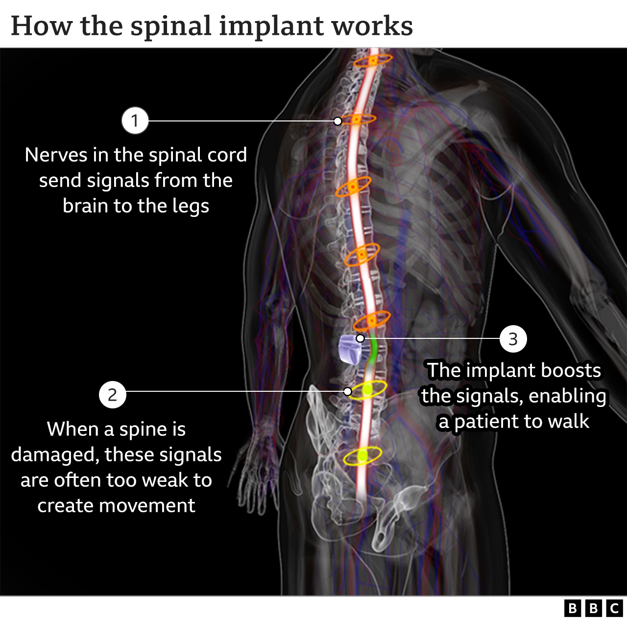
SharecloseShare pageCopy linkAbout sharingThis video can not be playedTo play this video you need to enable JavaScript in your browser.A paralysed man with a severed spinal cord has been able to walk again, thanks to an implant developed by a team of Swiss researchers.It is the first time someone who has had a complete cut to their spinal cord has been able to walk freely.The same technology has improved the health of another paralysed patient to the extent that he has been able to become a father.The research has been published in the journal Nature Medicine. Michel Roccati was paralysed after a motorbike accident five years ago. His spinal cord was completely severed – and he has no feeling at all in his legs.But he can now walk – because of an electrical implant that has been surgically attached to his spine. Someone this injured has never been able to walk like this before. The researchers stress that it isn’t a cure for spinal injury and that the technology is still too complicated to be used in everyday life, but hail it nonetheless as a major step to improving quality of life.I met Michel at the lab where the implant was created. He told me that the technology “is a gift to me”.”I stand up, walk where I want to, I can walk the stairs – it’s almost a normal life.”It was not the technology alone that drove Michel’s recovery. The young Italian has a steely resolve. He told me that from the moment of his accident, he was determined to make as much progress as he could.”I used to box, run and do fitness training in the gym. But after the accident, I could not do the things that I loved to do, but I did not let my mood go down. I never stopped my rehabilitation. I wanted to solve this problem.”The speed of Michel’s recovery amazed the neurosurgeon who inserted the implant and expertly attached electrodes to individual nerve fibres, Prof Jocelyne Bloch from the Laboratoire de Neurothérapies et Neuromodulation – LNTM.”I was extremely surprised,” she told me. “Michel is absolutely incredible. He should be able to use this technology to progress and be better and better.”‘I always dreamed of walking again’Doctors use deep-brain ultrasound therapy to treat tremorsParkinson’s results beyond researchers’ wildest dreamsThe research has been backed by Dr Ram Hariharan, a consultant at the Northern General Hospital in Sheffield who is independent of the research team and also speaks for the Spinal Injuries Association. “They have done something that has not been done before.”I have not heard of any study where they have put in an implant [into a patient with a complete spinal cord cut] and demonstrated muscle movements and improving balance, enough to stand and walk.”But he added more clinical trials needed to be carried out before he could be convinced that it was an effective treatment.”We need more numbers [of patients] to show that it is first safe and that it significantly enhances their lives. Only then can it be taken forward.”Nerves in the spinal cord send signals from the brain to the legs. Some people are paralysed when the nerves are damaged through injury.In Michel’s case there’s no signal at all to his legs because the spinal cord is completely severed. But the implant sends signals directly to his legs enabling him to walk, but only when the implant is on.This video can not be playedTo play this video you need to enable JavaScript in your browser.So far nine people have received the implant and regained the ability to walk. None of them use it to help them walk in their everyday lives – because it’s too complicated at this stage. Instead, they use it to practise walking – which exercises their muscles, improves their health – and often, restores a little bit of movement. David M’zee was one of the first patients to receive the implant. Like Michel, he was able to walk with the implant while using a walker. David’s health improved to such an extent he has was able to have a baby girl with his partner Janine, something that was not possible after his accident in 2010.His daughter, Zoe, is now one year old. When I was with them, she raced with her Dad with her baby walker and took great delight in beating him.”It’s really beautiful!” he beamed with fatherly pride.”It is great fun. It’s the first time I have been walking with her in that way – she with her baby walker, I with my walker.”Having a family has given David a huge amount of joy. And the implant has helped him in subtle, but important ways.”It helps with the hypertension. I had it for so long. At first I didn’t realise I had it. I was getting so tired from time to time. “Once we found out that the implant can increase the blood pressure it was like ‘Wow, that’s how life can be!'””It’s these small things that make a big difference,” he told me.There is still a long way to go before the technology can be used routinely to help paralysed people to walk, according to Prof Grégoire Courtine, who led the team that developed the technology at the École Polytechnique Fédérale de Lausanne (EPFL).”This is not a cure for spinal cord injury. But it is a critical step to improve people’s quality of life. We are going to empower people. We are going to give them the ability to stand, to take some steps. It is not enough, but it is a significant improvement.”A cure would require regeneration of the spinal cord, possibly with stem cell therapies, which are still at a very early stage of research. Prof Courtine believes that his implant technology could be used in conjunction with nerve regeneration treatments once they are ready.Follow Pallab on TwitterEPFLSpinal injuries associationThe BBC is not responsible for the content of external sites.

Read more →

Vaccine protection against SARS-CoV-2 infection wanes quickly but is better maintained against severe disease, study finds

The vaccine-induced protection against SARS-CoV-2 infection wanes within a couple of months, but at different speed according to vaccine type. However, protection against severe COVID-19 appears to be better maintained. This is shown in a nationwide, registry-based study performed by researchers at Umeå University, Sweden, that now is published in The Lancet.
“The bad news is that the protection against infection seems to be diminished by seven months after the second dose of vaccine,” says Peter Nordström, professor of geriatric medicine at Umeå University. “The good news, however, is that the protection against a severe infection that leads to hospitalization or death seems to be better maintained. Vaccination is therefore very wise and important.”
The study, which received much attention once first posted as a ‘preprint’ in October, has now been peer-reviewed and the results as such confirmed. Following extensive revision and additional analyses, leading to results that more clearly show that the protection against severe COVID-19 seems better maintained than that against infection, the study has now been published in The Lancet.
The study is a nationwide, observational study based on registry-data from the Public Health Agency of Sweden, the National Board of Health and Welfare, and Statistics Sweden. The main analysis included almost 1.7 million individuals, and the results were confirmed in an even larger population of almost 4 million individuals. The results showed that protection against infection of any severity waned progressively following the peak which occurred a month after the second dose.
Six months after vaccination, the remaining protection against infection was 29 per cent from two doses of Pfizer, and 59 per cent from two doses of Moderna. There was no remaining protection from months and onwards for AstraZeneca. With respect to infections that were severe enough to warrant a hospital stay, or where the individual died within 30 days of confirmed infection, the protection was better maintained.
Protection against severe disease was 89 per cent after one month and 64 per cent from four months an onwards during the rest of the maximum follow-up of nine months. There was some evidence to suggest a lower protection in the oldest individuals and in individuals with homemaker service.
“The results underscore and support the decision to offer a third dose,” says Marcel Ballin, doctoral student in geriatric medicine at Umeå University and co-author of the study. In particular, the results show that it was correct to prioritize the oldest and frailest individuals.”
Prior to this study, a few observational studies and follow-up studies of the clinical trials have investigated waning vaccine protection in other countries. However, these have mostly covered the initial four to six months, and for the Pfizer vaccine.
“What this study contributes with is the longer follow-up time and the fact that we were able to explore how well the protection is maintained according to different types of vaccines,” says Anna Nordström, adjunct professor in public health at Umeå University and co-author of the study.
“The strengths are that we have been able to do this in a real-world setting based on a population-based sample of the total population of Sweden. This increases the possibility to generalize the results to other countries with similar population structure as in Sweden.”
Story Source:
Materials provided by Umea University. Original written by Ola Nilsson. Note: Content may be edited for style and length.

Read more →

Trapping sperm in semen’s natural gel could lead to new contraceptive

A discovery that blocks the normal transition of semen from a thick gel to a liquid shows promise for development of a new form of non-hormonal, over-the-counter contraception.
A Washington State University-led research team recently showed that blocking prostate-specific-antigen in human ejaculate samples caused the semen to remain in its thick gel form, trapping the majority of the sperm. Normally, the semen will liquefy, allowing sperm to swim through the female reproductive system to fertilize an ovum or egg. The discovery is able to stop that process and detailed in the journal Biology of Reproduction.
“Our goal is to develop this into an easily accessible female contraceptive that would be available on-demand, meaning women could go buy it off the shelf,” said senior author Joy Winuthayanon, associate professor and director of WSU’s Center for Reproductive Biology. “It could be used in combination with a condom to lower the failure rate significantly.”
Currently over-the-counter contraceptives such as condoms and spermicides have an average 13% to 21% failure rate, the study authors noted. Hormonal-based contraceptives such as IUDs and birth control pills have lower failure rates, but they can have some side-effects and are not always easily available or affordable — which may be one reason why worldwide the unintended pregnancy rate is currently 48%, according to recent global health research.
The WSU team has been working on this contraceptive method since 2015 after it was accidentally discovered that some of the female mice in a different reproductive study could not get pregnant; upon investigating further, the researchers discovered the male’s semen was staying in solid form. The researchers then tried stopping the semen liquification process in mice on purpose, and using a non-specific protease inhibitor called AEBSF, they were able to disrupt sperm movement and reduce fertility in mice, detailing their results in an earlier Biology of Reproduction paper.
In the current study, the research team worked to see if they could translate those results to human samples. They found that AEBSF did have a contraceptive effect, but it was unclear whether this was simply due to its toxicity. They then used an antibody to target the prostate-specific antigen or PSA in human sperm. They chose PSA because it is the primary active protein in liquefication and secreted in large quantities from the prostate gland, which is present in humans but not in mice.
Typically, after ejaculation the PSA acts on the gel-forming proteins called semenogelins, explained first author Prashanth Anamthathmakula, who worked as a WSU post-doctoral fellow on the project.
“The semenogelins create a gel-like network with a fine mesh of proteins which traps the sperm. The PSA cleaves that mesh and the sperm become free,” said Anamthathmakula, who is now a senior research scientist at University of Missouri-Kansas City. “Using a PSA inhibitor, an antibody, we showed that we could block that liquefaction.”
The next step is to identify more specific small molecule inhibitors that would effectively prevent PSA’s ability to liquefy semen without any detrimental side effects. The researchers noted that current spermicides have been shown to lower the natural vaginal barriers against sexually transmitted diseases like HIV. By targeting the liquefaction process of the semen itself, this advance could avoid that type of toxicity, but more research needs to be done.
“It is a bit of a long process because we don’t want off-target effects,” said Winuthayanon. “If we are going to develop this into the contraceptive product, it may be something that women would use often, so we want something that is safe and has no unintended effects.”
Story Source:
Materials provided by Washington State University. Original written by Sara Zaske. Note: Content may be edited for style and length.

Read more →

Advanced prostate cancer antibody drug shows success in pet dogs

Dogs are proving to be a far better scientific model for study of prostate cancer than mice, the typical animal used in the lab for this type of research. In the first use of canines in an advanced prostate cancer study, scientists explored the pathways used by the cancer to evade the immune system and identified an antibody drug that significantly improves survival rates.
The findings are described in a paper appearing in the Journal for ImmunoTherapy of Cancer.
Prostate cancer is the most common cancer in men worldwide. At advanced stages, there exist treatments that are effective in most cases for a short while, but eventually the disease progresses past being susceptible to any curative treatment. The prognosis beyond this point is typically gloomy.
Researchers have hypothesized that a particular type of white blood cell — regulatory T cells, or Tregs — may be inhibiting the ability of the immune system to recognize and thereby attack prostate cancer cells (as well as some other types of cancer cells). Tregs play an essential role in the immune system, ensuring that white blood cells that recognize and attack foreign cells don’t mistake the body’s own cells as foreign and attack them. This is why Tregs are sometimes called “suppressor” T cells, as they suppress the immune system and prevent self-attack.
However, it is also thought that some types of cancer cells can trick Tregs into thinking that they also shouldn’t be attacked. Researchers have repeatedly found an excess of Tregs hanging around such cancers — a process they call Treg tumor infiltration, likely misdirecting the other T cells that would otherwise want to attack the tumor as “foreign.”
Activation (more formally, expression) of the gene that controls production of Forkhead box protein 3 (FOXP3) is necessary in turn for the production of Tregs. FOXP3 is a transcription factor, a type of protein that controls the rate of transcription of genes. Transcription is what we call the process of taking genetic information that exists in genes on DNA molecules and packaging that same information in messenger RNA, which then goes on to tell the protein factories in the cell to make a particular type of protein that corresponds to the original gene.

Read more →

Discovery of 29 new acne risk genes provides hope for new treatments

A study of the genetics of acne has identified 29 regions of the genome that influence the condition. These genetic insights offer potential new targets for treatment. They may also help clinicians identify individuals at high risk of severe disease.
The research, is the largest study of its kind, involving genetic data from over 20,000 individuals with acne. It was supported by the National Institute for Health Research Guy’s and St Thomas’ BRC. The study was led by investigators at the St John’s Institute of Dermatology at Guy’s and St Thomas’ NHS Foundation Trust and King’s College London and the QIMR Berghofer Medical Research Institute in Brisbane.
Acne is a common skin condition. Estimates indicate it affects 80% of teenagers. Spots and cysts, pigment changes and scarring are all common features. The face is the most common site, with the chest and back also frequently involved. The negative psychological consequences of acne are seen in all ages, but perhaps of particular concern for teenagers.
The research, published in Nature Communications, analysed nine genome wide association study datasets from patients around the world. These studies involved scanning the whole genomes 20,165 people who had acne and 595,231 people who did not. The study identified 29 new genetic variants that are more common in people with acne. It also confirmed 14 of the 17 variants already known to be associated with the condition. This brings the total number of known variants to 46.
Professor Catherine Smith is a Professor of Dermatology and Therapeutics at St John’s Institute of Dermatology at Guy’s and St Thomas’. She said: “Despite major treatment advances in other skin conditions, progress in acne has been limited. As well as suffering from the symptoms of acne, individuals describe consequent profound, negative impacts on their psychological and social wellbeing. It’s exciting that this work opens up potential avenues to find treatments for them.”
A number of genes were identified that are common in people with acne and are also linked to other skin and hair conditions. The team believe this will help to understand the causes of acne, which could be a mix of factors.
Professor Michael Simpson is Head of the Genomic Medicine Group at King’s College London. He said: “We know that the causes of acne are complicated, with a mix of biological factors such as genetics and hormones, and environmental factors. Understanding the genetics of the condition will help us to disentangle some of these causes, and find the best way to treat the condition. This is a really promising area for further study, and opens up a lot of avenues for research.”
The research also found a link between the genetic risk of acne and disease severity. Individuals who have the highest genetic risk are more likely to have severe disease. While further research is required, this finding raises the potential to identify individuals at risk of severe disease for early intervention.

Read more →