Can tech help revive India's 'crumbling' health system?
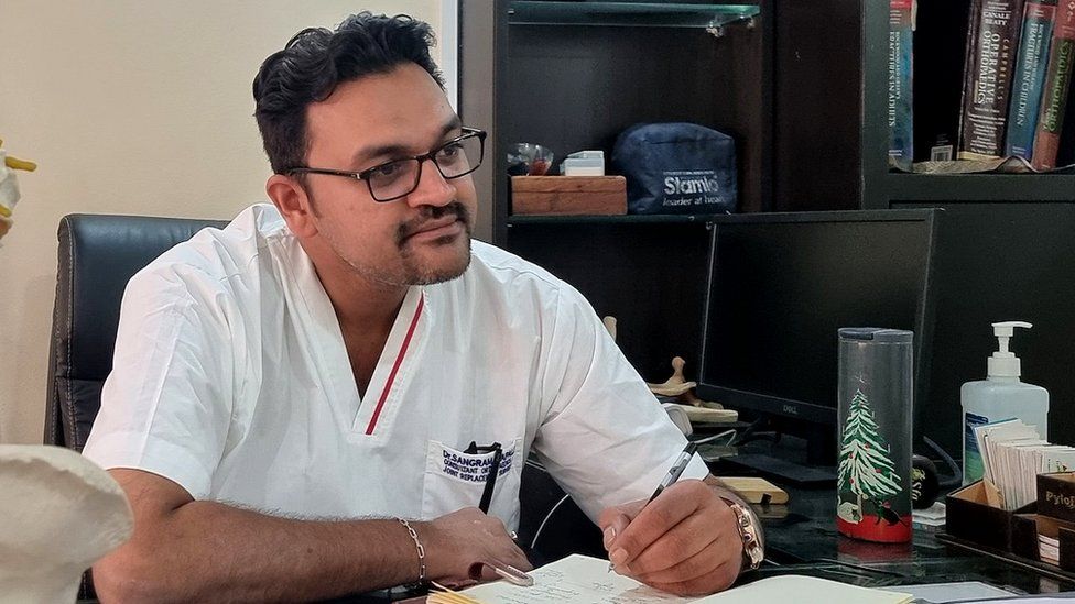
SharecloseShare pageCopy linkAbout sharingImage source, S KapaleThe first wave of Covid-19, that swept India in early 2020, was a gruelling experience for Dr Sangram Kapale and his colleagues.”The kind of mental pressure we were all going through is very difficult to put in words,” he says.”It was about saving lives with minimal resources. We had a lack of medicine, beds, oxygen.”The lack of skilled manpower, like paramedics, was another issue. We were forced to use medical students who had no practical experience of handling patients.”Dr Kapale was in charge of a temporary Covid treatment centre in Pune, Maharashtra which had 800 beds. The centre became overwhelmed with patients, and anxious family members gathered outside.”On one hand, we were doing everything within our capacity, or beyond, to fight Covid and save lives, and on the other hand we were accused of neglect by the relatives because they didn’t know what was happening inside.” Image source, Getty ImagesMuch of the chaos back then can be attributed to lack of resources, according to health care workers. Pre-pandemic, India reported one of the lowest levels of public spending on healthcare in the world.In 2019, spending on healthcare was equivalent to just 1.5% of India’s gross domestic product (GDP). By comparison, China spent 6.7% of its GDP on health in 2019, and in the UK that figure was 10.2%.Since then, the Indian government has ramped-up its spending and there is a target to spend 2.5% of GDP on health by 2025.Some are hopeful the devastating impact of the pandemic was a turning point for India’s entire healthcare system, with a future focus on technology and innovation.”All aspects of access to healthcare, diagnostics and life-sciences are moving …to low-cost and high-tech,” says Akriti Bajaj, an analyst who focuses on healthcare at Invest India, a government agency promoting investment.Image source, DozeeThere are thought to be more than 6,000 start-ups in the Indian healthcare sector, one of those is Dozee. The firm’s technology involves using a smart sensor underneath a patient’s bedsheet on a hospital bed. It tracks the micro-vibrations produced by the body when the heart pumps blood, and keeps track of a patient’s breathing and other movements. These observations are then translated in to data and processed by artificial intelligence-based algorithms which, if anything unusual is detected, can alert nurses and doctors at a central monitoring station.The company wants to install its technology in more than 1,000 hospitals and 5,000 beds by the end of 2022.”Our idea was to simplify the entire [monitoring] process, make it more ‘patient-centric’ and bring the technology to the wards, and even to patients’ homes,” says the company’s founder, Mudit Dandwate.Mr Dandwate believes technology could boost the quality of healthcare across India.”In India, the crumbling healthcare infrastructure was laid bare [by Covid] – poor hospital infrastructure; acute shortage of doctors; nursing staff and equipment and specialised treatment facilities, particularly in primary healthcare centres, in rural areas.He says government spending on healthcare has increased over the last two years and both the public and private sector will scale-up their facilities – with technology playing a “key role” in that transformation – in the coming years .Image source, G ManjunathAnother health tech entrepreneur, Dr Geetha Manjunath, founded Niramai in Bangalore, in 2016. She wanted to improve cancer screening, particularly in young women.”Unlike in Europe and the US, where early detection is enabled through regular, systematic screening programmes, India sees high mortality rates, due to late-stage detection,” she explains.Dr Manjunath says that in India, more than half of breast cancer cases are seen in women under 50 years of age and traditional X-ray detection has low accuracy among this group. So, her firm developed a technique for detecting early-stage breast cancer using a small, portable screening device.A high-resolution thermal sensor is used to measure the temperature variations on the patient’s chest, AI then analyses these 400,000 temperature points to generate a report and mark any abnormal regions. The thermal imaging is simple to use, meaning the test can be done by lower-skilled health workers, working in more remote areas of India. Dr Manjunath says the device makes a more systematic screening programme feasible and that earlier detection could, in turn, reduce treatment costs and save lives.Image source, Getty ImagesSo, what do doctors make of the new technology appearing on some wards?Dr Manjunth HG, head of the anaesthesia department at KR hospital in Mysore, has used Dozee’s system. He says that, while the technology is useful, in his opinion it has its limitations.”AI has a long way to go, although it is helping the medical fraternity in a great way, it cannot replace humans, ever. Even if AI is helpful, we need doctors and human presence in ICUs and hospitals. So, it’s just an assistance for us.” Back in Pune, Dr Sangram Kapale is just relieved that, after a second, devastating wave of Covid in 2021, a national vaccination programme appears to have brought the virus under control for the time-being.”After the vaccinations, the severity of the disease has definitely reduced. As a community, now we have to face whatever it throws at us in future.”

Read more →

Bunkers that save sight? Researchers take a close look

Chronically stressing the retina can weaken it and damage our ability to see. But retinal cells have a remarkable ability to wall off damage, a team of neuroscientists led by UConn Health reports in the 1 March issue of PNAS. The walling-off or “bunkering” of the damage may be key to preserving our eyesight.
The retina is a delicate tissue in the back of the eye that detects light and transmits images to the brain. Muller glia are very long cells that span the thickness of the retina and provide mechanical strength, supporting the neurons and light receptors that detect light, shape and color.
Muller glia are also involved in protein changes related to retinal injury, being the first cells to respond. UConn School of Medicine neuroscientist Royce Mohan and colleagues have discovered that the endfeet, a specialized zone in Muller cells, is where proteins become modified when the retina is under stress. These endfeet are at the opposite end of the retina, quite a distance from light receptors. The researchers propose in the paper that this segregation of the endfeet and light receptors may permit light detection to continue even as the retina responds to stress.
The modification of proteins Mohan’s lab has been studying is called citrullination. In citrullination, the amino acid arginine is changed into citrulline. Because in early stages of stress or disease, the citrullinated proteins stay sequestered in the Muller cells’ endfeet, Mohan calls this area the citrullination bunker. But if this bunker is chronically engaged, then the overabundance of citrullinated proteins reach other parts of the retina. Muller cells in human age-related macular degeneration (AMD) and mouse models of retinal degeneration reveal citrullinated proteins extending out of the endfeet and spreading throughout the cells.
Citrullination may have many effects on Muller glial cells which are only just being understood. For example, arginine is positively charged, while citrulline is not. The loss of the positive charges is permanent, and may irreversibly change the flexibility or other mechanical properties of the Muller glial cells. This may cause the Muller cells to become incapable of adapting to fluid build-up when the retina swells up under stress. Alternatively, it’s possible the citrullinated proteins could appear foreign to the body and draw the attention of the immune system, potentially beginning autoimmune disease. Turning off citrullination in the end feet bunker could delay or avoid these problems and preserve eyesight for longer.
This team has also identified that the endfeet citrullination process is controlled by an enzyme known as peptidyl arginine deiminase-4 (PAD4). Small molecule inhibitors of PAD4 have been developed for other types of citrullination-dependent diseases, such as rheumatoid arthritis. Mohan believes that such therapeutic agents could be applied to reduce citrullination at early stages of AMD and spare the retina of undesired responses to this protein modification.
Story Source:
Materials provided by University of Connecticut. Original written by Kim Krieger. Note: Content may be edited for style and length.

Read more →

Lead exposure in last century shrank IQ scores of half of Americans, study finds

In 1923, lead was first added to gasoline to help keep car engines healthy. However, automotive health came at the great expense of our own health and well-being.
A new study calculates that exposure to car exhaust from leaded gas during childhood stole a collective 824 million IQ points from more than 170 million Americans alive today, about half the population of the United States.
The findings, from Aaron Reuben, a PhD candidate in clinical psychology at Duke University, and colleagues at Florida State University, suggest that Americans born before 1996 may now be at greater risk for lead-related health problems, such as faster aging of the brain. Leaded gas for cars was banned in the U.S. in 1996, but the researchers say that anyone born before the end of that era, and especially those at the peak of its use in the 1960s and 1970s, had concerningly high lead exposures as children.
The team’s paper appeared the week of March 7 in the journal Proceedings of the National Academy of Sciences.
Lead is neurotoxic and can erode brain cells after it enters the body. As such, there is no safe level of exposure at any point in life, health experts say. Young children are especially vulnerable to lead’s ability to impair brain development and lower cognitive ability. Unfortunately, no matter what age, our brains are ill-equipped for keeping it at bay.
“Lead is able to reach the bloodstream once it’s inhaled as dust, or ingested, or consumed in water,” Reuben said. “In the bloodstream, it’s able to pass into the brain through the blood-brain barrier, which is quite good at keeping a lot of toxicants and pathogens out of the brain, but not all of them.”
One major way lead used to invade bloodstreams was through automotive exhaust.

Read more →

World Trade Center responders at higher risk for blood cancer-associated mutations, study finds

Scientists from Vanderbilt-Ingram Cancer Center (VICC) collaborated with researchers from New York to determine that 9/11 first responders to the World Trade Center have increased levels of mutations that escalate their risk for blood cancers or cardiovascular disease, according to a study published March 7 in Nature Medicine.
The researchers determined that a significantly higher percentage of World Trade Center responders have an increased mutational burden when compared to blood sample data from BioVU, Vanderbilt’s biorepository of DNA extracted from discarded blood collected during routine clinical testing. Among the World Trade Center firefighters, 10% had evidence of clonal hematopoiesis compared to 6.7% for firefighters who were not exposed to particulate matter from the burning skyscrapers. Clonal hematopoiesis is an age-associated phenomenon marked by mutations in commonly mutated genes within blood cells that provide those cells a competitive advantage and increases risk of blood cancer and cardiovascular disease.
The VUMC team was able to access the DNA of 203 Nashville firefighters from BioVU. The Vanderbilt team was able to use the de-identified, annotated data within the Synthetic Derivative (SD) to locate over 200 firefighters who were age, sex and smoking-status matched to first responders at the World Trade Center disaster. Combined with 52 firefighters recruited at the annual convention of the International Association of Firefighters, this control group was compared to those exposed to particulate matter at the World Trade Center disaster.
The VICC researchers were led by Michael Savona, MD, holder of the Beverly and George Rawlings Directorship in Hematology Research, professor of Medicine and Head of Hematology, Cellular Therapy and Stem Cell Transplantation at VICC.
“This is the first publication that I am aware of that successfully leveraged BioVU to measure somatic genetic changes to study clonal hematopoiesis,” said Savona, one of four corresponding authors on the study.
Alexander Silver, a MD/PhD candidate working in the Savona Lab, is one of seven lead authors of the study.
The research team included scientists and physicians from Vanderbilt, Albert Einstein College of Medicine, Montefiore Medical Center, the Fire Department of the City of New York Bureau of Health Services, Rutgers Cancer Institute of New Jersey, Weill Cornell Medicine, Memorial Sloan Kettering Cancer Center, Sylvester Comprehensive Cancer Center, New York University School of Medicine, Genoptix, The Leukemia Lymphoma Society and Dana Farber Cancer Center.
Researchers also exposed mice to World Trade Center particulate matter thought to be equivalent to what the first responders absorbed. The scientists observed a significant expansion of hematopoietic stem cells 30 days after exposure.
The researchers concluded that first responders to the World Trade Center have an increased mutational burden that puts them at greater risk for blood cancers beyond what normally occurs with aging, and further studies of the particulate matter and the mechanism of blood cancer development are under way.
The research work was supported by the National Institutes of Health, The Leukemia Lymphoma Society, EvansMDS (an initiative of the Edward P. Evans Foundation), the V Foundation for Cancer Research, the Adventure Alle Fund, The Biff Ruttenberg Foundation, the Beverly and George Rawlings Directorship, and a gift from the Dempsey and Leinbach Families.
Other Vanderbilt authors on the study included Cosmin “Adi” Bejan, PhD, assistant professor of Biomedical Informatics, and clinical fellows in Hematology/Oncology, Shannon Stockton, MD, and Travis Spaulding, MD.
Story Source:
Materials provided by Vanderbilt University Medical Center. Original written by Tom Wilemon. Note: Content may be edited for style and length.

Read more →

Predicting the progression of rheumatoid arthritis

Predicting the future may be beyond our grasp, but what about predicting disease progression? Researchers in Japan have delved into the human genome to investigate a predictive tool for the progression of rheumatoid arthritis (RA), an inflammatory autoimmune disease showing progressive joint damage.
In a new study published in Arthritis & Rheumatology, researchers led by Tokyo Medical and Dental University (TMDU) used data from a genome-wide association study (GWAS) of RA susceptibility to construct a polygenic risk score (PRS). They evaluated the PRS’s ability to predict radiographic progression — progressive anatomical damage assessed by radiographic imaging — in individuals with RA.
In a GWAS, genomic analysis of a group of individuals is performed to identify genetic variants that may be associated with a certain trait or disease. A PRS can be generated from a GWAS dataset and represents an individual’s risk of developing a specific disease based on a summation of the genetic variants associated with that disease. Previous studies have identified genetic factors associated with radiographic progression of RA, including the presence of anti-citrullinated protein antibodies (ACPAs) and variants located in the human leukocyte antigen (HLA) region of the human chromosome that contribute to regulating the immune system. However, the predictive accuracy of these factors is not robust. Therefore, the TMDU-led research team set out to evaluate the ability of the PRS to predict radiographic progression in people with RA.
“We generated the PRS using summary statistics from a GWAS analysis of RA susceptibility and evaluated radiographic joint damage retrospectively from patient medical records,” explains lead author Suguru Honda.
The researchers then conducted statistical analysis to assess whether there is an association between PRS and severity of radiographic progression. Additionally, the research team performed a multivariable analysis to evaluate the association between radiographic progression and the combination of PRS and other factors such as sex, age of onset, and presence of ACPAs or HLA region variants.
“Our analyses revealed an association between PRS and radiographic progression,” says senior author Yuta Kochi. “The PRS significantly differed between severe and non-severe progression groups.”
The researchers found that patients with a higher PRS had a higher risk of severe progression, particularly among younger-onset individuals. Furthermore, the multivariable analysis revealed that the association of the PRS with radiographic progression is not influenced by other clinical factors. Thus, PRS’s could be used to predict radiographic progression. These findings highlight the potential applications of genetic profiling in the development of precision medicine approaches for the treatment of RA.
Story Source:
Materials provided by Tokyo Medical and Dental University. Note: Content may be edited for style and length.

Read more →

New maps show airplane contrails over the U.S. dropped steeply in 2020

As Covid-19’s initial wave crested around the world, travel restrictions and a drop in passengers led to a record number of grounded flights in 2020. The air travel reduction cleared the skies of not just jets but also the fluffy white contrails they produce high in the atmosphere.
MIT engineers have mapped the contrails that were generated over the United States in 2020, and compared the results to prepandemic years. They found that on any given day in 2018, and again in 2019, contrails covered a total area equal to Massachusetts and Connecticut combined. In 2020, this contrail coverage shrank by about 20 percent, mirroring a similar drop in U.S. flights.
While 2020’s contrail dip may not be surprising, the findings are proof that the team’s mapping technique works. Their study marks the first time researchers have captured the fine and ephemeral details of contrails over a large continental scale.
Now, the researchers are applying the technique to predict where in the atmosphere contrails are likely to form. The cloud-like formations are known to play a significant role in aviation-related global warming. The team is working with major airlines to forecast regions in the atmosphere where contrails may form, and to reroute planes around these regions to minimize contrail production.
“This kind of technology can help divert planes to prevent contrails, in real time,” says Steven Barrett, professor and associate head of MIT’s Department of Aeronautics and Astronautics. “There’s an unusual opportunity to halve aviation’s climate impact by eliminating most of the contrails produced today.”
Barrett and his colleagues have published their results today in the journal Environmental Research Letters. His co-authors at MIT include graduate student Vincent Meijer, former graduate student Luke Kulik, research scientists Sebastian Eastham, Florian Allroggen, and Raymond Speth, and LIDS Director and professor Sertac Karaman.

Read more →

Traffic accidents significantly dropped during COVID-19 lockdown, study finds

Research from Rensselaer Polytechnic Institute shows that traffic accidents decreased by nearly half during the two-month period at the start of the COVID-19 pandemic when residents of most states were under a stay-at-home order.
Using a detailed data set from the Louisiana Department of Transportation and Development, Jason Huh, an assistant professor in the Department of Economics at Rensselaer, found that traffic accidents in Louisiana decreased by 47% during March and May 2020 when Louisiana Governor John Bel Edwards issued a series of orders that closed businesses and schools, limiting mobility throughout the state. Accidents involving injuries decreased by 46% and those where an ambulance was called fell by 41%.
The number of fatal accidents, however, showed no significant decrease.
Dr. Huh’s research also showed that the decline in accidents was not equal across all demographics. Traffic accidents involving males, nonwhite drivers, and individuals in the 25 — 64 age group all saw a smaller reduction in accidents.
“The number of cars on the roads was altered substantially due to these stay-at-home orders,” Dr. Huh said. “Differential effects on individual behaviors might stem from differences in compliance with the lockdown or job characteristics such as whether remote work is possible.”
Dr. Huh was able to calculate the dollar value of the reduction in car accidents by combining publicly available data from the National Highway Traffic Safety Administration with his findings of a 47% reduction to approximate that the COVID-19 lockdown led to a decrease in car crash costs of $21 billion nationally during the March to May 2020 time period.
“While it may seem obvious that when fewer people are driving due to a public health crisis there would be fewer accidents, the precise magnitude of the impact and who it is impacting most is important and useful information for the public, researchers, and policymakers alike,” he said.
Dr. Huh was joined in the research paper, — “COVID-19 Lockdown and Traffic Accidents: Lessons from the Pandemic” — by Stephen Barnes at the University of Louisiana at Lafayette, Louis-Philippe Beland at Carleton University (Ottawa), and Dongwoo Kim at Texas Christian University.
Story Source:
Materials provided by Rensselaer Polytechnic Institute. Original written by Jeanne Hedden Gallagher. Note: Content may be edited for style and length.

Read more →

A speed limit could be a breakthrough for stem cell therapy

Replacing sick or damaged cells with healthy cells: this is a major goal of regenerative medicine. One of the most promising approaches is cellular reprogramming, whereby one cell type in our body converts to another cell type. Research carried out at Helmholtz Munich and Ludwig-Maximilians-Universität München has discovered new ways to improve the cellular reprogramming efficiency, unlocking potential for cellular repair therapies.
Adult cells in our body can only give rise to the same cell type. For example, a skin cell cannot give rise to a muscle cell but to skin cells only. This limits the potential use of adult cells for therapy. During early development, however, the cells in the embryo have the capacity to generate all cell types of our body, including stem cells. This capacity, which is called totipotency, has served as an inspiration for researchers to find new ways to recapitulate totipotency through cellular reprogramming in the lab.
Totipotent cells have their own speed
Totipotent cells have many properties, but we do not know all of them yet. Researchers at Helmholtz Munich have now made a new discovery: “We found out that in totipotent cells, the mother cells of stem cells, DNA replication occurs at a different pace compared to other more differentiated cells. It is much slower than in any other cell type we studied,” says Tsunetoshi Nakatani, first-author of the new study.
DNA replication, in fact, is one of the most important biological processes. Throughout the course of our lives, each time that a cell divides it generates an exact copy of its DNA so that the resulting daughter cells carry identical genetic material. This fundamental principle enables faithful inheritance of our genetic material.
The researchers discovered that the speed of DNA replication is also low in totipotent-like cells, which scientists can culture in a petri dish. Tsunetoshi Nakatani adds: “This led us to the question: If we manage to change the speed at which DNA replicates, can we improve the reprogramming of cells into totipotent cells?”
Less speed, improved cellular reprogramming
In an outstanding experimental effort, the researchers observed indeed that slowing down the DNA replication speed — for example by limiting the substrate that the cells use for DNA synthesis — increases reprogramming efficiency, that is, the rate at which cells can convert to another cell type.
“This is amazing,” says Maria-Elena Torres-Padilla, the leader of the study. “Over the years, we have been studying totipotent cells in order to learn how nature has made them so incredibly capable of generating all cell types of our bodies. This is a fundamental strategy of our research towards regenerative medicine approaches. This new concept is very simple, yet extremely important and we believe that it is a huge advance for stem cell therapy.”
About the people
Prof. Maria-Elena Torres-Padilla is Head of the Stem Cell Center at Helmholtz Munich and leads the Institute for Epigenetics and Stem Cells. She is also Professor for Stem Cell Biology at Ludwig-Maximilans-Universität München (LMU). Tsunetoshi Nakatani is the first author of this study and is a postdoc in Torres-Padilla’s group at Helmholtz Munich.

Read more →

Researchers uncover how the human brain separates, stores, and retrieves memories

Researchers have identified two types of cells in our brains that are involved in organizing discrete memories based on when they occurred. This finding improves our understanding of how the human brain forms memories and could have implications in memory disorders such as Alzheimer’s disease. The study was supported by the National Institutes of Health’s Brain Research Through Advancing Innovative Neurotechnologies (BRAIN) Initiative and published in Nature Neuroscience.
“This work is transformative in how the researchers studied the way the human brain thinks,” said Jim Gnadt, Ph.D., program director at the National Institute of Neurological Disorders and Stroke and the NIH BRAIN Initiative. “It brings to human neuroscience an approach used previously in non-human primates and rodents by recording directly from neurons that are generating thoughts.”
This study, led by Ueli Rutishauser, Ph.D., professor of neurosurgery, neurology and biomedical sciences at Cedars-Sinai Medical Center in Los Angeles, started with a deceptively simple question: how does our brain form and organize memories? We live our awake lives as one continuous experience, but it is believed based on human behavior studies, that we store these life events as individual, distinct moments. What marks the beginning and end of a memory? This theory is referred to as “event segmentation,” and we know relatively little about how the process works in the human brain.
To study this, Rutishauser and his colleagues worked with 20 patients who were undergoing intracranial recording of brain activity to guide surgery for treatment of their drug-resistant epilepsy. They looked at how the patients’ brain activity was affected when shown film clips containing different types of “cognitive boundaries” — transitions thought to trigger changes in how a memory is stored and that mark the beginning and end of memory “files” in the brain.
The first type, referred to as a “soft boundary,” is a video containing a scene that then cuts to another scene that continues the same story. For example, a baseball game showing a pitch is thrown and, when the batter hits the ball, the camera cuts to a shot of the fielder making a play. In contrast, a “hard boundary” is a cut to a completely different story — imagine if the batted ball were immediately followed by a cut to a commercial.
Jie Zheng, Ph.D., postdoctoral fellow at Children’s Hospital Boston and first author of the study, explained the key difference between the two boundaries.

Read more →

Protect against aging and age-related disease with the MondoA protein

A step toward discovering the fountain of youth could involve protecting against the inevitable accumulation of “senescent” cells associated with aging and age-related diseases. Now, researchers from Japan have identified the Mondo A protein as key to protecting against the accumulation of senescent cells.
In a study published this month in Cell Reports, researchers led by Osaka University have shown that MondoA delays cellular senescence, and therefore promotes longevity, by activating autophagy. Autophagy is a process whereby cells undergo controlled breakdown and recycling of their components, which is important for maintaining stable conditions in the cellular environment and for enabling adaptation to stress. Activation of autophagy by MondoA partly involves suppressing a protein called Rubicon, which is a negative regulator of autophagy. Rubicon can increase with aging in various tissues and model organisms, which can cause the decline in autophagy seen with aging.
Furthermore, MondoA is also essential to maintaining stable conditions of parts of the cell called mitochondria, which are responsible for energy production. MondoA does this by regulating another molecule, Prdx3, which is involved in mitochondrial turnover. Mitochondria are constantly fusing and dividing, which is important for maintaining their health. Prdx3 is part of the process by which autophagy occurs in mitochondria, preventing senescence. The research team led by Osaka University concluded that MondoA plays a key role in the regulation of Prdx3 and therefore in maintaining mitochondrial stability.
Particularly dense accumulation of senescent cells has been observed in the kidney. The researchers therefore looked at ischemic acute kidney injury (AKI) in mice. “Mice with ischemic AKI and reduced levels of MondoA showed increased senescence,” explains lead author Hitomi Yamamoto-Imoto. “We also found that decreased MondoA in the nucleus correlated with human aging and ischemic AKI. MondoA therefore counteracts cellular senescence in aging and ischemic AKI in both mice and humans.”
Drugs that eliminate senescent cells, called senolytics, are currently being considered as treatment for age-associated diseases. However, senescent cells play important roles, and their complete removal may have considerable side effects. “Our work shows that the transcriptional activation of MondoA can protect against cellular senescence, kidney injury associated with aging, and organismal aging,” explains senior author Tamotsu Yoshimori. “Activation of MondoA and therefore autophagy could be a potentially safe therapeutic strategy.” This work could well open new and safer avenues for the treatment of aging and age-related diseases.
Story Source:
Materials provided by Osaka University. Note: Content may be edited for style and length.

Read more →