Piezo1 possible key to supporting muscle regeneration in Duchenne Muscular Dystrophy

One protein, Piezo1, is key to marshalling muscle stem cells’ unique shapes and response to injuries, but it is in low supply in those with Duchenne muscular dystrophy, according to a team at the Perelman School of Medicine at the University of Pennsylvania. However, when they re-activated Piezo1, it allowed muscle stem cells in mice to return to their normal, distinctly-shaped states so that they could repair broken down, dystrophic muscles. These findings, published in Science Advances, open the door to potential molecular-level treatments that may slow or even halt the progression of muscular dystrophy.
“We showed that muscle stem cells have a variety of extensions that are used to sense their environment to respond to injuries, all of which is controlled by the protein Piezo1,” said the study’s lead author, Foteini Mourkioti, PhD, an assistant professor of Orthopaedic Surgery. “This is in contrast to previous belief, which considered muscle stem cells to be simply round and dormant in undamaged muscles.”
The Piezo1 protein was identified just about a decade ago — and netted a Nobel Prize last year for those who uncovered it — but its function in skeletal muscles has been largely unknown. However, when Penn researchers examined it in muscle stem cells, they found that it governed how the cells formed and coordinated their response to muscle damage.
Typically, muscle stem cells are called in by the body to repair muscle tissue damage. Little has been known about how they actually do that because most research on them has been done through point-in-time observations in the lab. But Mourkioti and her team were able to observe mice and discovered that their muscle stem cells have protrusions they use to communicate with each other that make them look similar to neurons (nerve cells).
Muscle stem cells also were found by Mourkioti’s team to exist in a balance of three different categories: responsive (or active) cells, intermediate cells, and sensory cells (which are closer to unassigned stem cells). The more active the cell, the less protrusions they were likely to have, while the sensory stem cells had more protrusions, typically. Amid an injury, the researchers observed that muscle stem cells focus on a rapid reaction from their responsive cells. If an injury is big enough or takes enough time, intermediate cells and, eventually, sensory cells are brought in to address the damage and adjust their form accordingly.
In effect, if muscle stem cells were organized like an army, the responsive cells with fewer (or no) protrusions would be the shock troops and the sensory cells with four or more protrusions are equivalent to the reserves a general might call in if the front-line troops were being overwhelmed. As these reserve cells are called up, they shift form to have fewer protrusions.

Read more →

When the brain sees a familiar face

In a study led by Cedars-Sinai, researchers have uncovered new information about how the area of the brain responsible for memory is triggered when the eyes come to rest on a face versus another object or image. Their findings, published in the peer-reviewed journal Science Advances, add to scientific understanding of how memory works, and to evidence supporting a future treatment target for memory disorders.
While vision feels continuous, people move their eyes from one distinct spot to another three to four times per second. In this study, investigators found that when the eyes land on a face, certain cells in the amygdala, a part of the brain that processes social information, react and trigger memory-making activity.
“You could easily argue that faces are one of the most important objects we look at,” said Ueli Rutishauser, PhD, director of the Center for Neural Science and Medicine at Cedars-Sinai and senior author of the study. “We make a lot of highly significant decisions based on looking at faces, including whether we trust somebody, whether the other person is happy or angry, or whether we have seen this person before.”
To conduct their experiments, the investigators worked with 13 epilepsy patients who had electrodes implanted in their brains to help determine the focus of their seizures. The electrodes also allowed investigators to record the activity of individual neurons within the patients’ brains. While doing so, the researchers tracked the position of the subjects’ eyes using a camera to determine where on the screen they were looking.
The researchers also recorded the study participants’ theta wave activity. Theta waves, a distinct type of electrical brain wave, are created in the hippocampus and are key in processing information and forming memories.
Investigators first showed study participants groups of images that included human and primate faces and other objects, such as flowers, cars and geometric shapes. They next showed participants a series of images of human faces, some of which they had seen during the first activity, and asked whether or not they remembered them.

Read more →

Youngest brain tumor patients have significantly poorer outcomes than older pediatric patients

A University of Colorado (CU) Cancer Center researcher has found, through extensive data analysis, that the youngest patients with brain tumors — those ages birth to 3 months — have about half the five-year survival rate as children ages 1 to 19.
In research findings recently published in the Journal of Neuro-Oncology, Adam Green, MD, an associate professor of pediatric hematology/oncology in the CU School of Medicine, and his co-researchers analyzed population-based data for almost 14,500 children ages 0 to 19 who were diagnosed with brain tumors. They found significantly poorer outcomes among the youngest patients.
“It’s unusual to see infants or babies with brain tumors, but we do see them,” Green explains. “We generally just don’t have the same standards of treatment that we do for older children. We also know that infants can’t report their own symptoms like older kids often can.”
Analyzing nationwide cancer data
Green and his co-researchers used data from the National Cancer Institute’s Surveillance, Epidemiology, and End Results (SEER) Program, a national cancer registry that covers more than a quarter of the U.S. population and represents the extensive diversity of the country.
The researchers extracted SEER data relating to childhood brain tumors and divided it into three age groups — 0 to 3 months, 3 to 6 months, and 6 to 12 months. They compared data in these three groups with brain tumor data in people ages 1 to 19.

Read more →

Lung tissue from the lab

An international research team has found a simple method for growing lung tissue in the lab. These organoids could be used in diagnosis, drug development, and fundamental research.
Laboratory studies of lung tissue usually require the removal of large amounts of human or animal tissue. Now scientists from the University of Freiburg’s Faculty of Medicine have succeeded in collaboration with American researchers in generating tiny quantities of lung tissue, so-called organoids, from just a few body cells in the lab. The tissue forms a three-dimensional structure as it develops, complete with the tiny hairs on the surface typical of lung tissue. These organoids can play an important part in future research on lung diseases, drug development, or personalized medicine. The researchers published their method in the online version of the American Journal of Physiology.
“The method we developed for growing lung tissue is simple and inexpensive and is very good at reproducing important biological aspects,” says the Freiburg lead investigator Dr. Peter Walentek, Emmy Noether research group leader at the University of Freiburg’s Faculty of Medicine and scientist at the Medical Center — University of Freiburg. In addition, he is a member of the University of Freiburg’s Cluster of Excellence Centre for Integrative Biological Signalling Studies. The study was headed by scientists at the University of California in San Francisco, USA, in close collaboration with the Freiburg researchers. They combined lung cells with two messengers after two weeks of cultivation in the laboratory, whereupon the stem cells were positively influenced and the organoids formed. Until now, this process involved many steps. For example, cells that had been removed first had to be brought into an embryo-like state by means of complicated methods. Furthermore, until now the outside of the tissue in such organoids was always directed inward and was much less like the natural model.
Organoids enable individual planning of therapies
Cells from patients with the lung disease cystic fibrosis led to the development of characteristically altered organoids in the lab. “In the future, this uncomplicated method might even allow us to grow the tissue of individual lung patients in the lab, in order to hopefully test in advance whether a therapy is effective or not,” says Walentek. In addition, the scientists can use the organoid to study how healthy lung tissue develops and precisely how genetic changes affect, for example, the formation of the tiny hair-like structures known as cilia. “The malformation of these cilia leads not only to lung diseases but also, among other things, to genetic kidney diseases, which we are investigating at the Collaborative Research Center NephGen (SFB1453),” says Walentek.
Method offers alternative to animal models
Until now, the healthy development of lung tissue and genetic lung diseases were often studied in animal models. The newly established method should be able to replace some of these studies: “Growing tissue in its natural three-dimensional form in the lab is an important way to reduce the use of animals in research. This is another reason why this research is so important,” says Prof. Dr. Lutz Hein, Dean of the University of Freiburg Faculty of Medicine.
Story Source:
Materials provided by University of Freiburg. Note: Content may be edited for style and length.

Read more →

New strategy reduces brain damage in Alzheimer’s and related disorders, in mice

Alzheimer’s disease is the most common and best known of the tauopathies, a set of neurodegenerative brain diseases caused by toxic tangles of the protein tau. A study by researchers at Washington University School of Medicine in St. Louis has shown that targeting astrocytes — an inflammatory cell in the brain — reduces tau-related brain damage and inflammation in mice.
The findings, available online in Science Translational Medicine, highlight the pivotal role of astrocytes in driving brain damage in tauopathies, and open up new avenues toward better therapies for the group of devastating and difficult to treat conditions.
“Brain inflammation is emerging as a contributor to the development of Alzheimer’s disease, and that inflammation is driven by non-neuronal cells in the brain, including astrocytes,” said senior author Gilbert Gallardo, PhD, an assistant professor of neurology. “Our study highlights that inflamed astrocytes are contributing to tau-associated pathologies and suggests that suppressing their reactivity may be beneficial in reducing brain inflammation and delaying Alzheimer’s progression.”
Tau, normally found inside neurons in the brain, helps form internal scaffolding that gives neurons their shape. When tau gets tangled, though, it leads to brain inflammation, tissue damage and cognitive decline. Tau forms tangles in people who carry mutations in the tau gene or who have experienced assaults on the brain such as repeated concussions or exposure to neurotoxic chemicals. In Alzheimer’s, tau tangles take shape relatively late in the disease process, apparently triggered by earlier disease-related brain changes such as the buildup of plaques of the protein amyloid beta.
In many neurodegenerative conditions, so-called reactive astrocytes — astrocytes activated in such a way that they cause harm to, rather than protect, brain tissue — are plentiful at sites of neuronal damage. In previous work on amyotrophic lateral sclerosis (ALS), a neurodegenerative disease but not a tauopathy, Gallardo and colleagues identified an astrocyte protein that encouraged the cells to take on toxic characteristics and exacerbate brain inflammation. Gallardo suspected that the protein, named alpha2-Na+/K+ adenosine triphosphatase (alpha2-NKA), also may drive the toxicity of astrocytes in Alzheimer’s disease and other tauopathies.
Gallardo, first author Carolyn Mann, then a technician in Gallardo’s lab, and co-author Celeste Karch, PhD, an associate professor of psychiatry, obtained data on the expression level of the gene that codes for alpha2-NKA. They studied brain samples from 80 people who had died of Alzheimer’s; 82 who had of died of a tauopathy called progressive supranuclear palsy (PSP); and 76 who had died of causes unrelated to neurodegeneration. The researchers found that alpha2-NKA was highly expressed in people who had died of Alzheimer’s or PSP compared with those who had died of other causes, suggesting that the protein could be a contributor to brain damage in both conditions.
To further investigate the role of alpha2-NKA, the researchers turned to mice genetically engineered to start developing tau tangles by about 6 months of age. By 9 ½ months of age, such animals’ brains are damaged, atrophied and inflamed, and they have lost the ability to properly perform everyday tasks of rodent life such as building a nest. The researchers found that, like people with tauopathies, the genetically modified mice also had elevated levels of alpha2-NKA in their brains. The levels rose as the mice got older and the inflammation and brain damage worsened.
Digoxin, a drug used to treat heart conditions, interferes with the activity of alpha2-NKA. The researchers tested whether treating mice with digoxin could reduce tau tangles, brain shrinkage and inflammation, and behavioral changes. The drug worked, and moreover, it worked whether they gave the compound to mice under 6 months old, when the animals were just beginning to develop tau tangles, or at 8 months, when the tangles and damage already were established.
“The take-home message here is that suppressing the inflamed astrocytic state halts disease progression,” Mann said. “This is important because experimental therapeutics for Alzheimer’s and related tauopathies have focused largely on clearing pathological proteins that have been implicated in neuronal dysfunction and death. But our study gives evidence that targeting inflamed astrocytes and brain inflammation may be the key to successfully treating such conditions.”
While digoxin has been approved by the Food and Drug Administration for certain heart conditions, its effects on the brain must be studied more thoroughly before it can be evaluated as a potential therapy for Alzheimer’s and related tauopathies, Gallardo said.

Read more →

Natural COVID-19 antibodies lasts seven months for children, according to new study

Children previously infected with COVID-19 develop natural circulating antibodies that last for at least seven months, according to a new study led by researchers at UTHealth Houston.
The study was published today in Pediatrics.
Researchers examined data from 218 children across the state of Texas between the ages of 5 and 19 who were enrolled in the Texas CARES survey, which began in October of 2020 with the goal of assessing COVID-19 antibody status over time among a population of adults and children in Texas.
Volunteers who enrolled in the study provided researchers with three separate blood draws. Samples were collected before the vaccine rollout and during the Delta and Omicron variants. To date, investigators have completed three different phases of the study.
“This is the first study from the Texas CARES survey that includes data from all three time points in the survey,” said Sarah Messiah, PhD, MPH, corresponding author of the study and professor of epidemiology, human genetics, and environmental sciences at UTHealth School of Public Health Dallas campus. “These findings are important because the information we collected from children infected with COVID-19 didn’t differ at all by whether a child was asymptomatic, severity of symptoms, when they had the virus, were at a healthy weight or had obesity, or by gender. It was the same for everyone.”
While 96% of those infected with COVID-19 continued to have antibodies up to seven months later, well over half (58%) of the sample were negative for infection-induced antibodies at their third and final measurement. The findings do not include the impact of vaccine protection.
The results of Texas CARES, Messiah said, are just a step in understanding the virus’s impact on children. To date, 14 million kids in the U.S. have tested positive for the virus, she said.
“Adult literature shows us that natural infection, plus the vaccine-induced protection, gives you the best defense against COVID-19. There has been a misunderstanding from some parents who think just because their child has had COVID-19, they are now protected and don’t need to get the vaccine. While our study is encouraging in that some amount natural antibodies last at least six months in children, we still don’t know the absolute protection threshold. We have a great tool available to give children additional protection by getting their vaccine, so if your child is eligible, take advantage of it,” Messiah said.
The Texas CARES study is ongoing. To learn more about how to get involved visit: https://sph.uth.edu/projects/texascares/
Additional UTHealth Houston authors included Stacia DeSantis, PhD; Luis Leon-Novelo, PhD; Yashar Talebi, MS; Frances Brito, MSc; Harold W. Kohl, III, PhD, MS; Melissa Valerio-Shewmaker, PhD; Jessica Ross, BS; Michael D. Swartz, PhD; Ashraf Yaseen, PhD; Steven H. Kelder, PhD, MPH; Shiming Zhang, MS; Onyinye S. Omega-Njemnobi, MD, PhD; Michael O. Gonzalez, MPH; Leqing Wu, MS; and Eric Boerwinkle, PhD. Other authors included David Lakey, MD, with The University of Texas System; and Jennifer A. Shuford, MD, MPH and Stephen J. Pont, MD, MPH, with the Texas Department of State Health Services.
The study was funded and supported by the Texas Department of State Health Services (#HHS000866600001) and the University of Texas System.

Read more →

Little evidence on how psilocybin therapy interacts with existing psychiatric treatments, review finds

As Oregon embarks on a voter-approved initiative to permit psychoactive mushrooms in clinical use, a new systematic evidence review from Oregon Health & Science University reveals a lack of scientific research describing the interactions between widely used psychiatric medications and psychedelics like psilocybin and MDMA.
The scarcity of data is problematic for people believed to benefit most from psychedelics: those with mental health conditions such as depression, anxiety and post-traumatic stress disorder.
The review was published last week in the journal Psychopharmacology.
“There’s a huge deficit in the scientific literature,” said lead author Aryan Sarparast, M.D., assistant professor of psychiatry in the OHSU School of Medicine. “There’s a major incongruence between the public enthusiasm and exuberance with psychedelic substances for mental health issues — and what happens when they combine with the existing mental health treatments that we have now.”
The researchers decided to conduct the evidence review because they wanted to learn more about interactions between widely prescribed medications such as antidepressants and psychedelics, including MDMA and psilocybin, known colloquially as magic mushrooms.
They found a total of 40 studies dating back to 1958, including 26 from randomized controlled studies, 11 case reports and three epidemiologic studies.

Read more →

How Long Should It Take to Grieve? Psychiatry Has Come Up With an Answer.

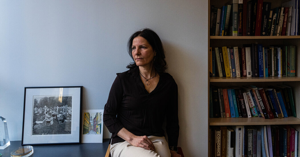
The latest edition of the DSM-5, sometimes known as “psychiatry’s bible,” includes a controversial new diagnosis: prolonged grief disorder.After more than a decade of argument, psychiatry’s most powerful body in the United States added a new disorder this week to its diagnostic manual: prolonged grief.The decision marks an end to a long debate within the field of mental health, steering researchers and clinicians to view intense grief as a target for medical treatment, at a moment when many Americans are overwhelmed by loss.The new diagnosis, prolonged grief disorder, was designed to apply to a narrow slice of the population who are incapacitated, pining and ruminating a year after a loss, and unable to return to previous activities.Its inclusion in the Diagnostic and Statistical Manual of Mental Disorders means that clinicians can now bill insurance companies for treating people for the condition.It will most likely open a stream of funding for research into treatments — naltrexone, a drug used to help treat addiction, is currently in clinical trials as a form of grief therapy — and set off a competition for approval of medicines by the Food and Drug Administration.Since the 1990s, a number of researchers have argued that intense forms of grief should be classified as a mental illness, saying that society tends to accept the suffering of bereaved people as natural and that it fails to steer them toward treatment that could help.A diagnosis, they hope, will allow clinicians to aid a part of the population that has, throughout history, withdrawn into isolation after terrible losses.“They were the widows who wore black for the rest of their lives, who withdrew from social contacts and lived the rest of their lives in memory of the husband or wife who they had lost,” said Dr. Paul S. Appelbaum, who is chair of the steering committee overseeing revisions to the fifth edition of the D.S.M.“They were the parents who never got over it, and that was how we talked about them,” he said. “Colloquially, we would say they never got over the loss of that child.”Throughout that time, critics of the idea have argued vigorously against categorizing grief as a mental disorder, saying that the designation risks pathologizing a fundamental aspect of the human experience.They warn that there will be false positives — grieving people told by doctors that they have mental illnesses when they are actually emerging, slowly but naturally, from their losses.And they fear grief will be seen as a growth market by drug companies that will try to persuade the public that they need medical treatment to emerge from mourning.“I completely, utterly disagree that grief is a mental illness,” said Joanne Cacciatore, an associate professor of social work at Arizona State University who has published widely on grief, and who operates the Selah Carefarm, a retreat for bereaved people.“When someone who is a quote-unquote expert tells us we are disordered and we are feeling very vulnerable and feeling overwhelmed, we no longer trust ourselves and our emotions,” Dr. Cacciatore said. “To me, that is an incredibly dangerous move, and short sighted.”“I completely, utterly disagree that grief is a mental illness,” said Joanne Cacciatore, an associate professor of social work at Arizona State University who operates the Selah Carefarm, a retreat for bereaved people.Adriana Zehbrauskas for The New York Times‘We don’t worry about grief’The origins of the new diagnosis can be traced back to the 1990s, when Holly G. Prigerson, a psychiatric epidemiologist, was studying a group of patients in late life, gathering data on the effectiveness of depression treatment.She noticed something odd: In many cases, patients were responding well to antidepressant medications, but their grief, as measured by a standard inventory of questions, was unaffected, remaining stubbornly high. When she pointed this out to psychiatrists on the team, they showed little interest.“Grief is normal,” she recalls being told. “We’re psychiatrists, and we don’t worry about grief. We worry about depression and anxiety.” Her response was, “Well, how do you know that’s not a problem?”Dr. Prigerson set about gathering data. Many symptoms of intense grief, like “yearning and pining and craving,” were distinct from depression, she concluded, and predicted bad outcomes like high blood pressure and suicidal ideation.Her research showed that for most people, symptoms of grief peaked in the six months after the death. A group of outliers — she estimates it at 4 percent of bereaved individuals — remained “stuck and miserable,” she said, and would continue to struggle with mood, functioning and sleep over the long term.“You’re not getting another soul mate and you’re kind of eking out your days,” she said.In 2010, when the American Psychiatric Association proposed expanding the definition of depression to include grieving people, it provoked a backlash, feeding into a broader critique that mental health professionals were overdiagnosing and overmedicating patients.“You’ve got to understand that clinicians want diagnoses so they can categorize people coming through the door and get reimbursement,” said Jerome C. Wakefield, a professor of social work at New York University. “That is a huge pressure on the D.S.M.”Still, researchers kept working on grief, increasingly viewing it as distinct from depression and more closely related to stress disorders, like post-traumatic stress disorder. Among them was Dr. M. Katherine Shear, a psychiatry professor at Columbia University, who developed a 16-week program of psychotherapy that draws heavily on exposure techniques used for victims of trauma.By 2016, data from clinical trials showed that Dr. Shear’s therapy had good results for patients suffering from intense grief, and that it outperformed antidepressants and other depression therapies. Those findings bolstered the argument for including the new diagnosis in the manual, said Dr. Appelbaum, who is chair of the committee in charge of revisions to the manual.In 2019, Dr. Appelbaum convened a group that included Dr. Shear, of Columbia, and Dr. Prigerson, now a professor at Weill Cornell Medical College, to agree on criteria that would distinguish normal grief from the disorder.The most sensitive question of all was this: How long is prolonged?Though both teams of researchers felt that they could identify the disorder six months after a bereavement, the A.P.A. “begged and pleaded” to define the syndrome more conservatively — a year after death — to avoid a public backlash, Dr. Prigerson said.“I have to say that they were kind of politically smart about that,” she added. The concern was that the public was “going to be outraged, because everyone feels because they still feel some grief — even if it’s their grandmother at six months, they are still missing them,” she said. “It just seems like you’re pathologizing love.”Measured at the year mark, she said, the criteria should apply to around 4 percent of bereaved people.The new diagnosis, published this week in the manual’s revised edition, is a breakthrough for those who have argued, for years, that intensely grieving people need tailored treatment.“It’s kind of like the bar mitzvah of diagnoses,” said Dr. Kenneth S. Kendler, a professor of psychiatry at Virginia Commonwealth University who has played an important role in the last three editions of the diagnostic manual.“It’s sort of an official blessing in the world,” he said. “If we were on the planetary committee of the American Astronomical Society deciding what is a planet or not — this one’s in, and Pluto we kicked out.”If the diagnosis comes into common use, it is likely to popularize Dr. Shear’s treatment and also give rise to a range of new ones, including drug treatments and online interventions.Dr. Shear said it was difficult to predict what treatments would emerge.“I don’t really have any idea, because I don’t know when the last time there was a really brand-new diagnosis,” she said.She added, “I really am in favor of anything that helps people, honestly.”Dr. M. Katherine Shear, a psychiatry professor at Columbia University and a founding director of the Center for Prolonged Grief, has been studying the condition since 1995.Yana Paskova for The New York TimesA loop of griefAmy Cuzzola-Kern, 54, said Dr. Shear’s treatment helped her break out of a terrible loop.Three years earlier, her brother had died suddenly in his sleep of a heart attack. Ms. Cuzzola-Kern found herself compulsively replaying the days and hours leading up to his death, wondering whether she should have noticed he was unwell or nudged him to go to the emergency room.She had withdrawn from social life and had trouble sleeping through the night. Though she had begun a course of antidepressants and seen two therapists, nothing seemed to be working.“I was in such a state of protest — this can’t be, this is a dream,” she said. “I felt like I was living in a suspended reality.”She entered Dr. Shear’s 16-session program, called prolonged grief disorder therapy. In sessions with a therapist, she would narrate her recollection of the day that she learned her brother had died — a painful process, but one that gradually drained the horror out of the memory. By the end, she said, she had accepted the fact of his death.The diagnosis, she said, mattered only because it was a gateway to the proper treatment.“Am I ashamed or embarrassed? Do I feel pathological? No,” she said. “I needed professional help.”Yet, others interviewed said they were wary of any expectation that grief should lift in a particular period of time.“We would never put a time frame around when someone should or shouldn’t feel that they have moved forward,” said Catrina Clemens, who oversees the victim services department of Mothers Against Drunk Driving, which provides services to bereaved relatives and friends. The organization encourages bereaved people to seek mental health care, but has no role in diagnosis, said a spokesperson.Filipp Brunshteyn, whose 3-year-old daughter died after an automobile accident in 2016, said grieving people could be set back by the message that their response was dysfunctional.“Anything we inject into this journey that says, ‘that’s not normal,’ that could cause more harm than good,” he said. “You are already dealing with someone very vulnerable, and they need validation.”To set a year as a point for diagnosis is “arbitrary and kind of cruel,” said Ann Hood, whose memoir, “Comfort: A Journey Through Grief,” describes the death of her 5-year-old daughter from a strep infection. Her own experience, she said, was “full of peaks and valleys and surprises.”The first time Ms. Hood walked into her daughter Grace’s room after her death, she saw a pair of ballet tights lying in a tangle on the floor where the little girl had dropped them. She screamed. “Not the kind of scream that comes from fright,” she later wrote, “but the kind that comes from the deepest grief imaginable.”She slammed the door, left the room untouched and eventually turned off the heat to that part of the house. At the one-year mark, a well-meaning friend told her it was time to clear out the room — “nothing worse than a shrine,” he told her — but she ignored him.Then one morning, three years after Grace’s death, Ms. Hood woke up and returned to the room. She sorted her daughter’s clothes and toys into plastic bins, emptied the bureau and closet and lined up her little shoes at the top of the stairs.To this day, she is not sure how she got from one point to the other. “All of a sudden, you look up,” she said, “and a few years have gone by, and you’re back in the world.”

Read more →

Jack Willis, TV Producer and Empathetic Filmmaker, Dies at 87

A survivor of a crippling accident, his documentaries and news coverage for public television focused on poverty, race and other social issues.Jack Willis, a journalist and television executive who won several Emmys and a Polk Award for his innovative films and news and documentary programming during the embryonic years of cable and public broadcasting, died on Feb. 9 in Zurich. He was 87.He underwent assisted suicide at a clinic there, his wife, Mary Pleshette Willis, said. He lived in Manhattan.When he was in his late 30s, Mr. Willis broke his neck in a body surfing accident that temporarily left him a quadriplegic before he miraculously recovered, his wife said, inspiring a television movie. But after a half century, the injuries were taking their toll. Six years ago, he broke his hip and began using a wheelchair, she said.From 1971 to 1973, Mr. Willis was director of programming and production for WNET, the public television station in New York, where he introduced innovative local news coverage as executive producer of “The 51st State,” a program that took its name from the 1969 campaign of the author Norman Mailer during his zany run for mayor in which he proposed that New York City secede from New York State.The program, which won an Emmy Award, focused on communities rather than the more traditional fare of the nightly local news.“He pioneered in-depth local coverage of New York’s outer boroughs on WNET, focusing on long-ignored and disenfranchised minorities and immigrants, often letting them speak for themselves,” said Stephen B. Shepard, former editor in chief of Business Week and founding dean of the City University of New York Graduate School of Journalism. “For Jack, it was always about the people affected by government decisions.”Mr. Willis was an executive producer of another Emmy-winning series, “The Great American Dream Machine,” a weekly 90-minute program on PBS. The television critic John J. O’Connor of The New York Times, writing in 1971, said the program had been conceived as “a free‐form program that could offer the viewer worthwhile bits and pieces of humor, controversy, entertainment, investigative reporting, opinion, documentary and theatrical sketches.”“It has been called a hodgepodge of the brilliant and the trite,” he added, but concluded that it was “one of the most exciting and imaginative segments of television to come along this season.”Looking back, Mr. Willis himself told The Times in 2020: “It was a great time in public television. If you thought it, you could do it.”In 1963, he directed his first documentary, “The Streets of Greenwood,” a 20-minute film about a voter-registration drive in the Mississippi Delta. Collaborating with two friends, Phil Wardenburg and John Reavis, Mr. Willis shot it with a camera he had borrowed from the folk singer Pete Seeger, whose concert in a cotton field was featured in the film.In 1979, Mr. Willis shared the George Polk Award for best documentary with Saul Landau for “Paul Jacobs and the Nuclear Gang.” The film focused on the journalist Paul Jacobs’s investigation of radiation hazards from atomic testing in Nevada in the 1950s and ’60s and the federal government’s efforts to suppress information on its threat to public health.Two other films he produced — “Lay My Burden Down” (1966), about the plight of tenant farmers in rural Alabama, and “Every Seventh Child” (1967), questioning tax subsidies and other government benefits for Catholic education — were shown at the New York Film Festival.Mr. Willis wrote, directed and produced “Appalachia: Rich Land Poor People” (1968), which exposed grinding poverty largely caused, the film argued, by corporate greed, racism and ineffective local government.Mr. Willis’s commitment to civil rights was reflected in his enduring friendship with the singer Harry Belafonte, an activist in the movement, who described Mr. Willis in an email as “a soul brother” whose “intellect and humor, combined with his courageousness, make him one of the most precious people I have ever known.”“For those on the political left,” Mr. Belafonte added, “he was living proof of the proverb, ‘You can cage the singer but not the song.’”Jack Lawrence Willis was born on June 20, 1934, in Milwaukee to Louis Willis, a manufacturer of women’s shoes, and Libbie (Feingold) Willis, a homemaker. The family moved to California when he was 9.He earned a bachelor’s degree in political science in 1956 from the University of California, Los Angeles, where he also played shortstop on the varsity baseball team. He liked to recall that he was recruited by a Boston Red Sox farm team.Mr. Willis dropped out of U.C.L.A. School of Law to serve in the Army for two years, then graduated in 1962 and moved to New York, where he hoped to connect with a job teaching in Africa or the Middle East.While waiting for a job abroad that never materialized, he worked briefly in television for Allen Funt’s “Candid Camera” and David Susskind’s “Open End.”He ran a movie production company in California, then was hired as vice president for programming and production at CBS Cable, a short-lived but well-received performing arts channel.From 1990 to 1997, Mr. Willis was president of KTCA, the public television station in Minneapolis-St. Paul, Minn., then returned to New York, where, working for George Soros’s Open Society Institute, he developed a media program. In 1999, he was a founder of Link TV, a nonprofit satellite TV network. He retired in 2011.In addition to his wife, he is survived by their two daughters, Sarah Willis and Kate Willis Ladell; three grandchildren; and his brother, Richard.Mr. Willis and his wife wrote a book, “… But There Are Always Miracles” (1974), about his body-surfing accident in 1969 off Southampton, N.Y. They had been planning to marry when a crashing wave broke his neck and left him paralyzed from the chest down. He was told he would never walk again.After two operations and six months of inpatient rehabilitation, he walked out of Rusk Institute of Rehabilitation Medicine in Manhattan. The couple married a year later.His story was adapted into a TV film, “Some Kind of Miracle” (1979), with a screenplay by the couple. They wrote and produced other films together.Shortly before he died, Ms. Willis said, her husband told her that the accident had “taught me to put everything in perspective — including the fear of failure.” He admitted to no regrets, she said, “except,” she quoted him as saying, “for taking that wave and turning down the Boston Red Sox.”

Read more →

Marijuana for medical use may result in rapid onset of cannabis use disorder

Obtaining a medical marijuana card (MMC) to use cannabis products to treat pain, anxiety, or depression symptoms led to the onset of cannabis use disorder (CUD) in a significant minority of individuals while failing to improve their symptoms, according to a study by Massachusetts General Hospital (MGH) researchers and published in JAMA Network Open. Researchers found that individuals at greatest risk of developing the addictive symptoms of CUD were those seeking relief from anxiety and depression, suggesting the need for stronger safeguards over the dispensing, use, and professional follow-up of people who legally obtain cannabis through MMCs.
“There have been many claims about the benefits of medical marijuana for treating pain, insomnia, anxiety and depression, without sound scientific evidence to support them,” says lead author Jodi Gilman, PhD, with the Center for Addiction Medicine at MGH. “In this first study of patients randomized to obtain medical marijuana cards, we learned there can be negative consequences to using cannabis for medical purposes. People with pain, anxiety or depression symptoms failed to report any improvements, though those with insomnia experienced improved sleep.” Particularly disturbing to Gilman was the fact individuals with symptoms of anxiety or depression — the most common conditions for which medical cannabis is sought — were most vulnerable to developing cannabis use disorder. CUD symptoms include the need for more cannabis to overcome drug tolerance, and continued use despite physical or psychological problems caused by the cannabis.”
“Medical” cannabis has surged in popularity as 36 states and the District of Columbia have commercialized its use (as of December 2021) for myriad health conditions through medical marijuana cards. These cards require written approval of a licensed physician who, under the current system, is typically not the patient’s primary care provider but a “cannabis doctor” who may provide authorization to patients with only a cursory examination, no recommendations for alternative treatments, and no follow-up. Indeed, the medical marijuana industry functions outside regulatory standards that apply to most fields of medicine.
MGH researchers began their trial in 2017 with 269 adults (average age of 37) from the greater Boston area who were interested in obtaining a medical marijuana card. One group was allowed to get MMCs immediately, while the second group, designed to serve as a control, was asked to wait 12 weeks before obtaining a card. Both groups were tracked over 12 weeks. The team found that the odds of developing CUD were nearly two times higher in the MMC cohort than in the wait list control group, and that by week 12, 10 percent of the MMC group had developed a CUD diagnosis, with the number rising to 20 percent in those seeking a card for anxiety or depression.
“Our study underscores the need for better decision-making about whether to begin to use cannabis for specific medical complaints, particularly mood and anxiety disorders, which are associated with an increased risk of cannabis use disorder,” says Gilman. Regardless of the specific health condition for which cannabis is sought, Gilman believes that regulation and distribution of cannabis to people with medical marijuana cards must be greatly improved. “There needs to be better guidance to patients around a system that currently allows them to choose their own products, decide their own dosing, and often receive no professional follow-up care.”
Gilman is associate professor of Psychiatry at Harvard Medical School (HMS). Senior author A. Eden Evins, MD, is the Cox Family Professor of Psychiatry at HMS.
The study was funded by the National Institute on Drug Abuse (NIDA).
Story Source:
Materials provided by Massachusetts General Hospital. Note: Content may be edited for style and length.

Read more →