How multiple genes impact vision development and result in rare sight disorder

An international team of health researchers have, for the first time, described how genetic defects influence the spectrum of vision development and cause problems in developing babies’ eyes.
Researchers from the University of Leicester led an international effort consisting of 20 expert centres in the largest study of its kind to date, examining the genes associated with arrested development of the fovea.
The fovea is part of the retina at the back of the human eye, and is the structure responsible for sharp, central vision. Arrested development of the fovea, or foveal hypoplasia, is rare, and is often caused by genetic changes. This lifelong condition can have serious consequences and can affect the individual’s ability to read, drive and complete other daily tasks.
There are currently no treatments available for this condition. Most often, during infancy, one of the first visible signs of a foveal problem is ‘wobbly eyes’. This is often seen in the first six months of life. There are large gaps in our knowledge about which genes control the development of the fovea and at what time points during development this occurs.
Now, in a study published in the journal Ophthalmology combining data from more than 900 cases across the world, researchers have been able to identify the spectrum of genetic changes behind these foveal defects and — crucially — at which point they occur in the development of the unborn baby.
Dr Helen Kuht is a research orthoptist and Wellcome Trust post-doctoral fellow within the Ulverscroft Eye Unit at the University of Leicester, and first author for the study. She said:
“This research has really helped to solve the puzzle of why some babies with these genetic changes present with varying severity of foveal hypoplasia. Thus allowing us to diagnose, predict future vision and help prioritise genetic testing, subsequent counselling, and support.”

Read more →

High performance microscopy for non-invasive conjunctival goblet cell examination

Conjunctival goblet cells (CGCs) are specialized epithelial cells secreting mucins to form the mucus layer of tear film. The mucus layer spreads the tear film on the ocular surface for protection. The dysfunction and death of CGCs causes tear film instability and is associated with various ocular surface diseases including the dry eye disease (DED). Because DED is a multifactorial disease with multiple causes, it is important to find the causes and disease status. Therefore, CGC examination is important for the precise diagnosis and effective treatment of ocular surface diseases; however, CGC examination has not been possible until now due to lack of non-invasive devices.
A POSTECH research team led by Professor Ki Hean Kim and Ph.D. candidates Jungbin Lee and Seonghan Kim (Department of Mechanical Engineering), in collaboration with professors Hong Kyun Kim and Byeong Jae Son (Department of Ophthalmology) of Kyungpook National University and Professor Chang Ho Yoon (Department of Ophthalmology) of Seoul National University, has developed a high-performance microscopy system for non-invasive CGC examination in patients. Recognized for technical advancement and potentials, this research has recently been published in IEEE Transactions on Medical Imaging, an international journal on medical imaging.
Earlier in 2019, the research team had discovered for the first time that moxifloxacin, an FDA-approved ophthalmic antibiotic, stains CGCs, and demonstrated high-contrast CGC imaging by using moxifloxacin as a cell labeling agent. However, CGC imaging in humans was impossible due to various limitations of conventional microscopy techniques such as shallow depth-of-fields (DOFs) and slow imaging speeds.
To overcome these limitations, the research team developed a high-speed extended DOF microscopy which had a 1 mm DOF (25x DOF extension) and 10 frames per second imaging speed. A deformable mirror was used in the system to axially sweep the imaging plane and to capture CGCs on the arbitrary tilted conjunctiva in single frames. The acquired images contained both in-focus and out-of-focus information, and the deconvolution was used to filter the in-focus information only. 
Professor Ki Hean Kim of POSTECH explained, “The newly developed imaging system can obtain high-resolution in-focus images of CGCs in live animal models and is also applicable to humans.” He added, “Going forward, we will develop a device for imaging patients and then run clinical trials to test the feasibility of non-invasive CGC examination in the diagnosis and treatment of ocular surface diseases.”
This study was conducted with the support from the Samsung Research Funding & Incubation Center (Project number SRFC-IT2101-05).
Story Source:
Materials provided by Pohang University of Science & Technology (POSTECH). Note: Content may be edited for style and length.

Read more →

Researchers rejuvenate mouse organs through cellular reprogramming

Many diseases, including cancer, are associated with ageing and they are becoming more prevalent as life expectancy increases. Therefore, studying and understanding these processes is crucial if we are to deal with these conditions and also promote healthier ageing.
One of the fields within research into ageing addresses rejuvenation, that is, the restoration of the characteristics of younger cells in aged cells or tissues.
Headed by ICREA researcher Dr. Manuel Serrano, scientists from the Cellular Plasticity and Disease lab at IRB Barcelona have managed to rejuvenate some mouse organs and tissues via cell reprogramming. Specifically, the scientists have observed significant changes in the pancreas, liver, spleen, and blood of the animals.
“This work sought to identify the initial processes of in vivo reprogramming and cell rejuvenation and pinpoint those that can be modified in future studies, whether by drugs or at a nutritional level,” explains Dr. Serrano.
The Yamanaka factors and cell reprogramming
All tissues in our bodies are characterised by having highly specialised cells, such as neurons or muscle cells, among many others. The identity of these cells was considered fixed and inflexible until the Japanese researcher Shinya Yamanaka found a way to alter their identity (that is to say, “reprogram them”) by introducing high levels of four proteins, called the “Yamanaka factors” (OCT4, SOX2, KLF4 and MYC), into them. Although these proteins can be found in some of our cells, it is the simultaneous presence of high levels of all four that can alter cell identity.

Read more →

Biodegradable gel boosts immune system's attack on several cancers in mice

A new biodegradable gel improves the immune system’s ability to keep cancer at bay after tumors are surgically removed.
The gel, tested in mice, releases drugs and special antibodies that simultaneously deplete immune-blocking cells called macrophages from the surgical site and activate T cells so they can attack cancer.
University of Wisconsin-Madison scientists tested the gel on mouse models of several cancers. They found that the gel effectively kept in check tumors that are known to respond well to this kind of immune therapy, like CT26 colon cancers. But the gel also worked well against B16F10 melanomas, S180 sarcomas and 4T1 triple negative breast cancers, which are less responsive to immune therapy and more prone to metastasizing.
These proof-of-concept experiments will support additional research on other animal models that could lead to future clinical trials in people.
The experiments were led by the lab of Quanyin Hu, a professor in the UW-Madison School of Pharmacy, with support from pharmacy professor Seungpyo Hong and colleagues in the UW School of Medicine and Public Health. The team published their findings April 6 in the journal Nature Communications.
“We are really glad to see that this local strategy can work against so many different kinds of tumors, especially these non-immunogenic tumors,” says Hu. “We are even more glad to see this local treatment can inhibit tumor metastasis.”
Surgery is an excellent treatment for many tumors, but small numbers of cancer cells that remain after the operation can allow tumors to grow back. To counteract this process, the researchers developed their gel to slowly release into the surgical site two key components.

Read more →

Century-old malaria parasite puzzle solved as ape origin traced

Scientists have solved a 100-year-old mystery about the evolutionary links between malaria parasites that infect humans and chimpanzees.
They have discovered that the parasite P. malariae – one of six species that spreads malaria among humans – originated in African apes before evolving to infect people.
While it is often associated with mild disease, if untreated P. malariae can cause long-lasting, chronic infections that may last a lifetime, researchers say.
The evolutionary puzzle has its origins in the 1920s when scientists identified chimpanzees infected by parasites that appeared identical to P. malariae under a microscope.
It was thought both parasites belonged to the same species, but – until now – this could not be verified as the genetic make-up of the chimpanzee strain had never been studied.
Now, scientists at the University of Edinburgh, in collaboration with colleagues at the University of Pennsylvania, USA, have used leading edge techniques to study the parasites’ DNA.

Read more →

Achilles’ heel of dangerous hospital pathogen

Each year, over 670,000 people in Europe fall ill through pathogenic bacteria that are resistant to antibiotics, and 33,000 die of the diseases they cause. In 2017, the WHO named antibiotic resistance as one of the greatest threats to health worldwide. Especially feared are pathogens that are resistant to several antibiotics. Among them, Acinetobacter baumannii stands out, a bacterium with an extraordinarily pronounced ability to develop multiresistance and, as a “hospital superbug,” dangerous above all for immunosuppressed patients. Acinetobacter baumannii is highly resilient because it can remain infectious for a long time even in a dry environment and thus endure on the keyboards of medical devices or on ward telephones and lamps. This property also helps the microbe to survive on dry human skin or in body fluids such as blood and urine, which contain relatively high concentrations of salts and other solutes.
The team from Research Unit 2251 of the German Research Foundation led by Goethe University has now shed light on a central mechanism via which Acinetobacter baumannii settles in such an adverse environment: like many bacteria as well as plants or fungi, Acinetobacter baumannii is able to synthesize the sugar alcohol mannitol, a substance excellent at binding water. In this way, Acinetobacter baumannii prevents desiccation.
Almost unique, however, is the way that Acinetobacter baumannii synthesizes mannitol: instead of two enzyme complexes as are common in most organisms, the two last steps in mannitol synthesis are catalysed by just one. A team of researchers led by Professor Beate Averhoff and Professor Volker Müller already discovered this “MtlD” enzyme with two catalytic activities back in 2018. The team headed by Professor Klaas Martinus Pos, who is also a member of the Research Unit, has now succeeded in shedding light on the enzyme’s spatial structure.
He explains: “We’ve discovered that the enzyme is usually present in the form of free monomers. Although these have the necessary catalytic activities, they are inactive. Only a dry or salty environment triggers what is known as ‘osmotic stress’ in the bacterium, after which the monomers aggregate as dimers. Only then does the enzyme become active and synthesize mannitol.” The researchers have also identified which parts in the structure are particularly important for the enzyme’s catalytic functions and for dimer formation.
Professor Volker Müller, spokesperson for Research Unit 2251, is convinced: “Our work constitutes an important new approach for fighting this hospital pathogen since we’ve identified a biochemically sensitive point in the pathogen’s metabolism. In the future, this could be the starting point for customized substances to inhibit the enzyme.”
Story Source:
Materials provided by Goethe University Frankfurt. Note: Content may be edited for style and length.

Read more →

The cacao tree enigma

Cacao has long been a sought-after raw material for the worlds food industry. At first glance, it therefore seems surprising that biology knows little about the pollination of the cacao tree — although it is precisely this process that is the basis for fruit set and ultimately for the yield.
At second glance, however, one quickly understands why the pollination of this tropical crop holds so many secrets: cacao flowers are very small and are usually found by the thousands on a tree. The insects that gather at the flowers are also tiny and very diverse in terms of species. All these factors make systematic observations difficult.
Study in the north and south of Peru
A new study now brings more clarity. It was conducted in Peru by an international research team at the Chair of Animal Ecology and Tropical Biology at Julius-Maximilians-Universität (JMU) Würzburg, Germany. The leading project organization was Bioversity International, the Deutsche Gesellschaft für internationale Zusammenarbeit (GIZ) funded the project.
South America is the native region of the cacao tree, which occurs there in the undergrowth of tropical rainforests. In agriculture, too, the cacao tree is planted in the shade of larger trees, in so-called agroforestry systems. The researchers applied glue to cacao flowers in 20 such systems in northern and southern Peru to investigate which animals visit the flowers. They also analysed the influence of the degree of shading and the distance to the nearest forest on visitor activity at the flowers.
The results have been published in the journal Ecological Solutions and Evidence. The first author is biologist Justine Vansynghel, who has been a doctoral student at JMU under Professor Ingolf Steffan-Dewenter since 2018.

Read more →

Laser light, a dye and a nonsurgical implant could help overcome obesity

When dieting and exercise aren’t enough, weight-loss surgery can be an effective obesity treatment. But people who don’t want surgery have other options, including insertion of an appetite-suppressing balloon or other implant in the stomach. Now, researchers report in ACS Applied Materials & Interfaces that they have augmented that procedure in laboratory animals by coating an implant with a laser-activated dye that kills cells producing ghrelin, the “hunger hormone.”
Implants can be inserted in the stomach through the mouth after local anesthesia. In 2019, Hwoon-Yong Jung, Jung-Hoon Park and colleagues designed a new type of implant. The “intragastric satiety-inducing device” (ISD) consists of a stent — which lodges in the lower esophagus — attached to a disk that rests in the opening to the stomach. The disk has a hole in the center to let food through. Tests in pigs showed that the ISD lowered food intake and weight gain by enhancing the feeling of fullness and reducing levels of ghrelin, which is produced by cells near the top of stomach. But the device caused complications, including acid reflux and migration into the stomach. In their latest project, Jung, Park, Kun Na and colleagues wanted to find out if they could suppress ghrelin even more by coating the ISD’s disk with a compound that, with a shot of laser light, could kill some of the ghrelin-producing cells. The implant could then be removed to avoid the side effects associated with the initial design.
In this preliminary study, the team coated ISDs with methylene blue — an FDA-approved drug — and then placed them in the stomachs of young pigs. When exposed to laser light, the coating released singlet oxygen, an energized form of oxygen that killed nearby ghrelin-producing cells in the pigs’ stomachs and then rapidly disappeared. After one week, the treatment reduced ghrelin levels and body weight gain by half compared with an untreated pig, though these differences declined in the following weeks unless the light treatment was repeated. With further development, the simple procedure could become a new type of minimally invasive treatment to help obese patients lose weight, the researchers say.
The authors acknowledge support from the Korea Medical Device Development Fund grant funded by the Korean government (Project number: KMDF_PR_20200901_0036).
Story Source:
Materials provided by American Chemical Society. Note: Content may be edited for style and length.

Read more →

A second booster helped protect older people from Omicron infection, but waned quickly, an Israeli study says.

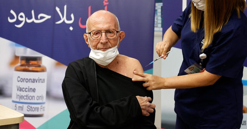
A second booster shot of the Pfizer-BioNTech Covid vaccine provides additional short-term protection against Omicron infections and severe illness among older adults, according to a large new study from Israel.But the booster’s effectiveness against infection in particular wanes after just four weeks and almost disappears after eight weeks. Protection against severe illness did not ebb in the six weeks after the extra dose, but the follow-up period was too short to determine whether a second booster provided better long-term protection against severe disease than a single booster.The study focused on adults ages 60 and older, and did not provide data on the effectiveness of a second booster in younger populations.The findings, published on Tuesday in the New England Journal of Medicine, suggest that additional boosters are likely to provide fleeting protection against Omicron infections in older recipients, and are consistent with evidence that vaccine effectiveness against infection wanes faster than against severe disease.“For confirmed infection, a fourth dose appeared to provide only short-term protection and a modest absolute benefit,” the researchers wrote.The results come in the midst of a debate over whether and when Americans might need additional boosters. The Food and Drug Administration is convening a panel of outside advisers on Wednesday for discussion on the broader U.S. booster strategy.The rapid spread of the highly transmissible Omicron variant, which can evade some of the body’s immune defenses, has intensified the discussion of whether second boosters are broadly necessary.Last month, the F.D.A. authorized second booster shots of the Pfizer-BioNTech and Moderna vaccines for adults ages 50 and older, as well as immunocompromised people ages 12 and older. The agency also authorized an mRNA booster for adults who have already received two doses of the Johnson & Johnson vaccine.It’s likely to be a tough sell: While 66 percent of Americans have been vaccinated, just 30 percent have received a booster shot.It is clear that the Omicron variant has blunted the effectiveness of Covid vaccines, but data on the benefits of a second booster remains limited. A previous study from Israel, which has not yet been published in a scientific journal, found that older adults who received a second booster were 78 percent less likely to die of Covid-19 than those who had received just one booster shot.But scientists criticized the study’s methodology, and the benefits of a second booster for young, healthy adults are less clear. Some experts note that most adults who have been vaccinated and boosted once are already likely to be protected from severe illness and death.On Jan. 2, Israel authorized a fourth dose of the Pfizer-BioNTech vaccine for adults ages 60 and older and members of other high-risk populations who had received their third shots at least four months earlier. Israel’s vaccination campaign has relied heavily on the Pfizer-BioNTech vaccine.The new study is based on records from the Israeli Ministry of Health on more than 1.2 million older adults who were eligible for the fourth shot between Jan. 10 and March 2, when Omicron was the dominant variant in the country.The researchers compared the rate of confirmed virus infections and cases of severe Covid-19 among those who had received a fourth dose to those who had received just three doses.Protection against infection appeared to peak four weeks after the fourth shot: the rate of confirmed infections was twice as high in the three-dose group as in the four-dose group. By eight weeks after the fourth shot, however, the additional protection against infection had almost disappeared, the researchers found.Rates of severe disease were 3.5 times higher in the three-dose group than the four-dose group four weeks after the booster shot, the researchers found. That protection did not appear to wane and actually ticked up slightly by the sixth week after the shot, when rates of severe disease were 4.3 times higher in the three-dose group.But the study covered a relatively short period, and whether the benefits against illness hold up over the longer term remains unknown. The study did not report data on deaths.

Read more →

In India, Parents of Children with Rare Disease Plea for Help Online

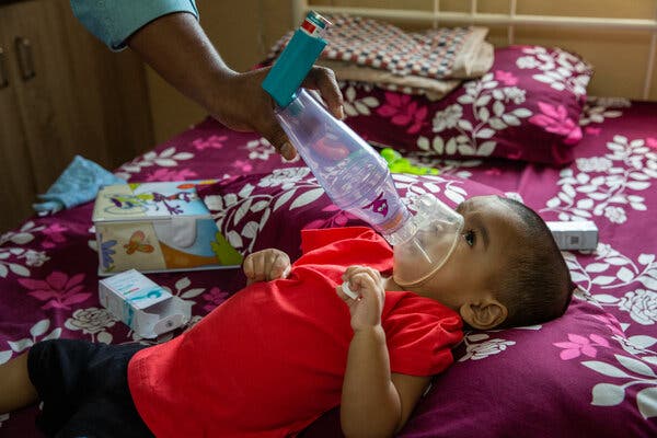
ELURU, India — When her baby started struggling to breathe, Stella Praveen had a terrible feeling that something was gravely wrong with her 14-month-old daughter, Ellen.She ran barefoot to a nearby clinic, but the doctors there said the child needed to see a specialist right away. Without an ambulance, she jumped on the back of a motorcycle and rode 35 miles to a children’s hospital in another town, where Ellen remained in intensive care for 12 days.Two weeks later, Ms. Praveen learned that her daughter, who had never been able to lift her neck nor roll over, was suffering from spinal muscular atrophy, a rare condition often fatal by age 2.“We had not even heard of this disease,” Ms. Praveen said as tears rolled down her face. “She was misdiagnosed many times.”The Praveen family was momentarily heartened when it learned that a promising gene therapy treatment was available, but was quickly crestfallen when it heard the cost: $2.1 million.In India, and in many of the world’s poorer countries, the pharmaceutical industry’s latest advances for rare diseases are often agonizingly out of reach, impossible for almost all but the wealthiest families to afford and not covered by health insurance.In desperation — and encouraged by the occasional success story — families are turning to social media to raise funds.Every morning, Ellen’s father, Rayapudi Praveen, sends hundreds of emails on five crowdfunding websites like ImpactGuru and GoFundMe, asking people to contribute money to save his daughter’s life.“Dear Sir, my daughter Ellen is suffering,” every email begins. “Can you help us?”With only four months left before Ellen turns 2, time is running out — and the family is still far from its goal.Spinal muscular atrophy is an inherited neuromuscular disease that kills more infants worldwide than any other genetic disorder. In India, one study put its prevalence at one in every 7,744 live births, or about 3,200 Indian babies each year. Symptoms for all four types of the condition emerge at different stages. Infants like Ellen with Type 1, the most severe, show symptoms within their first six months of life: struggling to move their limbs, to swallow, to suck and eventually to breathe. They usually do not live past 2.Ellen being given an inhaler by her father in February, when she was 18 months old. She has Type 1 spinal muscular atrophy and has trouble breathing.Ellen’s parents looking at her medical records. Her ailment was diagnosed in October.Across India, pediatric neurologists said, rising awareness among parents about the disease is leading to more patients being identified.In recent years, India has established a reputation as a low-cost manufacturing hub for multinational pharmaceuticals, and the drugs made here are often substantially cheaper than imported ones, thanks in part to government price caps.But the therapies for many rare diseases are still typically imported, forcing patients and parents to confront an excruciating truth: India’s status as a rising pharmaceutical superpower is of no help to them.In 2019, the U.S. Food and Drug Administration approved the gene therapy Zolgensma, which alters the underlying genetic cause of spinal muscular atrophy and may permanently stop the disease’s progression.At $2.1 million, the Zolgensma therapy from the pharmaceutical firm Novartis is believed to be the highest price ever set for a one-time treatment.Spinraza, another drug, costs $750,000 in the first year and $375,000 a year after that, and needs to be taken for a lifetime.Neither Zolgensma nor Spinraza, made by Biogen, is manufactured in India or approved for use here, so parents import them with the help of their doctors, a process that involves special government approvals.The only drug approved for the condition in India is Evrysdi, manufactured by Roche. It’s the cheapest among the three treatments, but it still costs $53,000 to $80,000 a year, and that’s a discounted price for India, negotiated with Roche by the government.None of these drugs are covered by insurance in India, so families face a wrenching choice: Raise the necessary money or see their children waste away.So far, the Praveens have raised just over $100,000 for Ellen’s treatment, but they’re not giving up hope, and their optimism isn’t entirely unfounded.Since May 2019, when Zolgensma was introduced, the parents of at least 10 children have succeeded in raising the $2.1 million through crowdfunding.Last year, Yogesh Gupta started a crowdfunding campaign and sent emails to anyone he knew asking for help for his son, Ayaansh, who has Type 1. Soon a team of 125 friends, colleagues and relatives began sending messages on social media platforms to politicians and Bollywood stars. Moved by the plight of the child, the officials and celebrities not only donated money themselves but also helped spread the word. After three and a half months, Mr. Gupta said he raised $2.1 million.“There is a lot of improvement,” Mr. Gupta said of his son after he received the Zolgensma treatment. “He can slightly lift his legs and neck control is far better.”Raman Nagumantri is more than halfway there, having raised $1.6 million for his 19-month-old daughter, Khyati.Khyati Nagumantri, a few months shy of her second birthday, at home in Bangalore, India, in February. She has Type 1 spinal muscular atrophy, and her parents estimate she is on oxygen for more than 18 hours a day.Raman Nagumantri, Khyati’s father, has raised $1.6 million on crowdfunding sites for his daughter’s treatment.  “We don’t remember a day when we have slept for the whole night since she was diagnosed,” Mr. Nagumantri said. “But we are close, and I can do anything, anything, to get those required funds in these four months.”For almost all the world’s children with Type 1, their best chance of survival may lie with the Global Managed Access Program, or gMAP, which provides Zolgensma for free to a select number of eligible patients under the age of 2 in countries where the gene therapy has not received regulatory approval or is not covered by insurance.Representatives from Novartis said more than 250 children from around the world had received the therapy free through gMAP.Novartis declined to share the total number of patients from India, but Dr. Ann Mathew, a leading pediatric neurologist, who has over 400 spinal muscular atrophy patients, said 40 children had received the treatment in the past year across India, the majority through gMAP. Nineteen of her patients have taken Zolgensma in the past 13 months, 16 free and three paid.Biogen said 200 patients in India had received Spinraza for free.Patient advocacy groups are pushing for government intervention to negotiate better prices with pharmaceutical companies.“When the government intervenes, the prices will automatically go down,” said Alpana Sharma, co-founder of Cure SMA, a parent-led advocacy group. “This is what happened with cancer and other rare diseases like hemophilia.”While the parents of children with Type 1 face a harrowingly short time period for a cure, the treatments for Type 2, which has debilitating effects but is not typically fatal before adulthood, are also far beyond the means of most caregivers.In the coastal state of Goa, Ruby Borges and her husband, Benedict Borges, were devastated when their 5-year-old son, Dylan, was diagnosed three years ago with S.M.A.-Type 2. In most cases, Type 2 symptoms arrive between six and 18 months, and children suffering from it cannot walk.Dylan, who was diagnosed with S.M.A.-Type 2 three years ago, receiving treatment at home in Goa, India.Dylan’s mother says she has seen a 20 percent improvement in his condition because of the daily physiotherapy and the drug he takes, Evrysdi.At the time of Dylan’s diagnosis, Spinraza was the only treatment available.After he didn’t get into the compassionate access program, his parents turned to crowdfunding. Months of appeals made through church groups in their community helped collect $57,000. At that rate, it would take years to source the money to pay for Spinraza, and in the meantime, Dylan was growing weaker as his muscles atrophied.Doctors advised Dylan’s parents to start him on Evrysdi. They managed to buy enough supply of that drug to last through the end of the year. Dylan’s mother feels that the medication and intense physiotherapy are helping, and she said she had seen a 20 percent improvement in his condition. But she worries about how long she can keep relying on the generosity of strangers to keep her son alive.“People laugh when they hear the price of the medicine,” Ms. Borges said. “They wonder if I’m going to spend it on a car or a big house.”The parents of Ellen need even more money and have even less time.On a recent afternoon, her father walked down a dirt road toward a nearby highway, where he hitched a ride to the city of Vijayawada. Hours later, he arrived at the large house of a businessman-philanthropist he hoped could help.But it was not to be.“Accept your fate and move on,” the businessman told him.Mr. Praveen looked through a window onto the businessman’s sprawling lawn and vowed not to give up.“I will fight to her last breath,” he said.Besant Road, a market area in Vijayawada, the city where Ellen’s condition was diagnosed. One study found the prevalence of the disease in India to be one in every 7,744 live births.

Read more →