How T cell-derived interleukin-22 promotes antibacterial defense of colonic crypts

Intestinal epithelial cells line the inner wall of the gut, creating a barrier to dangerous bacteria like enteropathogenic E. colithat seek to attach and efface that barrier, causing diarrhea. Such pathogens pose significant risks to human health and cause infant death in developing countries.
In a study published in the journal Immunity, Carlene L. Zindl, Ph.D., and Casey T. Weaver, M.D., of the University of Alabama at Birmingham Department of Pathology show how two types of immune cells — one a part of the innate immune system and the other a part of the adaptive immune system — play distinct and indispensable roles to defend that barrier.
“In this study, we define a nonredundant role for interleukin-22-producing T cells in antibacterial defense of colonic crypts,” Weaver said. “Our findings address a central, unresolved issue regarding the coordination of innate and adaptive immunity and specialization of innate lymphoid cells, or ILCs, and CD4 T cells. Since the discovery of ILC subsets and appreciation of their functional parallel with T cell subsets, it has been unclear what functions are unique to each immune cell population.”
The study used mice with bacterial infection of the colon by Citrobacter rodentium, which models human disease caused by enteropathogenic and enterohemorrhagic E. coli. Colons of mice and humans have surface intestinal epithelial cells, or surface IECs, that face the lumen of the colon and line the mouths of colonic crypts. The colonic crypts are the numerous tiny indentations in the colon that are shaped like thick-walled test tubes; at the bottom of each crypt are stem cells that give rise to all new IEC subsets.
Each crypt is only about 75 to 110 cells deep and 23 cells in circumference, and the human colon has about 9,950,000 crypts. Crypt IECs line each crypt.
Interleukin-22, or IL-22, is a cytokine signaling protein produced by cells to initiate an immune response. The UAB researchers developed mice that have a reporter gene in IL-22, so they could tell which cells produced IL-22. They also were able to target a deficiency of IL-22 to different immune cell populations, to learn the effect of that loss of IL-22 production in a subset of cells upon the progress of C. rodentium infections.

Read more →

Having Covid Can Be Confusing. Here’s What to Expect

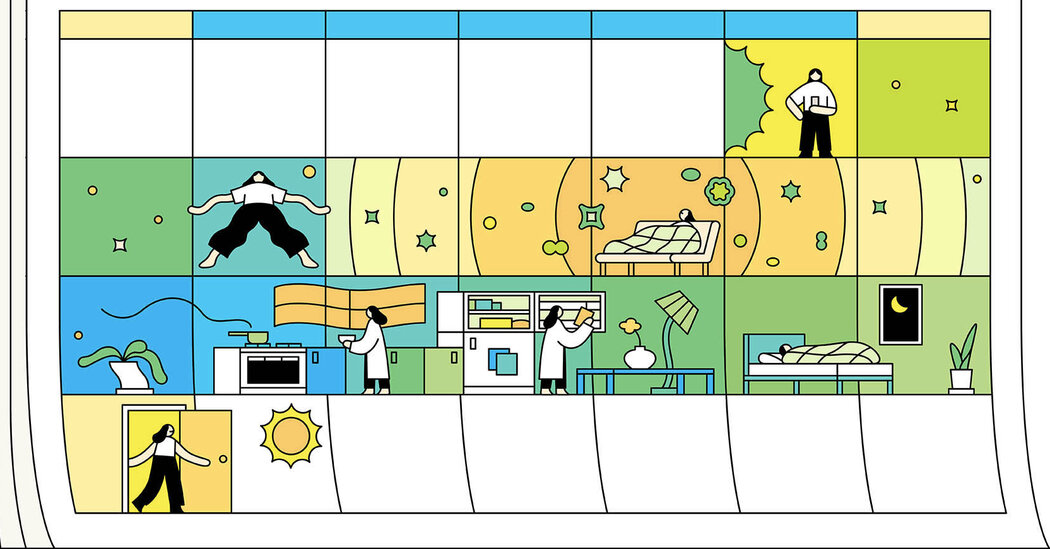
Having Covid can be a wildly confusing experience. But you can still make a plan to get through the course of illness.The highly contagious subvariant of Omicron, known as BA.2, has prompted concern among health officials as it becomes the dominant version of the coronavirus around the world. So far, signs suggest that BA.2 is 30 to 80 percent more transmissible than the original version of Omicron, and cases are already going up in several states across the United States.While we have tools to prepare for a spike in cases — vaccination, frequent testing, high-quality masks and social distancing — experts worry that the public’s capacity to keep up with precautionary measures is waning. And it can still be confusing to know what to expect with a Covid infection. When do you need to test? How long will your infection last? Like previous coronavirus variants, BA.2 can be wildly unpredictable in its timeline and range of symptoms.To make matters even trickier, you won’t know for sure if you’re dealing with BA.2 or the original Omicron subvariant. “It’s not something that’s reported clinically,” said Dr. Stuart Ray, an infectious diseases specialist at Johns Hopkins University School of Medicine in Baltimore. But regardless of which subvariant you have, you can apply the same course of action, Dr. Ray said. You should mark your calendar and test at the first sign of illness, track your oxygen levels with a pulse oximeter and be on the lookout for signs that your infection is becoming more serious, like difficulty breathing or chest pains.Early evidence indicates that BA.2 does not make people more sick than the previous version of Omicron, which itself was less severe than the Delta variant. But every patient is different, Dr. Ray said, and while most have mild illness and recover in about a week, it is possible to get really sick from BA.2. Like the original, BA.2 is adept at sneaking past immune defenses, even if you are vaccinated and boosted.Here’s what you need to know at every stage of an infection.When — and how often — to take a Covid-19 testLike the previous Omicron variant, BA.2 moves fast and people who do develop symptoms, may start feeling sick two to three days after an exposure to the coronavirus, said Aubree Gordon, an epidemiologist at the University of Michigan. Some of the early symptoms may be very similar to a cold or flu, and include a sore throat, nasal congestion, cough or fever. Some people also report a loss of taste or smell, muscle aches, headaches, gastrointestinal issues and skin rashes. “I would definitely test as soon as I had any symptoms,” Dr. Gordon said.If you use a home test and get a negative result, you should continue taking precautions and test again 24 to 48 hours later, Dr. Gordon said. It could be that the virus simply hasn’t ramped up to levels detectable on a rapid test yet. If symptoms persist and you still test negative at home a few days later, you may want to get a lab-based P.C.R. test, which is more sensitive at detecting traces of the coronavirus.Even if you’re already vaccinated and boosted, your protective antibodies can wane over time, making you vulnerable to an infection. The Food and Drug Administration has authorized second boosters for older adults and those with underlying medical conditions that put them at high risk for severe disease. And while a recent bout with Omicron may provide some immunity, it is possible to be reinfected with the new version. Testing positive on a rapid antigen test can provide important information about when you’re contagious to others. If you’re at high risk, it’s also critical to test and consult with a doctor early in the course of illness in order to be eligible for antiviral pills or monoclonal antibody therapy, which need to be taken within five days of symptom onset.How long it will take to recoverWhile early Covid-19 symptoms remain pretty similar across different variants, what has changed is the course of illness, according to Dr. Roy Gulick, chief of infectious disease at NewYork-Presbyterian/Weill Cornell Medical Center. Some patients never develop more than mild symptoms, while others see their fever or other symptoms start to improve about five to six days after they first get sick, he said. The period between days 5 and 10 is critical when you have Covid-19 because some people may experience a turn for the worse right around then.“The main reason that people are hospitalized is shortness of breath and low oxygen in the blood,” Dr. Gulick said. If you notice either of these symptoms, especially about a week after you get sick, seek medical care immediately.Fortunately, people infected with Omicron are less likely to need hospitalization than in previous coronavirus waves, Dr. Gulick said. “If someone is hospitalized, we’re seeing that they tend to be hospitalized with milder illness and stay fewer days in the hospital,” he said. “And also the risk of progression while in the hospital is lower compared to previous variants.”That being said, Dr. Gulick reiterated that high risk patients consult with their provider early on after testing positive, before they develop any difficulty breathing, because they may be able to take medications to prevent the progression of symptoms.After a week, a small subset of people might take turn for the worse despite feeling like their symptoms were clearing up. Researchers have found that this second phase of illness is somewhat unique to Covid-19, said Dr. Chaz Langelier, an expert on respiratory infections at the University of California at San Francisco. In the first phase of illness, your body is actively dueling with high levels of virus and you may get a fever — an outward symptom that your immune system is mounting a big fight. People who experience a second phase of Covid-19 no longer have virus in their body, but their immune response has created a domino effect of inflammation in their lungs and the damage may lead to outward signs of extreme fatigue, chest pain, shortness of breath or blue finger tips or lips.The second phase of illness has become less common with Omicron and the BA.2 subvariant, Dr. Langelier said. Because of the immunity from vaccines and boosters, as well as previous infections, most people are able to ramp up an immune response to the virus without wreaking later havoc on the rest of the body. This combination of previous immunity and milder subvariants means that most people should fully recover from their coronavirus infection in two weeks.When it’s safe to go out and be with other peopleIf you don’t have symptoms any more or have been fever-free for 24 hours and other signs of your illness have been consistently improving, the Centers for Disease Control and Prevention says that you can leave isolation after five days. But you should keep wearing a mask around others for an additional five days.The caveat is that this advice is based on older coronavirus variants . And some researchers worry that it may lead to people ending isolation too early. Data from the original Omicron variant suggests that as many as half of Covid-19 patients will still be potentially infectious on day five.Dr. Gordon and other experts recommend “testing out” of your illness to be on the safe side. “Try testing on day five, and if you’re still positive then wait and test at day seven again,” Dr. Gordon said. Rapid home tests correspond pretty well to when your viral load is high and when you’re actually contagious.Once you get a negative rapid test and you meet the C.D.C. criteria of decreasing symptoms, you can consider yourself in the clear, though it may still be a good idea to take it easy when returning to your normal activity levels.

Read more →

Why a Coronavirus-Flu ‘Twindemic’ May Never Happen

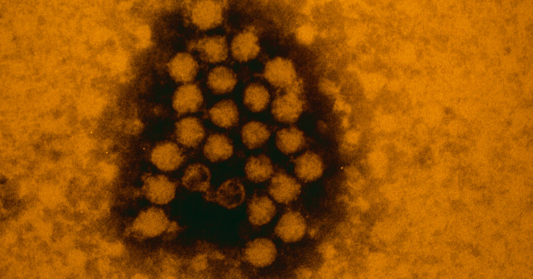
Scientists are exploring a theory suggesting that exposure to one respiratory virus helps the body fend off competing pathogens.An intriguing theory may help explain why the flu and Covid-19 never gripped the nation simultaneously — the so-called twindemic that many public health experts had feared.The idea is that it wasn’t just masks, social distancing or other pandemic restrictions that caused flu and other respiratory viruses to fade while the coronavirus reigned, and to resurge as it receded.Rather, exposure to one respiratory virus may put the body’s immune defenses on high alert, barring other intruders from gaining entry into the airways. This biological phenomenon, called viral interference, may cap the amount of respiratory virus circulating in a region at any given time.“My gut feeling, and my feeling based on our recent research, is that viral interference is real,” said Dr. Ellen Foxman, an immunologist at the Yale School of Medicine. “I don’t think we’re going to see the flu and the coronavirus peak at the same time.”At an individual level, she said, there may be some people who end up infected with two or even three viruses at the same time. But at a population level, according to this theory, one virus tends to edge out the others.Still, she cautioned, “The health care system can become overburdened well before the upper limit of circulation is reached, as the Omicron wave has shown.”Viral interference may help explain patterns of infection seen in large populations, including those that may arise as the coronavirus becomes endemic. But the research is in its early days, and scientists are still struggling to understand how it works.Before the coronavirus became a global threat, influenza was the among the most common severe respiratory infections each year. In the 2018-2019 season, for example, the flu was responsible for 13 million medical visits, 380,000 hospitalizations and 28,000 deaths.The 2019-2020 flu season was winding down before the coronavirus began to rage through the world, so it was unclear how the two viruses might be influencing each other. Many experts feared that the viruses would collide the next year in a twindemic, swamping hospitals.Those worries were not realized. Despite a weak effort to ramp up flu vaccinations, cases remained unusually low throughout the 2020-2021 flu season, as the coronavirus continued to circulate, according to the Centers for Disease Control and Prevention.Only 0.2 percent of samples tested positive for influenza from September to May, compared with about 30 percent in recent seasons, and hospitalizations for flu were the lowest on record since the agency began collecting this data in 2005.Many experts attributed the flu-free season to masks, social distancing and restricted movement, especially of young children and older adults, both of whom are at the highest risk for severe flu. Flu numbers did tick upward a year later, in the 2021-2022 season, when many states had dispensed with restrictions, but the figures were still lower than the prepandemic average.So far this year, the nation has recorded about five million cases, two million medical visits, and fewer than 65,000 hospitalizations and 5,800 deaths related to the flu.Instead, the coronavirus has continued to dominate the winters, much more common than the flu, respiratory syncytial virus, rhinovirus and common cold viruses.A colored electron micrograph of rhinovirus, one of the causes of the common cold.A. Barry Dowsett/Science SourceThe respiratory syncytial virus, or R.S.V., usually surfaces in September and peaks in late December to February, but the pandemic distorted its seasonal pattern. It lay low through all of 2020 and peaked in the summer of 2021 — when the coronavirus had plummeted to its lowest levels since the pandemic’s beginning.The notion of that there is a sort of interplay between viruses first emerged in the 1960s, when vaccinations for polio, which contain weakened poliovirus, significantly cut the number of respiratory infections. The idea gained new ground in 2009: Europe seemed poised for a surge in swine flu cases late that summer, but when schools reopened, rhinovirus colds seemed somehow to interrupt the flu epidemic.“That prompted a lot of people at that time to speculate about this idea of viral interference,” Dr. Foxman said. Even in a typical year, the rhinovirus peaks in October or November and then again in March, on either end of the influenza season.Last year, one team of researchers set out to study the role of an existing immune response in fending off the flu virus. Because it would be unethical to deliberately infect children with the flu, they gave children in Gambia a vaccine with a weakened strain of the virus.Infection with viruses sets off a complex cascade of immune responses, but the very first defense comes from a set of nonspecific defenders called interferons. Children who already had high levels of interferon ended up with much less flu virus in their bodies than those with lower levels of interferon, the team found.The findings suggested that previous viral infections primed the children’s immune systems to fight the flu virus. “Most of the viruses that we saw in these kids before giving the vaccine were rhinoviruses,” said Dr. Thushan de Silva, an infectious disease specialist at the University of Sheffield in England, who led the study.This dynamic may partly explain why children, who tend to have more respiratory infections than adults, seem less likely to become infected with the coronavirus. The flu may also prevent coronavirus infections in adults, said Dr. Guy Boivin, a virologist and infectious disease specialist at Laval University in Canada.Recent studies have shown that co-infections of flu and the coronavirus are rare, and those with an active influenza infection were nearly 60 percent less likely to test positive for the coronavirus, he noted.“Now we see a rise in flu activity in Europe and North America, and it will be interesting to see if it leads to a decreased in SARS-COV-2 circulation in the next few weeks,” he said.Advances in technology over the past decade have made it feasible to show the biological basis of this interference. Dr. Foxman’s team used a model of human airway tissue to show that rhinovirus infection stimulates interferons that can then fend off the coronavirus.“The protection is transient for a certain period of time while you have that interferon response triggered by rhinovirus,” said Pablo Murcia, a virologist at the MRC Center for Virus Research at the University of Glasgow, whose team found similar results. But Dr. Murcia also discovered a kink in the viral interference theory: A bout with the coronavirus did not seem to prevent infection with other viruses. That may have something to do with how adept the coronavirus is at evading the immune system’s initial defenses, he said.“Compared to influenza, it tends to activate these antiviral interferons less,” Dr. de Silva said of the coronavirus. That finding suggests that in a given population, it may matter which virus appears first.Dr. de Silva and his colleagues have gathered additional data from Gambia — which had no pandemic-related restrictions that might have affected the viral patterns they were observing — indicating that rhinovirus, influenza and the coronavirus all peaked at different times between April 2020 and June 2021.That data has “made me a bit more convinced that interference could play a role,” he said.Still, the behavior of viruses can be greatly influenced by their rapid evolution, and by societal restrictions and vaccination patterns. So the potential impact of viral interference is unlikely to become apparent till the coronavirus settles into a predictable endemic pattern, experts said.R.S.V., rhinovirus and flu have coexisted for years, noted Dr. Nasia Safdar, an expert on health-care-associated infections at the University of Wisconsin—Madison.“Eventually that’s what will happen with this one, too — it will become one of many that circulate,” Dr. Safdar said of the coronavirus. Some viruses may attenuate the effects of others, she said, but the patterns may not be readily apparent.Looking at common-cold coronaviruses, some researchers have predicted that SARS-CoV-2 will become a seasonal winter infection that may well coincide the flu. But the pandemic coronavirus has already shown itself to be different from its cousins.For example, it is rarely seen in co-infections, while one of the four common-cold coronaviruses is frequently seen as a co-infection with the other three.“That’s the kind of interesting example that makes one sort of hesitate to make generalizations across multiple viruses,” said Jeffrey Townsend, a biostatistician at the Yale School of Public Health who has studied the coronavirus and its seasonality. “It seems to be somewhat virus-specific how these things occur.”

Read more →

What a Negative Result Means on At-Home Covid Tests

If you have symptoms but get a negative home test result, you may need to keep taking precautions and test again (and again).What does a negative result on a home Covid-19 test really mean?That’s the question that has confounded many people who have reached for a home test because they have a sore throat, cough or runny nose. After swabbing their nose and waiting an anxious 15 minutes, the result is negative.While there’s relief in getting a negative result, there’s also uncertainty. Am I really free of Covid? Or did the test just not detect it? Should I test again? Can I spend time with other people?The confusion is justified, say testing and public health experts. It stems from a lack of understanding about how the tests work. Rapid home antigen tests look for pieces of viral proteins from a swab of your nose, and they are designed to identify whether you have an infectious level of the virus. But a negative test is not a guarantee you don’t have Covid.It could be that your symptoms are an immune response signaling the arrival of Covid or another invader. The harder your immune system is working to tamp down the virus, particularly an immune system supercharged by vaccine antibodies, the more likely you are to get an early negative result on a rapid test, even if you’re infected.“It may be that the virus in your body is having a tug of war with your immune system,” said Dr. Michael Mina, chief science officer for eMed, a company that helps rapid test users get treatment from home. “If you test negative and you have symptoms, don’t assume you’re negative. Assume that the virus has not had an opportunity to grow up yet. The symptoms might mean your immune system is just triggering a very early warning.”Dr. Mina advises people to take a rapid test on the first day of symptoms. A positive result means you almost certainly have Covid. If the result is negative and your symptoms continue, you should still take precautions, wear a mask and avoid close contact with other people. If you can’t test daily, then wait 48 hours and test again. If you’re still negative but your symptoms persist or are getting worse, you should take another test on Day 4. Or you may want to go to a testing center to take a P.C.R. test, which can sometimes detect Covid a little sooner than a home test, although you may have to wait a day or two for the results.Experts say that if you have symptoms and continue to get negative results on home tests, it may be that your immune system is doing a good job beating the virus. Or it could be that you have another illness. Either way, you should try to avoid infecting others.“If you have symptoms and continue to test negative, the chances that you’re infectious with Covid have gone down a lot,” said Dr. Robert Wachter, the chair of the medicine department at the University of California, San Francisco. “But you probably should wear a mask that day because you have something.”And, remember, the result of your home test is just one piece of information. If you haven’t left the house in weeks, your negative result after a few tests is probably accurate. If you have symptoms and you’ve been spending time in bars or a family member has been exposed to Covid, you should be more cautious, even if the initial results are negative. It may be that you tested too early and that your viral load isn’t high enough to be detected.When Dr. Jillian Horton, an internal medicine doctor in Winnipeg, started feeling ill, she was pretty sure she had Covid. Her husband had been exposed and had symptoms, too. She decided to conduct an experiment of one, testing herself several times over the course of a few days to track the dynamics of the virus. “With my husband testing positive and myself very symptomatic, I was sure I had Covid,” Dr. Horton said. “I was curious to see what I could pinpoint in terms of when I might flip positive.”Dr. Horton’s husband became ill on a Friday night, and that evening she tested negative. On Saturday, she began to feel sick and tested herself three times throughout the day. All three results were negative.By Sunday morning, she woke up and was feeling worse. At 6 a.m. she tested and saw a faint line on the test — what she called a “weak positive.” She took two more tests on Sunday and both were negative.On Monday morning, she tested again, and the test rapidly turned positive.What is notable about Dr. Horton’s experiment is that if she had tested at a different time on Sunday, she may never have discovered the weak positive. Her immune system was clearly battling the virus, as evidenced by her two negative test results later in the day.Dr. Horton noted that testing at the right time to catch a high viral load was similar to putting a net in a stream. If the fish aren’t there, you won’t catch anything. But if you time it so that the fish are plentiful, you’ll catch your dinner.Dr. Horton said she was concerned that too many people think the tests aren’t working when, in fact, they are a useful tool if you understand how to use them. They are ideal for “ruling in” Covid, but you have to consider more information when evaluating a negative test.“So often I hear people say, ‘The test is useless,’” Dr. Horton said. “What my experience illustrated is that when you have symptoms, the tests are really ‘rule-in’ tests. I think of those two days when I was so symptomatic. I had one positive test and five negative tests. There was only one moment in there where I was more infectious.”Linsey Marr, a professor of civil and environmental engineering at Virginia Tech and one of the world’s leading experts on viral transmission, said she assumed her daughter had Covid even after a rapid test came back negative. The child had a fever and sore throat, and she had been exposed to Covid through her gymnastics team.But testing proved useful for knowing that her daughter wasn’t highly contagious, which helped Dr. Marr’s family know how to manage the risk. “We knew we needed to be careful,” Dr. Marr said. “But we didn’t have to totally put her in jail. The test told us that the viral load was not high enough that we had to lock her in her room and be that worried about all of us getting it.” Instead, the family wore masks and opened windows to improve ventilation.Kristina Kasparian, who works from home in Montreal, believes she may have gotten Covid from her husband, who is a schoolteacher. They disagreed on whether a home test he took showed a faint positive. But a few days later she woke up with tightness in her chest and a sore throat. Her test was positive, and her husband has continued to test negative.“It’s great to have this tool, but it’s such a variable snapshot in time,” she said.Dr. Mina said that despite the limitations, people would benefit from frequent testing any time they suspect they have been exposed, have symptoms or want to be sure they are not infectious before spending time with a person at high risk. He also recommends testing after you recover to be sure you’re not still infectious when you start interacting with others.“These are tools that have massive benefit during a pandemic like this,” Dr. Mina said. “They will catch you when you’re most infectious. They will even catch you most of the time when you’re just slightly infectious. They will catch almost everyone when they have a high enough viral load to spread. But it won’t be perfect.”

Read more →

CRISPR gene editing reveals biological mechanism behind common blood disorder

UNSW researchers have used CRISPR gene editing — a type of ‘molecular scissors’ — to understand how deletions in one area of the genome can affect the expression of nearby genes. The work, led by UNSW Associate Professor Kate Quinlan and Professor Merlin Crossley, together with collaborators from the US, will help researchers investigate new therapeutic approaches for one of the world’s most devastating genetic blood disorders — sickle cell disease.
Asymptomatic sickle cell disease patients actually lack a tiny part of the genome, scientists have shown.
The team’s findings are published today in academic journal Blood. (Just last week, A/Prof. Quinlan and Prof. Crossley received a $412,919 ARC linkage grant to fund a collaboration between UNSW Sydney and CSL that follows on from the work described in this paper.)
“Sickle cell disease and beta thalassemia, a closely related disease, are inherited genetic conditions that affect red blood cells. They are fairly common worldwide — over 318,000 infants with these conditions are born every year, and haemoglobin disorders cause 3 per cent of deaths in children aged under five years worldwide,” says co-lead author A/Prof. Quinlan.
Genetic mutations — specifically, a defect in the adult globin gene — are responsible for the disorders. The mutant genes affect the production of haemoglobin, the protein in red blood cells that carries oxygen around our bodies.
“Interestingly, when children are born, they don’t show disease symptoms at first, even if they have the mutations, because at that stage, they’re still expressing foetal globin and not yet adult globin. That’s because we have different haemoglobin genes that we express at different stages of development,” says A/Prof. Quinlan.

Read more →

Dengue detection smartphone tech shows new hope for low-cost diagnostics

Accurate home testing could be used for a wider range of illnesses, as new research shows the capability of smartphone-powered tests for Dengue Fever.
In a paper published in PLOS Neglected Tropical Diseases today, biomedical technology researchers from the University of Reading used a new diagnostic kit called Cygnus to detect Dengue Fever with significantly improved rates over lateral flow testing kits.
Working with academics and clinicians in Thailand, the team trialled the tests alongside already established alternatives in and found the new tests showed 82% clinical sensitivity, beating lateral flow testing (74% sensitivity) and matching hospital-based lab diagnostics (83% sensitivity). At the same time, these devices make 10 measurements allowing us to identify which of the 4 different dengue virus types caused the infection.
Dr Sarah Needs, Postdoctoral Research Associate in Microfluidic Antimicrobial Resistance Testing from the University of Reading is lead author of the paper.
Dr Needs said:
“The paper shows exciting potential for the use of the microfluidic ‘lab on a strip’ tests that can used in conjunction with a smartphone and are more powerful than LFT testing in this case. As well as being cheap to produce, the lab on a strip technology allows users to test many different targets at once in one single sample, so it could be useful to detect multiple diseases not just one.

Read more →

Crowning a quest into a very well-guarded secret: Structure of the kinetochore corona finally revealed

Cell division builds our bodies, supplying all cells in our tissues and organs, from the skin to the intestine, from the blood to the brain. It not only allows these organs to grow, but also to regenerate with fresh cells when required. Cell division starts with the replication of chromosomes, the carriers of the three billion nucleotides of the human genome.The replicated chromosomes are then distributed to the daughter cells in a process named mitosis. During mitosis, a network of thread-like structures named the mitotic spindle initially captures the chromosomes. After positioning them in a highly choreographed process, the spindle separates the chromosomes in opposite direction, so that when two cells form out of one, each inherits an exact copy of the genome. Even the smallest errors in this process will have dire physiological consequences.
A multilayered challenge
The kinetochore is the point of contact of chromosomes with the spindle, and is therefore crucially involved in the process of chromosome alignment and partition. It is a complicated multilayered protein complex. “Understanding kinetochores is a tremendous challenge, as they consist of several layers, each made of many interacting building blocks” says Musacchio. “The outermost layer, the corona, has retained some of the most interesting secrets of the kinetochore. Its assembly is particularly interesting, since the complex has a brief lifetime that ends right before the critical steps of chromosome alignment and segregation.”
In a series of previous studies, Musacchio’s laboratory made fundamental inroads into the structure and function of the different layers of kinetochores and how they connect chromosomes to microtubules. To gain this knowledge, the group adopted a reductionist approach named biochemical reconstitution. They produced the individual components of the protein networks outside the cell, in a test tube. They then reassembled them piece by piece to form an almost complete kinetochore that they could study in isolation, in a controlled and simplified environment that contrasts with the enormously complex, buzzing interior of a cell.
Applying the same strategy, the skilled team of two postdocs, Tobias Raisch and Giuseppe Ciossani, two PhD students, Ennio d’Amico and Verena Cmentowski, and other co-workers has now been able to rebuild the kinetochore corona. They showed that only two components are sufficient for that: the ROD-Zwilch-ZW10 (RZZ) protein complex and the protein Spindly, which plays an essential role in the interaction of the kinetochore with the microtubules. The corona assembles exclusively on kinetochores, and the mechanisms that limit its growth to these structures had remained a crucial unresolved question. By reconstituting the process in vitro, the scientists were able to identify an enzyme, the kinase MPS1, as the essential catalyst of RZZ corona assembly at the kinetochore.
One step closer to the crown
Electron microscopy (EM) has accompanied the study of kinetochores since the 1960’s, but it wasn’t until recently, that burgeoning methodological developments made this technique able to visualize the building blocks at the atomic scale. “In 2017, we generated the first ever 3D structural model of the RZZ complex by cryo-EM,” says Raunser. “However, at the 1 nm resolution of this initial model, it was impossible to observe the finest molecular details responsible for biological function.”
The new structural analysis improved the resolution to the point that atomic details emerged, finally explaining how interactions of RZZ components with themselves and with Spindly promote corona assembly into a large polymer that surrounds the kinetochore. “Our work crowns a succession of previous studies on the kinetochore corona, now providing us with a framework to understand the critical moment of cell division when the attachment of chromosomes to microtubules becomes essentially irreversible” concludes Musacchio. The team’s future studies will try to integrate the corona into reconstituted kinetochores, moving a new important step towards the reconstitution of chromosome segregation in vitro, a goal of extraordinary ambition that will shed light on the basis of a most fundamental process of life.
Story Source:
Materials provided by Max Planck Institute of Molecular Physiology. Note: Content may be edited for style and length.

Read more →

Old skins cells reprogrammed to regain youthful function

Research from the Babraham Institute has developed a method to ‘time jump’ human skin cells by 30 years, turning back the ageing clock for cells without losing their specialised function. Work by researchers in the Institute’s Epigenetics research programme has been able to partly restore the function of older cells, as well as rejuvenating the molecular measures of biological age. The research is published today in the journal eLife and whilst at an early stage of exploration, it could revolutionise regenerative medicine.
What is regenerative medicine?
As we age, our cells’ ability to function declines and the genome accumulates marks of ageing. Regenerative biology aims to repair or replace cells including old ones. One of the most important tools in regenerative biology is our ability to create ‘induced’ stem cells. The process is a result of several steps, each erasing some of the marks that make cells specialised. In theory, these stem cells have the potential to become any cell type, but scientists aren’t yet able to reliably recreate the conditions to re-differentiate stem cells into all cell types.
Turning back time
The new method, based on the Nobel Prize winning technique scientists use to make stem cells, overcomes the problem of entirely erasing cell identity by halting reprogramming part of the way through the process. This allowed researchers to find the precise balance between reprogramming cells, making them biologically younger, while still being able to regain their specialised cell function.
In 2007, Shinya Yamanaka was the first scientist to turn normal cells, which have a specific function, into stem cells which have the special ability to develop into any cell type. The full process of stem cell reprogramming takes around 50 days using four key molecules called the Yamanaka factors. The new method, called ‘maturation phase transient reprogramming’, exposes cells to Yamanaka factors for just 13 days. At this point, age-related changes are removed and the cells have temporarily lost their identity. The partly reprogrammed cells were given time to grow under normal conditions, to observe whether their specific skin cell function returned. Genome analysis showed that cells had regained markers characteristic of skin cells (fibroblasts), and this was confirmed by observing collagen production in the reprogrammed cells.

Read more →

Enhancing deep sleep

Researchers have developed a wearable device that plays specific sounds to enhance deep sleep. The first clinical study has now shown that the device is effective, but not at the same level of effectiveness for everyone.
Many people, especially the elderly, suffer from abnormal sleep. In particular, the deep sleep phases become shorter and shallower with age. Deep sleep is important for the regeneration of the brain and memory, and also has a positive influence on the cardiovascular system.
Researchers have shown that the brain waves characterizing deep sleep, so-​called slow waves, can be improved by playing precisely timed sounds through earphones while sleeping. While this works well in the sleep laboratory under controlled conditions, there has thus far been no at home solution that can be used longer than just one night.
SleepLoop to the rescue
As part of the SleepLoop project, researchers at ETH Zurich have developed a mobile system that can be used at home and aims to promote deep sleep through auditory brain stimulation.
The SleepLoop system consists of a headband that is put on at bedtime and worn throughout the night. This headband contains electrodes and a microchip that constantly measure the brain activity of the person sleeping. Data from this is analysed autonomously in real-time on the microchip using custom software. As soon as the sleeping person shows slow waves in the brain activity characterising deep sleep, the system triggers a short auditory signal (clicking). This helps to synchronise the neuronal cells and enhance the slow waves. What makes the solution unique is that the person sleeping is not conciously aware of this sound during deep sleep.

Read more →

Human Lung Chip reveals the effects of breathing motions on lung immune responses

The average person will take more than 600 million breaths over the course of their life. Every breath stretches the lungs’ tissues with each inhale and relaxes them with each exhale. The mere motions of breathing are known to influence vital functions of the lungs, including their development in babies, the production of air-exchange-enhancing fluid on their inner surfaces, and maintenance of healthy tissue structure. Now, new research from the Wyss Institute at Harvard University has revealed that this constant pattern of stretching and relaxing does even more — it generates immune responses against invading viruses.
Using a Human Lung Chip that replicates the structures and functions of the lung air sac, or “alveolus,” the research team discovered that applying mechanical forces that mimic breathing motions suppresses influenza virus replication by activating protective innate immune responses. They also identified several drugs that reduced the production of inflammatory cytokines in infected Alveolus Chips, which could be useful in treating excessive inflammation in the lung. Based on these studies, one of those drugs was licensed to Cantex Pharmaceuticals for the treatment of COVID-19 and other inflammatory lung diseases. Data from the research were recently included in the company’s Investigational New Drug (IND) application to the FDA to initiate a Phase 2 clinical trial for COVID-19.
“This research demonstrates the importance of breathing motions for human lung function, including immune responses to infection, and shows that our Human Alveolus Chip can be used to model these responses in the deep portions of the lung, where infections are often more severe and lead to hospitalization and death,” said co-first author Haiqing Bai, Ph.D., a Wyss Technology Development Fellow at the Institute. “This model can also be used for preclinical drug testing to ensure that candidate drugs actually reduce infection and inflammation in functional human lung tissue.” The results are published today in Nature Communications.
Creating a flu-on-a-chip
As the early phases of the COVID-19 pandemic made painfully clear, the lung is a vulnerable organ where inflammation in response to infection can generate a “cytokine storm” that can have deadly consequences. However, the lungs are also very complex, and it is difficult to replicate their unique features in the lab. This complexity has hindered science’s understanding of how the lungs function at the cell and tissue levels, in both healthy and diseased states.
The Wyss Institute’s Human Organ Chips were developed to address this problem, and have been shown to faithfully replicate the functions of many different human organs in the lab, including the lung. As part of projects funded by the NIH and DARPA since 2017, Wyss researchers have been working on replicating various diseases in Lung Airway and Alveolus Chips to study how lung tissues react to respiratory viruses that have pandemic potential and test potential treatments.

Read more →