What will it take to transform obesity care for all?

When a person with diabetes, high blood pressure, or osteoarthritis goes to their primary care clinic, they expect treatment that’s grounded in modern medical evidence.
That might mean nutrition and exercise recommendations, prescriptions for medications, regular measurement of their progress and referrals for visits to specialists or even surgery.
But if they have obesity, they may not get the same level of evidence-based care for that chronic condition, recent studies have shown.
Why not? Like obesity itself, the answer is complicated, say Michigan Medicine primary care physicians with advanced training in obesity medicine.
The reasons include health insurance rules that exclude obesity medications and behavioral therapy, lack of training among primary care clinicians on best practices for treating obesity such as prescribing newer medications, and societal stigma against people with obesity.
And unlike those other chronic health conditions, few outside forces have driven primary care clinics to focus on providing high-quality, personalized obesity care for all.

Read more →

Nerve stimulation promotes resolution of inflammation

The nervous system is known to communicate with the immune system and regulate inflammation in the body. Researchers at Karolinska Institutet in Sweden now show how electrical activation of a specific nerve can promote healing in acute inflammation. The finding, which is published in the journal PNAS, opens new ways to accelerate resolution of inflammation.
The way the body regulates inflammation is only partly understood. Previous research by Peder Olofsson’s group at Karolinska Institutet and other research groups has shown that electrical stimulation of the vagus nerve can reduce inflammation. Such nerve stimulation has been used with encouraging results in clinical studies of patients with inflammatory bowel disease and rheumatoid arthritis. However, how nerve signals regulate active resolution of inflammation was unclear.
“We have now studied effects of signals between nerves and immune cells at the molecular level,” says April S. Caravaca, a researcher in Peder Olofsson’s group at the Department of Medicine, Solna, Karolinska Institutet and the Stockholm Center for Bioelectronic Medicine at MedTechLabs. “A better understanding of these mechanisms will allow for more precise applications that harness the nervous system to regulate inflammation.”
The researchers showed that electrical stimulation of the vagus nerve in inflammation shifts the balance between inflammatory and specialised anti-inflammatory molecules, which promotes healing.
“Inflammation and its resolution plays a key role in a wide range of common diseases, including autoimmune diseases and cardiovascular diseases,” says Peder Olofsson. “Our findings provide insights on how the nervous system can accelerate resolution of inflammation by activating defined signalling pathways.”
The researchers will continue to study how nerves regulate the healing of inflammation in more detail.
“The vagus nerve is only one of many nerves that regulate the immune system. We will continue to map the networks of nerves that regulate inflammation at the molecular level and study how these signals are involved in disease development,” says Dr Olofsson. “We hope that this research will provide a better understanding of how pathological inflammation can resolve, and contribute to more effective treatments of the many inflammatory diseases, such as atherosclerosis and rheumatism.”
The study was supported by grants from the Knut and Alice Wallenberg Foundation, the Swedish Research Council, the Swedish Heart-Lung Foundation, MedTechLabs and Novo Nordisk. Peder Olofsson holds shares in Emune AB. Co-author Jesmond Dalli is the founder of and head of research at Resolomics Ltd.
Story Source:
Materials provided by Karolinska Institutet. Note: Content may be edited for style and length.

Read more →

Mobile app provides effective support for children with obesity

A mobile app that shows a child’s weight development in real-time for children with obesity provides greater weight loss compared to conventional care. The fact that both families and healthcare professionals can follow the same data facilitates individualised extra support when needed. This is shown by a study from Karolinska Institutet in Sweden published in the International Journal of Obesity.
The care of children and adolescents with obesity needs to be improved. International studies show that frequent follow-up visits, every two weeks, are most effective in maintaining behavioural changes.
“But it’s not feasible, neither for the families nor the healthcare system. We, therefore, need to find new ways to provide more support,” says Emilia Hagman, a researcher at the Department of Clinical Science, Intervention and Technology who in a study evaluated a new digital tool, a mobile app from the company Evira.
The study is a so-called pragmatic study, which means that the participants were not randomised, but rather, the treatments were evaluated in a real-life clinical setting.
Results improved by 100%
100 children who attended the Martina Children’s hospital (Martina Centrum för Vikthälsa) in Stockholm were able to try the digi-physical treatment concept over a period of one year. The app is connected to a scale that has no numbers on it, which the child would stand on each day. In the app, the family sees their child’s weight development as a curve that should be within a green weight target development curve. The target curve was determined individually and updated during physical visits every three months.

Read more →

Too much self-confidence can endanger health

Older people who overestimate their health go to the doctor less often. This can have serious consequences for their health, for example, when illnesses are detected too late. By contrast, people who think they are sicker than they actually are visit the doctor more often. This is what a new study by Sonja Spitzer from the Institute for Demography at the University of Vienna and Mujaheed Shaikh from the Hertie School in Berlin found based on data from over 80,000 Europeans aged 50 and older. The results were published in The Journal of the Economics of Aging.
Our confidence affects our behaviour. People who overestimate their abilities earn more, invest their money differently, and are more likely to be leaders. But they also act riskier, have more accidents, and live less healthy by drinking more alcohol, eating less healthily, and sleeping too little.
How people perceive the state of their health can also have consequences for their own health decisions — like whether to visit a doctor or not. A new study by Sonja Spitzer of the University of Vienna and Hertie School Professor Mujaheed Shaikh finds that individuals who overestimate their health visit the doctor 17.0% less often than those who correctly assess their health, which is crucial for preventive care such as screenings. Similar results were found for dentist visits.
The perception of one’s own health has, however, no effect on the number and duration of hospital stays; presumably because hospital stays are more regulated and often require a doctor’s referral.
Those who think they are sicker than they are visit the doctor more often
The authors also found that individuals who underestimate their health visit the doctor 21% more frequently. On the one hand, there is the disadvantage that these additional visits could cause unnecessary costs, which is relevant given population ageing and the associated high public health expenditure. On the other hand, people who underestimate their health and therefore pay close attention to it may be particularly fit in the long term, which could have a positive impact on society. Overall, it is difficult for outsiders to assess which visits are justified and which are not.
For their study, the researchers analysed data from over 80,000 Europeans aged 50 and older, using statistical methods. The data were collected as part of the SHARE study (Survey of Health, Aging and Retirement in Europe) between 2006 and 2013. First, the participants were asked how they assessed their health, for example, whether they had problems getting up from a chair after sitting for a long period. Then, the participants had to actually get up from a chair during a test — this way it can be determined whether someone overestimates, underestimates, or correctly assesses their health. The researchers also took misjudgments related to memory and mobility into account. Overall, the majority of survey participants correctly assess their health (79%), 11% overestimate, and 10% underestimate themselves.
Who knows about their health?
With their new study, the researchers built on a previous study that showed that the perception of health differs greatly depending on age, nationality, and education. The older people are, the more often they overestimate their health. The researchers also found large regional differences: according to the analysis, people in Southern Europe tend to overestimate their health, while people in Central and Eastern Europe often underestimate their health. Educated people are also more likely to correctly assess their health. The scientists’ appeal: Focus more on health education and health literacy. How healthy we feel can influence how healthy we actually are in the long term.
Story Source:
Materials provided by University of Vienna. Note: Content may be edited for style and length.

Read more →

Discovery could lead to better cancer immunotherapy

A type of white blood cell previously known only as a helper in the immune system appears also to be the instigator of the body’s defenses against cancerous tumors. The discovery could lead to more effective cancer immunotherapy, a promising treatment which uses the body’s own immune system, rather than radiation, to attack cancer cells.
In an animal study, Washington State University researchers found that a population of T cells called CD4-positive helper T cells helped start a chain of antitumor immunity defenses that allow killer cells to better infiltrate melanoma and breast cancer tumors. T cells belong to a class of white blood cells known as lymphocytes which move throughout the body’s lymphatic system.
Many previous studies, and current immunotherapies, have focused primarily on the role of one-type of killer cells, called CD8-positive T cells. Yet, less than 20% of patients respond to those therapies and the initiating role of the CD4-positive helper cells could make those therapies better, said Hui Zhang, lead author on the study published in the Journal of Immunology.
“One of the most challenging parts of current cancer immunotherapy is the low response rate,” said Zhang, a WSU assistant professor of pharmaceutical sciences. “The lack of knowledge of how to enhance lymphocyte infiltration into the tumor hampers the success of improving the response rate to cancer immunotherapy. Our finding shows promise in solving this problem.”
Cancer is the second leading cause of death both nationally and worldwide. Currently, surgery, chemotherapy and radiation therapy are the conventional approaches to cancer treatment. However, those approaches cannot cure many cancers because some become metastatic, spreading from the primary tumor throughout the body, and certain cancer stem cells can become resistant to chemotherapy and radiation.
A relatively new treatment, immunotherapy has shown promise in curing a range of cancers, but only a relatively low number of patients respond to it. Zhang’s research team hopes to change that with the knowledge of the mechanisms that help start the body’s immune defenses.

Read more →

Scientists solve long-standing mystery: Why do some asthma patients respond poorly to treatment?

Patients with the most severe form of asthma produce special substances in their airways when taking medicine during an asthma attack that block the treatment from working, according to a study where Rutgers scientists collaborated with researchers at Genentech, a member of the Roche Group.
Reporting in the journal Science Translational Medicine, scientists said two different so-called growth factors – naturally occurring substances that stimulate cell proliferation – activate in the airways of severe asthma patients as they inhale corticosteroids used as an emergency treatment during an asthma attack.
The discovery was made as researchers investigated an enduring mystery in asthma treatment: Why do some patients who suffer the most from the disease often have the least success with conventional rescue treatments?
Of the more than 25 million people in the U.S. with asthma, between 5 percent and 10 percent suffer from severe asthma, according to the American Lung Association. The corticosteroids used to decrease swelling and irritation in the airways of people with moderate asthma often fail to work in those with severe asthma. Severe asthma patients experience more frequent bouts of breathing problems than others.
Researchers found inhaled steroids in severe asthma patients promote the secretion of growth factors — fibroblast growth factor (FGF) and granulocytic colony forming growth factor (G-CSF) — in airway lining cells known as the epithelium.
“We believe this response explains why patients with severe asthma are unresponsive to such conventional therapy,” said author Reynold Panettieri Jr., a professor of medicine at Rutgers Robert Wood Johnson Medical School and vice chancellor of Clinical and Translational Science.
Researchers compared samples of bronchial airway epithelial cells (BAECs) that had been exposed to inhaled corticosteroids and were collected from three groups: those with severe asthma, those with moderate asthma and healthy volunteers.
By conducting a genetic analysis to determine what genes had been turned “on” in the BAECs, the scientists were able to see that the FGF and G-CSF growth factors had been expressed only in the cells of the patients with severe asthma.
Growth factors are important for regulating a variety of cellular processes, Panettieri said. In the case of an asthma attack in patients with severe asthma, the growth factors identified in the cells that line the major connecting airways work directly against the action of the corticosteroids. Findings from the study suggest different cellular pathways are at work in the cells of patients with severe asthma, particularly those involved in inflammation.
Here’s how the researchers envision a new medicine may work: In a study in mice, scientists found if they blocked the cascade of chemicals that ultimately triggers the growth factors to be secreted, corticosteroids effectively reversed airway inflammation and even prevented scarring of tissue.
“Our study has uncovered a potential mechanism to explain why patients with severe asthma are unresponsive to conventional therapy,” Panettieri said. “If we could uncover new approaches to treatment that directly affect that mechanism, we may be able to restore a sensitivity to the steroid and improve outcomes.”
Other Rutgers researchers on the Genentech-led paper include Cynthia Koziol-White, an assistant professor in the department of pharmacology at Robert Wood Johnson Medical School, and William Jester Jr., the chief operating officer of the Institute for Translational Medicine and Science. Additional authors on the paper are from Genentech of South San Francisco, Calif., and Texas A&M University in Houston, Texas.
Story Source:
Materials provided by Rutgers University. Original written by Kitta MacPherson. Note: Content may be edited for style and length.

Read more →

Monkeypox infections rise as guidance advises cases to abstain from sex

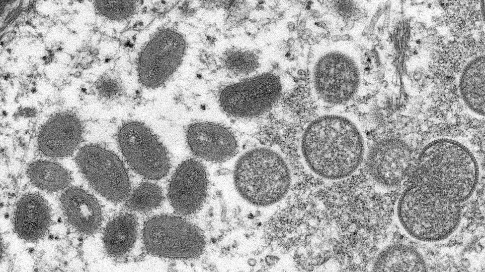
SharecloseShare pageCopy linkAbout sharingImage source, ReutersAnother 71 cases of monkeypox have been identified in England over the weekend bringing the UK total to 179, the UK Health Security Agency (UKHSA) says.New guidance is advising anyone with the virus to abstain from sex while they have symptoms.They are also told to use condoms for eight weeks after an infection as a precaution.The risk to the population is low, but people should be alert to new rashes or lesions, the UKHSA says.In total, 172 cases have been confirmed in England, with four in Scotland, two in Northern Ireland and one in Wales.What is monkeypox and how do you catch it?The Covid conspiracies being recycled for monkeypoxHow UK hospitals are tackling monkeypox outbreakCondom useNew guidance for doctors and other healthcare workers to help control the latest outbreak has now been agreed by the four health authorities in England, Wales, Scotland and Northern Ireland. People who have tested positive for the virus and their close contacts are being told to isolate at home for 21 days. They should avoid contact with other people until all lesions – or blisters – have healed and scabs have dried off. Anyone with a confirmed infection is now being advised to abstain from sex while they have symptoms.The guidance says that while there is currently no available evidence that monkeypox can be spread in sexual fluids, people confirmed to have the virus are advised to use condoms for eight weeks after infection as a precaution. Monkeypox: Handing out health advice without stigmaConfirmed cases and their close contacts should take extra care if they need to leave the house to see a doctor or other health worker. This means making sure any lesions are covered by clothes, wearing a face covering and avoiding public transport where possible.Protection for staffDr Ruth Milton, monkeypox strategic response director, at UKHSA said: “The highest risk of transmission is through direct contact with someone with monkeypox. “The risk to the UK population remains low and anyone with unusual rashes or lesions on any part of their body should immediately contact NHS 111 or their local sexual health service.”The new guidance also says healthcare workers who are pregnant and people with severely weakened immune systems should not care for anyone who is suspected or confirmed to have monkeypox.Staff working with confirmed cases should wear personal protective equipment, including FFP3 respirators, aprons, eye protection and gloves as a minimum, it adds.Those working with possible cases are recommended to wear surgical face masks which are fluid repellent, gowns, gloves and eye protection.UK health officials have purchased over 20,000 doses of a smallpox vaccine called Imvanex.It is being offered to close contacts of those diagnosed with the virus to try and reduce the risk of symptomatic infection and severe illness.More on this storyMonkeypox: Handing out health advice without stigmaMonkeypox: Time to worry or one to ignore?Got a new rash? How to tell if it is monkeypox

Read more →

Ultrasound-guided microbubbles boost immunotherapy efficacy

Researchers at The University of Texas MD Anderson Cancer Center have developed an ultrasound-guided cancer immunotherapy platform that generates systemic antitumor immunity and improves the therapeutic efficacy of immune checkpoint blockade. The findings from the preclinical study were published today in Nature Nanotechnology.
As the first-of-its-kind platform, the Microbubble-assisted UltraSound-guided Immunotherapy of Cancer (MUSIC) approach employs nanocomplexes combined with microbubbles to effectively deliver cyclic guanosine monophosphate-adenosine monophosphate (cGAMP), an immunotransmitter involved in anticancer immunity, into antigen-presenting cells (APCs). Inside the APCs, the microbubbles release cGAMP to activate the GMP-AMP synthase (cGAS)-stimulator of interferon genes (STING) pathway, which stimulates type I interferon responses that are essential for priming tumor-specific T cells.
In the preclinical study, the MUSIC strategy demonstrated a complete tumor eradication rate of 60% when administered as monotherapy in breast cancer models. When combined with an anti-PD-1 antibody, MUSIC significantly improved antitumor responses with minimal toxicity effects, including enhanced primary tumor control and decreased systemic disease progression. In addition, the combination therapy demonstrated superior survival benefit, with a 76% increase in median survival compared to either therapy alone.
“By investigating the mechanisms of action in producing a robust STING activation, we identified a new strategy to activate both the innate and adaptive antitumor immune responses,” said Wen Jiang, M.D., Ph.D., assistant professor of Radiation Oncology and the study’s co-senior author. “Our findings show that the MUSIC strategy is capable of paving the way toward novel image-guided strategies for targeted cancer immunotherapy.”
Immunotherapy has transformed cancer treatment, offering clinical benefits for patients with treatment-refractory metastatic cancers such as melanoma, non-small cell lung cancer and renal cell cancer. However, not all patients respond to immune checkpoint blockade. Therefore, developing a more effective immunotherapy strategy to benefit larger numbers of cancer patients with localized and metastatic disease remains an unmet clinical need.
“Although the majority of cancer immunotherapies have focused on boosting the adaptive branch of the body’s immune system, there has been a growing realization that both the innate and adaptive branches of the body’s immune system need to be engaged to generate optimal antitumoral immunity,” Jiang said. “This understanding has led to the development of new immunotherapies that target the regulators of innate immune systems, including the cGAS-STING pathway.”
Natural agonists, such as cyclic dinucleotides, activate the cGAS-STING pathway, but concerns over poor cytosolic entry, serum stability and systemic toxicity have been major limitations for clinical translation. To overcome these challenges, Jiang and his collaborators developed MUSIC as the first-ever image-guided cancer immunotherapy strategy that uses antibody targeting to activate STING in APCs through delivery of molecular drugs.
Because the microbubbles also serve as contrast agents for ultrasound, the researchers use ultrasound scanners to image the tumor and to precisely identify the location where the microbubbles have accumulated. After confirming the microbubbles are bound to the tumors, the researchers activate ultrasonic frequencies, which cause the microbubbles to oscillate and burst, creating transient pores in the cell membrane that allow nucleic acids to be transferred directly into the cell cytosol. This technique, called sonoporation, previously has been used on tumor cells, but the MUSIC platform is the first to bind nanocomplexes to microbubbles to deliver cGAMP immunotransmitters directly into APCs.
“The beauty of our platform is that ultrasound machines are already clinically available in many outpatient settings and microbubbles are FDA-approved contrast agents for ultrasound imaging,” Jiang said. “Therefore, we expect there is a very real opportunity to translate MUSIC into a clinic application to benefit cancer patients.”
The same concept and design principle behind the MUSIC platform’s microbubble technology could be readily translated to nanoscale systems for targeted systemic delivery and activation of innate immune sensors under image guidance for cancer immunotherapy applications, Jiang added.
“Our MUSIC platform is exciting because it provides a new framework for developing image-guided immunotherapy by using acoustically responsive biomaterials to enable efficient, targeted and robust immune activation to produce potent antitumor effects while minimizing systemic toxicity,” Jiang said. “The versatility of the MUSIC platform could potentially be applied to targeted delivery of other immune-stimulating agents, such as nucleotide-based vaccines, mRNAs and other gene therapies for multiple human diseases.”
The study was supported by grants from the Cancer Prevention and Research Institute of Texas (CPRIT), Department of Defense and the National Cancer Institute. The study’s co-corresponding author is Jacques Lux, Ph.D., assistant professor of Radiology at The University of Texas Southwestern Medical Center.

Read more →

Engineers boost signals from fluorescent sensors

Fluorescent sensors, which can be used to label and image a wide variety of molecules, offer a unique glimpse inside living cells. However, they typically can only be used in cells grown in a lab dish or in tissues close to the surface of the body, because their signal is lost when they are implanted too deeply.
MIT engineers have now come up with a way to overcome that limitation. Using a novel photonic technique they developed for exciting any fluorescent sensor, they were able to dramatically improve the fluorescent signal. With this approach, the researchers showed they could implant sensors as deep as 5.5 centimeters in tissue and still get a strong signal.
This kind of technology could enable fluorescent sensors to be used to track specific molecules inside the brain or other tissues deep within the body, for medical diagnosis or monitoring drug effects, the researchers say.
“If you have a fluorescent sensor that can probe biochemical information in cell culture, or in thin tissue layers, this technology allows you to translate all of those fluorescent dyes and probes into thick tissue,” says Volodymyr Koman, an MIT research scientist and one of the lead authors of the new study.
Naveed Bakh SM ’15, PhD ’20 is also a lead author of the paper, which appears today in Nature Nanotechnology. Michael Strano, the Carbon P. Dubbs Professor of Chemical Engineering at MIT, is the senior author of the study.
Enhanced fluorescence
Scientists use many different kinds of fluorescent sensors, including quantum dots, carbon nanotubes, and fluorescent proteins, to label molecules inside cells. These sensors’ fluorescence can be seen by shining laser light on them. However, this doesn’t work in thick, dense tissue, or deep within tissue, because tissue itself also emits some fluorescent light. This light, called autofluorescence, drowns out the signal coming from the sensor.

Read more →

The paired perils of breast cancer and diabetes

Breast cancer and type 2 diabetes would seem to be distinctly different diseases, with commonality only in their commonality. Breast cancer is the second most diagnosed malignancy after some types of skin cancer; approximately 1 in eight U.S. women will develop invasive breast cancer over the course of their lifetime. More than 10 percent of the U.S. population has diabetes, with an estimated 2 in 5 Americans expected to develop the chronic disease during their lifetime.
However, past research has uncovered associations between the two diseases. Women with diabetes, for example, have a 20 to 27 percent increased risk of developing breast cancer. Insulin resistance — a key characteristic of diabetes — has been associated with breast cancer incidence and poor survival. Population studies suggest diabetes risk begins to increase two years after a breast cancer diagnosis, and by 10 years post-diagnosis, the risk is 20 percent higher in breast cancer survivors than in age-matched women without breast cancer.
But these epidemiological linkages are not clear-cut or definitive, and some studies have found no associations at all. In a new paper, publishing May 30, 2022 in Nature Cell Biology, a research team led by scientists at University of California San Diego School of Medicine describe a possible biological mechanism connecting the two diseases, in which breast cancer suppresses the production of insulin, resulting in diabetes, and the impairment of blood sugar control promotes tumor growth.
“No disease is an island because no cell lives alone,” said corresponding study author Shizhen Emily Wang, PhD, professor of pathology at UC San Diego School of Medicine. “In this study, we describe how breast cancer cells impair the function of pancreatic islets to make them produce less insulin than needed, leading to higher blood glucose levels in breast cancer patients compared to females without cancer.”
Wang said the study was inspired by early work and guidance from Jerrold Olefsky, MD, professor of medicine and associate dean for scientific affairs in the Division of Endocrinology and Metabolism at UC San Diego School of Medicine. Olefsky is co-senior author of the study with Wang.
The culprit, according to Wang and Olefsky, are extracellular vesicles (EV) — hollow spheres secreted or shed by cells that transport DNA, RNA, proteins, fats and other materials between cells, a sort of cargo communication system.
In this case, the cancer cells were found to be secreting microRNA-122 into the vesicles. Wang said when vesicles reach the pancreas, they can enter the islet cells responsible for insulin production, dispense their miR-122 cargo and damage the islets’ critical function in maintaining a normal blood glucose level.
“Cancer cells have a sweet tooth,” Wang said. “They use more glucose than healthy cells in order to fuel tumor growth, and this has been the basis for PET scans in cancer detection. By increasing blood glucose that can be easily used by cancer cells, breast tumors make their own favorite food and, meanwhile, deprive this essential nutrient from normal cells.”
The research was conducted using mouse models, which found that slow-releasing insulin pellets or a glucose-lowering drug known as an SGLT2 inhibitor restored normal control of glucose in the presence of a breast tumor, which in turn suppressed the tumor’s growth.
“These findings support a greater need for diabetes screening and prevention among breast cancer patients and survivors,” said Wang, noting that an inhibitor of miR-122, developed by Regulus Therapeutics Inc. in San Diego, is currently in clinical trial as a potential treatment for chronic hepatitis C. It has been found to be effective in restoring normal insulin production and suppressing tumor growth in mouse models of breast cancer.
“These miR-122 inhibitors, which happen to be the first miRNA-based drugs to enter clinical trials, might have a new use in breast cancer therapy,” Wang said.
Co-authors include: Minghui Cao, Roi Isaac, Wei Yan, Xianhui Ruan, Li Jiang, Yuhao Wan, Jessica Wang, Christine Caron, Donald P. Pizzo, Xuxiang Liu, Andrew R. Chin, Miranda Y. Fong, Oluwole Fadare, Richard B. Schwab, Wei Ying and Jack D. Bui, all at UC San Diego; Dorothy D. Sears, Arizona State University; Steven Neben and Denis Drygin, Regulus Therapeutics, Inc., San Diego; Xiwei Wu, Joanne Mortimer, Yuan Yuan and Susan E. Yost, all at City of Hope, Duarte, CA; Ziting Gao, Kaizhu Guo and Wenwan Zhong, all at UC Riverside.

Read more →