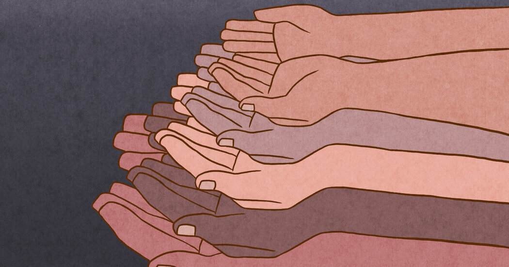
Navigating Fatherhood as a Black Man

The editor of a new book of essays shares how Black men can attend to their mental health while growing their families.This year Father’s Day will fall on June 19, or Juneteenth, a federal holiday commemorating the emancipation of enslaved Black people in the United States after the Civil War. And for Michael D. Hannon, an associate professor of counseling at Montclair State University in Montclair, N.J., that is “an awesome coincidence.”“We can celebrate Black fathers who are doing their best to protect, provide and prepare their families for success, while also acknowledging the spirit and the resilience and the pursuit of freedom among Black people in this country,” he said.Dr. Hannon, the self-described father of “two dope Black children” — an 18-year-old son and a 19-year-old daughter — has been counseling Black fathers for the last 10 years. And as the editor of the new book “Black Fathering and Mental Health,” he now seeks to elevate the voices of Black fathers — and aspiring ones, too — who also happen to be mental health counselors. Through a series of essays, each writer offers unique perspectives on the needs, challenges and victories of Black fathering in an “anti-Black world.”The book can serve as a resource for other counselors to help them provide culturally affirming and relevant support to Black fathers, but the personal stories in the collection are also meant for a general audience, who may identify with many of the joys and difficulties presented within.“It should not be this hard, am I right?” asked one of the essayists, S. Kent Butler, a professor of counselor education and school psychology at the University of Central Florida. “No, I am not right. When it comes to our Blackness, very little is easy about self-acceptance and others’ acceptance. So, where does the strength and resilience come from? What makes it all right? I believe it is my tribe.”Questions and answers have been edited and condensed for clarity.What inspired you to create this book? And why now?Much of the research I do is about Black fathers. So this has, quite frankly, been a long time coming. I really wanted to do at least three things.The first was to amplify the voices of Black fathers. Period.Second, I wanted for other people to be able to read and hear these voices in ways that maybe they hadn’t before.And then third, all of the people who wrote chapters in this book are mental health professionals. I asked them to answer some very specific questions: What might be useful for mental health professionals who are treating or serving Black father clients? What influenced their fathering practice? Did they seek counseling support if and when they confronted challenges and obstacles? And if they did, what did they learn? And if they didn’t, what stopped them?One of the essayists, Linwood G. Vereen, an associate professor of counseling education at Shippensburg University in Pennsylvania who has fathered five biracial children, wrote: “What I have learned in my journey through counseling is that my needs are valid. I have learned that it’s OK to release the unrealistic expectations of others that hurt my soul, and that my Black life matters. I have learned that as much as my children need to see success in life, they must also learn humility through seeing their father show humility.”Tell me more about why it was particularly important for you to feature the voices of Black fathers.It’s very easy to consume content about Black men that focuses on some of the challenges that have been systemically placed before us.You know the stereotype of the absentee Black father, or the overrepresentation of Black men who are incarcerated. But there’s a much more nuanced, rich and complex set of experiences that Black men have. There’s so much to know and understand and appreciate about who Black men are in the context of their communities and how they serve their biological children, and their fictive kin — or the children for whom they are “play uncles” and “play cousins.”And that’s important because we’re all subject to stereotyping and having prejudiced viewpoints, and no one deserves that. Things like going to the pediatrician with your child and the medical professionals telling you that they’re surprised to see you. Or going to another specialist appointment, maybe with your partner, and the medical professional or the specialist not even addressing any questions to you. Custody cases can transpire in the court systems, as well, that may position Black fathers to not be able to be as engaged as they may want to be.Are there gems of wisdom from the book that may be helpful to Black fathers?We are socialized to be protectors of our families, protectors of our partners; to provide for our children and families; and prepare them for success. And that’s a lot of pressure. And many times that ability has been influenced by somebody’s socioeconomic profile. What we know now is that fathers, and Black fathers in particular, are contributing in ways much broader than financial provision, and finding ways to emotionally provide for their children. I can’t overstate how important those things are.“My children are the poster examples of strong, graceful, resilient, fearless and powerful, and most days they use their agency in an unapologetic manner,” Dr. Vereen wrote. “My greatest hope as their father is that they will always do this.”How can Black fathers protect their mental health?It’s not easy. What I would remind all Black fathers, and people in general, is that we have to find people and spaces that allow us to be as transparent as possible. We have to find community.For me, personally, my professional network — whether they’re counselors or my fraternity brothers — there are groups of men to whom I can go and be as brutally honest and as vulnerable as I need to be. It allows me to share all of the victories and the things that I want to celebrate — and it allows me also to share the most challenging, the most vulnerable parts of my experiences, hopefully without fear of judgment.If you just hit a wall, and you can’t get past or over the wall, maybe it would be helpful to talk to a professional counselor to help set a goal and reach that goal, because you haven’t been able to do it otherwise.“I have definitely sought counseling when needed and sometimes not sought it when I needed it,” Dr. Butler, the University of Central Florida professor, wrote in his essay.“I did seek family counseling services to help me support my stepson, which was tremendously useful for us as a family and for me as a father figure to him,” he said. “I was reminded that I did not have all of the answers, nor should I expect to have them all.”

Read more →

Biochemistry researchers repair and regenerate heart muscle cells

Researchers at the University of Houston are reporting a first-of-its-kind technology that not only repairs heart muscle cells in mice but also regenerates them following a heart attack, or myocardial infarction as its medically known.
Published in the Journal of Cardiovascular Aging, the groundbreaking finding has the potential to become a powerful clinical strategy for treating heart disease in humans, according to Robert Schwartz, Hugh Roy and Lillie Cranz Cullen Distinguished Professor of biology and biochemistry at the UH College of Natural Sciences and Mathematics.
The new technology developed by the team of researchers uses synthetic messenger ribonucleic acid (mRNA) to deliver mutated transcription factors — proteins that control the conversion of DNA into RNA — to mouse hearts.
“No one has been able to do this to this extent and we think it could become a possible treatment for humans,” said Schwartz, who led the study with recent Ph.D graduate Siyu Xiao and Dinakar Iyer, a research assistant professor of biology and biochemistry.
Synthetic mRNA Contributes to Stem Cell-Like Growth
The researchers demonstrated that two mutated transcription factors, Stemin and YAP5SA, work in tandem to increase the replication of cardiomyocytes, or heart muscle cells, isolated from mouse hearts. These experiments were conducted in vitro on tissue culture dishes.

Read more →

Genetic screening algorithm could identify people with kidney disease risk

A new algorithm developed by researchers at Columbia University can analyze thousands of variants across the genome and estimate a person’s risk of developing chronic kidney disease — and it works in people of African, Asian, European, and LatinX descent.
“With this polygenic method, we can identify individuals at risk decades before the onset of kidney disease, and those with high risk might adopt protective lifestyle changes to reduce that risk,” says Krzysztof Kiryluk, MD, associate professor of medicine and a physician-scientist in the division of nephrology at Columbia University Vagelos College of Physicians and Surgeons. (Diabetes, high blood pressure, obesity, and certain medications, such as NSAIDs, are known to increase the risk of kidney disease.)
Early detection of kidney disease could prevent many cases of kidney failure and reduce the need for transplant or dialysis, but the disease is often silent until it has caused significant kidney damage.
Genetic testing could offer a way to predict a person’s risk of kidney disease well before symptoms appear, but thousands of inherited variants are likely involved and most have only small effects. Adding to the complexity, certain genetic variants are more common in some ethnicities than others.
“In most populations, we can’t just look at one or two genetic variants and tell you what your risk is,” says Kiryluk. “Thousands of variants are likely contributing.”
In a new study published online in Nature Medicine, Kiryluk and his team described their method and tested it on 15 different groups of people, including those of European, African, Asian, and LatinX descent. The algorithm analyzes variants of a gene called APOL1 — known to be a common cause of kidney disease in people of African descent — and thousands of other kidney disease variants found in people of all ancestries.
Across all ancestries, people with the highest scores (in the top 2%) had triple the risk of kidney disease as the general population, equivalent to having a family history of kidney disease.
The study also confirmed that APOL1 was an important risk factor in people of African descent. But even when APOL1 is present in an individual, other genes can increase or decrease the risk of developing chronic kidney disease. “For people of African ancestry, APOL1 is an important part of the picture, but not the only part,” Kiryluk says. This information may be significant when new drugs being developed specifically for people with APOL1 become available.
“Individuals with APOL1 but low polygenic risk may not need specific interventions, since their risk could be comparable to that of the general population,” Kiryluk says. “In contrast, individuals with the highest genetic risk — those with APOL1 and a high polygenic risk — may benefit the most from lifestyle changes or drug treatment.”
More testing of the new prediction method is needed before it can be used in clinical settings, Kiryluk adds.
The method is being tested in a large national study, called eMERGE-IV, that will screen participants and offer additional follow-up and lab testing for people at high genetic risk. The study will determine if genetic testing for the new risk score affects clinical outcomes, including lifestyle changes and rates of new kdiney disease diagnoses.

Read more →

Researchers develop a comprehensive atlas of disease mortality

A detailed collection of mortality estimates for diseases, disorders and external factors is publishing on June 16 in the open access journal PLOS Medicine. The study provides a resource to estimate reductions in life expectancy for a comprehensive range of disorders. The atlas will be a useful tool for clinicians, academics, and policymakers looking into links between disorders and mortality estimates, as well as for researchers studying specific diseases.
Mortality metrics can help with decision-making and prioritizing healthcare resources, but studies looking at mortality and various disorders have typically not been comprehensive. They often focus on either comparing relative as opposed to absolute measures of mortality of people with a disorder to the general population, or crude estimates of life expectancy, not considering variation in the age of onset.
Oleguer Plana-Ripoll and colleagues at Aarhus University, Denmark created a comprehensive atlas of mortality-related estimates, based on de-identified data on 7,378,598 people living in Denmark from 2000 to 2018. They used national registers to examine individuals diagnosed with 1,803 specific categories of disorders and generated a panel of epidemiological and mortality metrics, including incidence rates, age-of-onset distributions, and comparison of mortality estimates for people with the disorder with the general population, as well as life years lost for each one. The team prepared an interactive data visualization to optimize the interrogation of their findings (http://nbepi.com/atlas).
A limitation of the study is the possibility that the association between the different disorders and mortality could be explained by other underlying factors associated with both the disorder and mortality.
Detailed and accessible data like this allows a fine-grained analysis of the associations between a comprehensive set of disorders and mortality-related estimates, and the findings can guide health research as well as serve as a benchmark to evaluate future health interventions.
Plana-Ripoll adds, “Most previous papers have used relative measures of mortality or crude estimates of life expectancy. Here, we use a new method that more accurately captures premature mortality for more than 1,800 different health conditions.”
Story Source:
Materials provided by PLOS. Note: Content may be edited for style and length.

Read more →

Computer tool can track stroke rehabilitation to boost recovery

A sensor-equipped computer program can accurately identify and count arm movements in people undergoing stroke rehabilitation, a new study shows. Now that it can do so, the next step, say the study authors, is to use the tool to define the intensity of movements that bring about the greatest recovery in patients’ ability to move independently and take care of themselves after a stroke.
The urgency of the work proceeds from the fact that arm mobility (as well as mobility in other limbs) is seriously reduced in more than half of stroke survivors. Each year, nearly 800,000 Americans suffer a stroke, according to estimates from the U.S. Centers for Disease Control and Prevention.
Led by researchers at NYU Grossman School of Medicine, the study showed that the tool, developed at New York University and called PrimSeq, was 77% effective in identifying and counting the number of arm motions prescribed during rehabilitation exercises for stroke patients. Sensors strapped to the arms and back were used to track movements in three dimensions. The developers say they plan further testing on more stroke patients to refine their computer model, cut down on the number of sensors needed, and then develop a smaller prototype device that could be worn on the arm and upper body.
“Our study demonstrates that a digital tool, which is being designed to serve the same function as a smartwatch, is highly accurate in tracking the intensity of patients’ movements during stroke rehabilitation therapy,” says co-senior investigator Heidi Schambra, MD, an associate professor in the Department of Neurology and the Department of Rehabilitation Medicine at NYU Langone.
“Such an aid is desperately needed because counts made from video recordings or other wearable sensors do not offer standardized measures of precisely how much rehabilitation exercise each patient is receiving,” says Schambra. “Any improvements in exercise ‘dose’ received must be based on accurate, automated measures of the type and number of arm movements involved in a given exercise.”
Previous research in animals suggests that intense exercise of the upper body can promote recovery after stroke. However, research in humans shows that stroke patients receive on average one-tenth of the exercise training proven effective in animals. This, researchers say, is mostly because there was no easy way — until the development of PrimSeq — to accurately track their arm movements.

Read more →

Diversity of centrosomes delivers new clues for neurological diseases

Internationally renowned neurobiologist Magdalena Götz is pursuing important leads in her quest to elucidate the causes of neurological diseases. Together with her team at Helmholtz Munich and Ludwig-Maximilians-Universität München, she has gained new insights into the human centrosome, whose malfunction is linked to many neurodevelopmental disorders.
The centrosome is the organelle responsible for the organization of the cytoskeleton during cell division, an essential function in organisms from yeast to humans. Until now, scientists assumed that the centrosome was very similar in all cells due to its general tasks. However, Magdalena Götz and her team evaluated this notion in neurons and their developmental precursors, so-called neuronal stem cells. “There is so much we don’t yet know about these cells, including how the centrosomes of neurons compare to those of neural stem cells and other cell types,” Götz says. Their subsequent discoveries now fundamentally challenge the assumption that all centrosomes are created equal.
Centrosomes are in fact not “one type fits all”
In close collaboration with the Helmholtz Munich Proteomic Core Facility led by Stefanie Hauck, the researchers found that the composition of proteins in centrosomes differs profoundly depending on the cell type. “We were surprised not only by the unexpected high degree of heterogeneity of the centrosomes, but also by the discovery of many unexpected proteins associated with them — for example, RNA-binding proteins and even proteins responsible for splicing (the processing of RNA), which normally takes place in the nucleus,” Götz explains:
The location of centrosome-associated proteins is crucial for disease
The scientists discovered that a specific protein (the ubiquitously expressed splicing protein PRPF6) is enriched at the centrosome in neural stem cells, but not in neurons. A mutation of the protein found in patients with brain malformation periventricular heterotopia also leads to a similar phenotype in animal models. Magdalena Götz concludes, “This means that the location of a protein is crucial for a disease. With our centrosome analysis, we now have an important resource to test further associations with neuronal diseases. In particular, our research can explain for the first time why a protein that is present in all cells, after mutation, causes a phenotype only in the brain, but not in other organs. This will allow further insights into disease mechanisms — and thus get one step closer to their treatment.”
About the people
Professor Magdalena Götz is a director at Helmholtz Munich since 2004, where she heads the Institute of Stem Cell Research. She also holds the Chair of Physiological Genomics at Ludwig-Maximilians-Universität München. In 2019, she received the Mendel Medal of the National Academy of Sciences Leopoldina for her scientific achievements. She conducted the current study together with first authors Adam C. O’Neill, Fatma Uzbas, Giulia Antognolli and Florencia Merino, among others.
Story Source:
Materials provided by Helmholtz Munich. Note: Content may be edited for style and length.

Read more →

Scientists unravel the mystery of genes that are key to brain development

Scientists are starting to understand the precise workings of a type of gene that, unlike other genes, does not code for proteins — the building blocks of life.
New research led by the University of Bath shows the mechanism by which genes coding for a subset of long non-coding RNA (lncRNA) interact with neighbouring genes to regulate the development and function of essential nerve cells.
Despite their prevalence on genes coding for lncRNA in the genome (estimates range from 18,000-60,000 lncRNA genes in the human genome compared to 20,000 protein-coding genes), these segments of DNA were once written-off as junk precisely because the information contained within them does not result in the production of a protein. However, it is now clear that some lncRNAs are anything but scrap, and these may come to play a key role in restoring physical function in people who have suffered serious nerve damage.
Although the function of most lncRNA genes remains a mystery, a subset are co-expressed in the brain along with neighbouring genes that code for proteins involved in gene expression control. In other words, genes for these lncRNAs and their protein-coding neighbours work as a pair. Together, they regulate the development and function of essential nerve cells, particularly in the brain during embryonic development and in early life.
The new study describes the regulatory pathway involved in controlling the levels of one of these gene pairs. Their location and quantity in the genome need to be carefully coordinated, as does the timing of their activity.
“We previously defined one of the most profound functions for lncRNA in the brain and our new study identifies an important signalling pathway that acts to coordinate the expression of this lncRNA and the key protein coding gene that it is paired with,” explains Dr Keith Vance, lead author of the study from the Department of Biology & Biochemistry at Bath.
“This new research takes us closer to understanding the basic biology of nerve cells and how they are produced. Regenerative medicine is the end-game and with further research we hope to develop a deeper understanding of how lncRNA genes operate in the brain.”
He adds: “This knowledge could be important for scientists looking for ways to replace defective neurons and restore nerve function — for instance in people who have had strokes.”
The research was funded by the Biotechnology and Biological Sciences Research Council (BBSRC) and is published today in PLOS Genetics.
Story Source:
Materials provided by University of Bath. Note: Content may be edited for style and length.

Read more →

To treat or to tolerate (pathogens), that is the question

Why do some people seem to never get sick while others consistently fall prey to viruses and bacteria? How can the spouse of a sick person avoid catching their partner’s bug despite sleeping next to them every night? Questions like these have become top-of-mind for many people during the COVID-19 pandemic, and scientists are now a big step closer to answering them thanks to some aquatic helpers: tadpoles.
Researchers at the Wyss Institute for Biologically Inspired Engineering at Harvard University have discovered genetic and biological mechanisms that enhance disease tolerance — the ability of cells and tissues to resist damage in the presence of invading pathogens — in developing tadpoles of Xenopus laevis frogs, and identified drugs that can keep the tadpoles alive even in the presence of lethal bacteria. Many of the same mechanisms are also found in mammals, suggesting that infections in humans and other animals could one day be treated by increasing their tolerance to pathogens.
“The standard approach to treating infections for the last 75 years has been to focus on killing the pathogen, but the overuse of antibiotics in livestock and in humans has led to the emergence of antibiotic-resistant bacteria that we are having a harder and harder time killing. Our research has shown that focusing on modifying a host’s response to a pathogen rather than killing the pathogen itself could be an effective way to prevent death and disease without exacerbating the problem of antibiotic resistance,” said first author Megan Sperry, Ph.D., a Postdoctoral Fellow at the Wyss Institute who is co-mentored by Wyss faculty members Michael Levin, Ph.D. and Donald Ingber, M.D., Ph.D.
The research is published today in Advanced Science.
Mapping the tadpole tolerance network
The phenomenon of some hosts being tolerant of infectious pathogens that should sicken them has been well-documented in science over the last few decades. Mice, for example, can harbor pneumonia-causing Pneumococcus bacteria in their nasal passageswithout displaying signs of illness, and African and Asian monkeys are known to be less susceptible to certain pathogens than humans and our close ape relatives.

Read more →

Us versus them: Harming the 'outgroup' is linked to elevated activity in the brain's reward circuitry

Humans tend to form groups, which often find themselves in conflict with rival groups. But why do people show such a ready tendency to harm people in opposing groups?
A new study led by researchers at Virginia Commonwealth University used functional brain imaging technology to reveal a potential answer: It increases activity in the brain’s reward network.
“At a time of deepening political divisions and global conflict, it is crucial for us to understand why people divide each other up into ‘us’ and ‘them’ and then show a profound willingness to harm ‘them,'” said corresponding author David Chester, Ph.D., an associate professor in the Department of Psychology in the College of Humanities and Sciences. “Our findings advance this understanding by suggesting that harming outgroup members is a relatively rewarding experience.”
The researchers had 35 male college students complete a competitive, aggressive task against either a student from their university or from what they were told was a rival university. In reality, participants unknowingly played against a computer program, and no real people were harmed.
They found that participants who were more aggressive against outgroup members (students from a rival university) versus ingroup members (students from their own university) exhibited greater activity in core regions of the brain’s reward circuit — the nucleus accumbens and ventromedial prefrontal cortex — while they decided how aggressive to be.
Both before and after outgroup exclusion, aggression toward outgroup members was positively associated with activity in the ventral striatum during decisions about how aggressive to be toward their outgroup opponent. Aggression toward outgroup members was also linked to greater post-exclusion activity in the rostral and dorsal medial prefrontal cortex during provocation from their outgroup opponent. These altered patterns of brain activity suggest that frontostriatal mechanisms may play a significant role in motivating aggression toward outgroup members.

Read more →

Drilling for rare disease therapeutics

To cure rare genetic diseases, from cystic fibrosis to Niemann-Pick, scientists at Scripps Research have turned to a computational approach usually used to pinpoint the best spot for an oil well. By using the method to analyze the spatial relationships between different variants of a protein — instead of the relationships between test wells across a landscape — the researchers can obtain valuable information on how disease affects a protein’s underlying shape and how drugs can restore that shape to normal.
The new method, detailed today in the journal Structure, requires only a handful of gene sequences, collected from people with disease. Then, it determines how the structure of each corresponding variant protein is associated with its function, and how this functional structure can affect pathology and be repaired by therapeutics. To show its utility, the Scripps Research team used the method to show why existing drugs for cystic fibrosis fall short of curing the disease.
“This is an important step forward for treating rare diseases,” says senior author William Balch, PhD, professor of Molecular Medicine at Scripps Research. “The fact that we can get so much information from a few gene sequences is really unprecedented.”
Studies on inherited diseases often rely on techniques that determine the precise three-dimensional shape of a protein affected by disease. But genetic diseases can be caused by dozens — or even hundreds or thousands — of different changes to the same gene, called variants. Some of these variants destabilize or change the protein shape in ways that make isolating the protein for further investigation much more difficult than usual.
Balch, with Scripps Research senior staff scientist Chao Wang and staff scientist Frédéric Anglés, instead wanted to use natural variation to their advantage. For most genes in the human genome, numerous variants exist in the human population; some of these variants cause disease and others have little impact on biology and go unnoticed. So the group developed a method called variation-capture (VarC) mapping to analyze this natural array of gene sequences and determine the mechanism by which they each changed a protein’s structure to cause disease.
Balch’s group integrated a handful of machine learning and statistical tools into VarC, including the methods that oil companies use to draw inferences about the location of an oil reservoir using only a small number of test wells. With only a few gene sequences this let the researchers determine the most likely structural mechanisms driving function for each variant leading to disease, as well as model how drugs impacted those structural functions.

Read more →