Higher protein intake while dieting leads to healthier eating

Eating a larger proportion of protein while dieting leads to better food choices and helps avoid the loss of lean body mass, according to a Rutgers study.
An analysis of pooled data from multiple weight-loss trials conducted at Rutgers shows that increasing the amount of protein even slightly, from 18 percent of a person’s food intake to 20 percent, has a substantial impact on the quality of the food choices made by the person. The study was published in the medical journal Obesity.
“It’s somewhat remarkable that a self-selected, slightly higher protein intake during dieting is accompanied by higher intake of green vegetables, and reduced intake of refined grains and added sugar,” said Sue Shapses, author of the study and a professor of nutritional sciences at the Rutgers School of Environmental and Biological Sciences (SEBS). “But that’s precisely what we found.”
In addition, the researchers found a moderately higher intake of protein provided another benefit to the dieters: a reduced loss of lean body mass often associated with weight loss.
Weight-loss regimens that employ calorie restrictions can often spur dieters to reduce the intake of healthy foods that contain micronutrients such as iron and zinc. Ingesting higher levels of proteins is often associated with healthier outcomes, but the link between protein intake and diet quality is poorly understood, according to researchers.
“The impact of self-selected dietary protein on diet quality has not been examined before, to our knowledge, like this,” said Anna Ogilvie, co-author of the study and a doctoral student in the Department of Nutritional Sciences at Rutgers SEBS. “Exploring the connection between protein intake and diet quality is important because diet quality is often suboptimal in the U.S., and higher-protein weight loss diets are popular.”
The data was collected from more than 200 men and women participating in clinical trials at Rutgers funded by the National Institutes of Health over the past two decades. The analysis of food records and diet quality for this study was funded by the Institute for the Advancement of Food and Nutrition Sciences in Washington, D.C. Participants were between the ages of 24 and 75 and registered a body mass index that categorized them as either overweight or obese. All participants were encouraged to lose weight by following a 500-calorie-deficit diet and met regularly for nutrition counseling and support over a six-month period.

Read more →

New genetic associations in pediatric NAFLD affect both risk and severity

In a pair of overlapping studies, a diverse team of researchers, led by scientists at University of California San Diego School of Medicine, have deepened investigations into the genetic origins of nonalcoholic fatty liver disease (NAFLD) in children, describing multiple gene variants (including some previously unknown) that contribute to the risk of developing NAFLD and gene variants associated with the severity of the liver disease.
The findings published in the June 25, 2022 online issue of the journal Hepatology.
NAFLD is a condition in which liver cells inappropriately store large droplets of fat, which interferes with the normal functioning of the liver. It can progress to liver inflammation and a disease called nonalcoholic steatohepatitis (NASH), followed by cirrhosis and liver cancer.
The American Liver Foundation estimates 10 percent of all children in the U.S. have NAFLD, and because they will have the disease for most of their lives, they are at greater risk for complications, such as requiring a liver transplant as young adults. Pediatric NAFLD is also associated with an increased risk of cardiovascular disease, type 2 diabetes and adult mortality.
Past research has identified some individual genomic variations of NAFLD associated with single points in a DNA sequence, known as single nucleotide polymorphisms or SNPs. However, the work has been limited. The new studies, dubbed the Genetics of Obesity Associated Liver Steatosis (GOALS), were designed to probe more deeply and broadly for relevant SNPs in children.
In the first study assessing genetic risk of transmission of NAFLD, researchers looked at 252 family trios (mother, father and child with biopsy-proven NAFLD). In the second, researchers examined 822 children with biopsy-proven NAFLD to investigate the association of SNPs with disease severity.
The authors confirmed that the PNPLA3 gene is associated with the greatest risk for having NAFLD. A different SNP in the gene TM6SF2 was most strongly associated with what percentage of liver cells was storing fat droplets in children with NAFLD.
“We knew that NAFLD is a genetic disease. Now, thanks to the hundreds of children and families that participated in GOALS, we were able to fill in key details about which genes contribute to having NAFLD and which genes contribute to how severe the disease will be for an individual child,” said senior author Jeffrey Schwimmer, MD, professor of pediatrics at UC San Diego School of Medicine and director of the Fatty Liver Clinic at Rady Children’s Hospital-San Diego.
Liver scar tissue (fibrosis) is a key determinant of who will suffer long-term consequences of their liver disease. Liver fibrosis results in diminished blood flow throughout the organ, causing loss of function. Untreated, it can progress to cirrhosis, liver failure and liver cancer. The researchers found that the PARVB rs6006473 SNP was highly and significantly associated with liver fibrosis, a new disease severity association in children with NAFLD. By combining this SNP information from other genes, the investigators were able to develop a model that could help to predict the severity of liver fibrosis in children with NAFLD.
Additionally, the PNPLA3 SNP was significantly enriched in children with borderline zone 1 NASH, a manifestation of disease commonly found in children but not adults. “Children with this type of NASH may respond differently to medications, and thus it is worth identifying this genotype in children for clinical trials for NASH,” said lead author Nidhi Goyal, MD, MPH, assistant clinical professor of pediatrics at UC San Diego School of Medicine and Rady Children’s Hospital-San Diego.
“This reinforces that NAFLD in children may be a distinct disease compared to adults,” Schwimmer said. The presence of novel SNPs in children with NAFLD and how they differentially affect disease outcomes may help guide future treatments. “These genetic associations may be pivotal to creating future therapeutics in pediatric NAFLD where currently treatment options are limited.”
Co-authors include: Kimberly P. Newton, UC San Diego and Rady Children’s Hospital-San Diego; Sara B Rosenthal, Chanod Nasamran, Jorge E. Angeles, Patricia Ugalde-Nicalo and Kathleen M. Fisch, all at UC San Diego; Cynthia A. Behling, Sharp Memorial Hospital; Mark H. Fishbein, Northwestern University; Kathryn E. Harlow, Indiana University Health; Ajay K. Jain, St. Louis University, Missouri; Jean P. Molleston, Indiana University; Stavra A. Xanthankos, University of Cincinnati College of Medicine; Katherine Yates, Johns Hopkins University, Maryland; and Nicholas J. Schork, Translational Genomics Research Institute, Arizona and City of Hope National Medical Center, California.

Read more →

The Many Uses of CRISPR: Scientists Tell All

Smartphones, superglue, electric cars, video chat. When does the wonder of a new technology wear off? When you get so used to its presence that you don’t think of it anymore? When something newer and better comes along? When you forget how things were before?Whatever the answer, the gene-editing technology CRISPR has not reached that point yet. Ten years after Jennifer Doudna and Emmanuelle Charpentier first introduced their discovery of CRISPR, it has remained at the center of ambitious scientific projects and complicated ethical discussions. It continues to create new avenues for exploration and reinvigorate old studies. Biochemists use it, and so do other scientists: entomologists, cardiologists, oncologists, zoologists, botanists.For these researchers, some of the wonder is still there. But the excitement of total novelty has been replaced by open possibilities and ongoing projects. Here are a few of them.BotanyThe Tomato QueenDr. Martin in her office at the John Innes Center.Elizabeth Dalziel for The New York TimesCathie Martin, a botanist at the John Innes Centre in Norwich, England, and Charles Xavier, founder of the X-Men superhero team: They both love mutants.But while Professor X has an affinity for superpowered human mutants, Dr. Martin is partial to the red and juicy type. “We always craved mutants, because that allowed us to understand functionality,” Dr. Martin said of her research, which focuses on plant genomes in the hopes of finding ways to make foods — especially tomatoes in her case — healthier, more robust and longer lasting.When CRISPR-Cas9 came along, one of Dr. Martin’s colleagues offered to make her a mutant tomato as a gift. She was somewhat skeptical, but, she told him, “I would quite like a tomato that produces no chlorogenic acid,” a substance thought to have health benefits; tomatoes without it had not been found before. Dr. Martin wanted to remove what she believed was the key gene sequence and see what happened. Soon a tomato without chlorogenic acid was in her lab.Instead of looking for mutants, it was now possible to create them. “Getting those mutants, it was so efficient, and it was so wonderful, because it gave us confirmation of all these hypotheses we had,” Dr. Martin said.Most recently, researchers at Dr. Martin’s lab used CRISPR to create a tomato plant that can accumulate vitamin D when exposed to sunlight. Just one gram of the leaves contained 60 times the recommended daily value for adults.Understand Sickle Cell DiseaseThe rare blood disorder, which can cause debilitating pain, strokes and organ failure, affects 100,000 Americans and millions of people globally, mostly in Africa.The Global Epicenter: In Nigeria, where 150,000 babies are born each year with sickle cell disease, the effects of the condition are pervasive and devastating. On the Edge of Fear: A cure for the disease, which in the United States mostly affects Black people, seems near. For some, it may come too late.Preventing Complications: A legacy of neglect toward Americans with sickle cell means that patients may not receive the treatments needed to stave off the disease’s risks. A Haunting Memory: The Times reporter Gina Kolata shares her experience reporting on the inequities in access to medical advances in the treatment of the disease.Dr. Martin explained that CRISPR could be used across a broad spectrum of food modifications. It could potentially remove allergens from nuts and create plants that use water more efficiently.“I don’t claim that what we did with vitamin D will solve any food insecurity problems,” Dr. Martin said, “but it’s just a good example. People like to have something that they can hang on to, and this is there. It’s not a promise.”Infectious DiseaseBringing Testing to Remote Parts of AfricaChristian Happi directs the African Centre of Excellence for Genomics of Infectious Diseases in Nigeria.Fikayo OyewaleChristian Happi, a biologist who directs the African Centre of Excellence for Genomics of Infectious Diseases in Nigeria, has spent his career developing methods to detect and contain the spread of infectious diseases that spread to humans from animals. Many of the existing ways to do so are costly and inaccurate.For instance, in order to perform a polymerase chain reaction, or PCR, test, you need “to go extract RNA, have a machine that’s $60,000 and hire someone who is specially trained,” Dr. Happi said. It’s both costly and logistically implausible to take this kind of testing to most remote villages.Recently, Dr. Happi and his collaborators used CRISPR-Cas13a technology (a close relative of CRISPR-Cas9) to detect diseases in the body by targeting genetic sequences associated with pathogens. They were able to sequence the SARS-CoV-2 virus within a couple of weeks of the pandemic arriving in Nigeria and develop a test that required no on-site equipment or trained technicians — just a tube for spit.“If you’re talking about the future of pandemic preparedness, that’s what you’re talking about,” Dr. Happi said. “I’d want my grandmother to use this in her village.”The CRISPR-based diagnostic test functions well in the heat, is quite easy to use and costs one-tenth of a standard PCR test. Still, Dr. Happi’s lab is continually assessing the accuracy of the technology and trying to persuade leaders in the African public health systems to embrace it.He called their proposal one that “is cheaper, faster, that doesn’t require equipment and can be pushed into the remotest corners of the continent. This would allow Africa to occupy what I call its natural space.”Hereditary IllnessSearching for a Cure for Sickle Cell DiseaseGang Bao, a biochemical engineer at Rice University, is working on a treatment for sickle cell disease using CRISPR.Rice UniversityIn the beginning there was zinc finger nuclease.That was the gene-editing tool that Gang Bao, a biochemical engineer at Rice University, first used to try to treat sickle cell disease, an inherited disorder marked by misshapen red blood cells. It took Dr. Bao’s lab more than two years of development, and then zinc finger nuclease would successfully cut the sickle cell sequence only around 10 percent of the time.Another technique took another two years and was only slightly more effective. And then, in 2013, soon after CRISPR was used to successfully edit genes in living cells, Dr. Bao’s team changed tack again.“From the beginning to having some initial results, CRISPR took us like a month,” Dr. Bao said. The method successfully cut the target sequence around 60 percent of the time. It was easier to make and more effective. “It was just amazing,” he said.The next challenge was to determine the side effects of the process. That is, how did CRISPR affect genes that weren’t being purposefully targeted? After a series of experiments in animals, Dr. Bao was convinced that the method would work for humans. In 2020 the Food and Drug Administration approved a clinical trial, led by Dr. Matthew Porteus and his lab at Stanford University, that is ongoing. And there is also hope that with CRISPR’s versatility, it might be used to treat other hereditary diseases. At the same time, other treatments that have not relied on gene editing have had success for sickle cell.Dr. Bao and his lab are still trying to determine all the secondary and tertiary effects of using CRISPR. But Dr. Bao is optimistic that a safe and effective gene-editing treatment for sickle cell will be available soon. How soon? “I think another three to five years,” he said, smiling.CardiologyLooking Into the Secrets of the HeartDr. Joseph Wu, director of the Stanford Cardiovascular Institute.Nina Riggio for The New York TimesDr. Wu pointed to a beating human heart stem cell on a screen in his lab.Nina Riggio for The New York TimesIt is hard to change someone’s heart. And that’s not just because we are often stubborn and stuck in our ways. The heart generates new cells at a much slower rate than many other organs. Treatments that are effective in other parts of the human anatomy are much more challenging with the heart.It is also hard to know what is in someone’s heart. Even when you sequence an entire genome, there are often a number of segments that remain mysterious to scientists and doctors (called variants of uncertain significance). A patient might have a heart condition, but there’s no way to tie it definitively back to their genes. “You are stuck,” said Dr. Joseph Wu, director of the Stanford Cardiovascular Institute. “So traditionally we would just wait and tell the patient we don’t know what’s going on.”But over the past couple of years, Dr. Wu has been using CRISPR to see what kind of effects the presence and absence of these befuddling sequences have on heart cells, simulated in his lab with induced pluripotent stem cells generated from the blood. By cutting out particular genes and observing the effects, Dr. Wu and his collaborators have been able to draw links between the DNA of individual patients and heart disease.It will be a long time before these diseases can be treated with CRISPR, but diagnosis is a first step. “I think this is going to have a big impact in terms of personalized medicine,” said Dr. Wu, who mentioned that he found at least three variants of uncertain significance when he got his own genome sequenced. “What do these variants mean for me?”BiotechnologyReinventing CerealKaren Massel, a biotechnologist at the University of Queensland in Australia.David Kelly for The New York TimesSorghum is used in bread, alcohol and cereal all over the world. But it hasn’t been commercially engineered to the same degree as wheat or corn, and, when processed, it often isn’t as tasty.Karen Massel, a biotechnologist at the University of Queensland in Australia, saw quite a bit of room for improvement when she first started studying the plant in 2015. And because millions of people eat sorghum worldwide, “if you make a small change you can have a huge impact,” she said.She and her colleagues have used CRISPR to try to make sorghum frost tolerant, to make it heat tolerant, to lengthen its growth period, to change its root structure — “we use gene editing across the board,” she said.Not only could this lead to more delicious and healthier cereal, but it could also make the plants more resistant to the changing climate, she said. But it is still no small task to accurately edit the genomes of crops with CRISPR.“Half the genes that we knock out, we just have no idea what they do,” Dr. Massel said. “The second we try to get in there and play God, we realize we’re a bit out of our depth.” But, using CRISPR combined with more traditional breeding techniques, Dr. Massel is optimistic, despite being a self-described pessimist. And she hopes that further advances will lead to commercializing gene-edited foods, making them more accessible and more acceptable. OncologyA New Treatment for CancersDr. Robert Stadtmauer, a hematologist-oncologist at Penn Medicine. “Even though it’s really sort of science fiction-y biochemistry and science, the reality is that the field has moved tremendously,” he said.Hannah Yoon for The New York TimesIn 2012, a 6-year-old girl was suffering from acute lymphoblastic leukemia. Chemotherapy had been unsuccessful, and the case was too advanced for a bone-marrow transplant. There didn’t seem to be any other options, and the girl’s physicians told her parents to go back home.Instead, they went to the Children’s Hospital of Philadelphia, where doctors used an experimental treatment called chimeric antigen receptor (CAR) T-cell therapy to turn the girl’s white blood cells against the cancer. Ten years later, the girl is cancer free.Since then, Dr. Carl June, a medical professor at the University of Pennsylvania who helped develop CAR T-cell therapy, and his collaborators, including Dr. Ed Stadtmauer, a hematologist-oncologist at Penn Medicine, have been working to improve it. That includes using CRISPR, which is the simplest and most accurate tool to edit T-cells outside the body. Dr. Stadtmauer, who specializes in dealing with various types of blood and lymph system cancers, said that “the last decade or so has just seen a revolution of treatment of these diseases; it’s been rewarding and exciting.”Over the past couple of years, Dr. Stadtmauer helped run a clinical trial in which T-cells that underwent significant CRISPR editing were inserted into patients with treatment-resistant cancers. The results were promising.“Patients that had very dismal prognoses are now doing much better, and some are being cured,” Dr. Stadtmauer said. He has continued to monitor the patients, and has found that the edited T-cells are still present in the blood, ready to attack tumor cells in the case of a relapse.The real benefit is that scientists now know that CRISPR-aided treatments are possible.“Even though it’s really sort of science fiction-y biochemistry and science, the reality is that the field has moved tremendously,” Dr. Stadtmauer said. He added that he was less excited by the science than how useful CRISPR had become. “Every day I see maybe 15 patients who need me,” he said. “That’s what motivates me.”

Read more →

CRISPR, 10 Years On: Learning to Rewrite the Code of Life

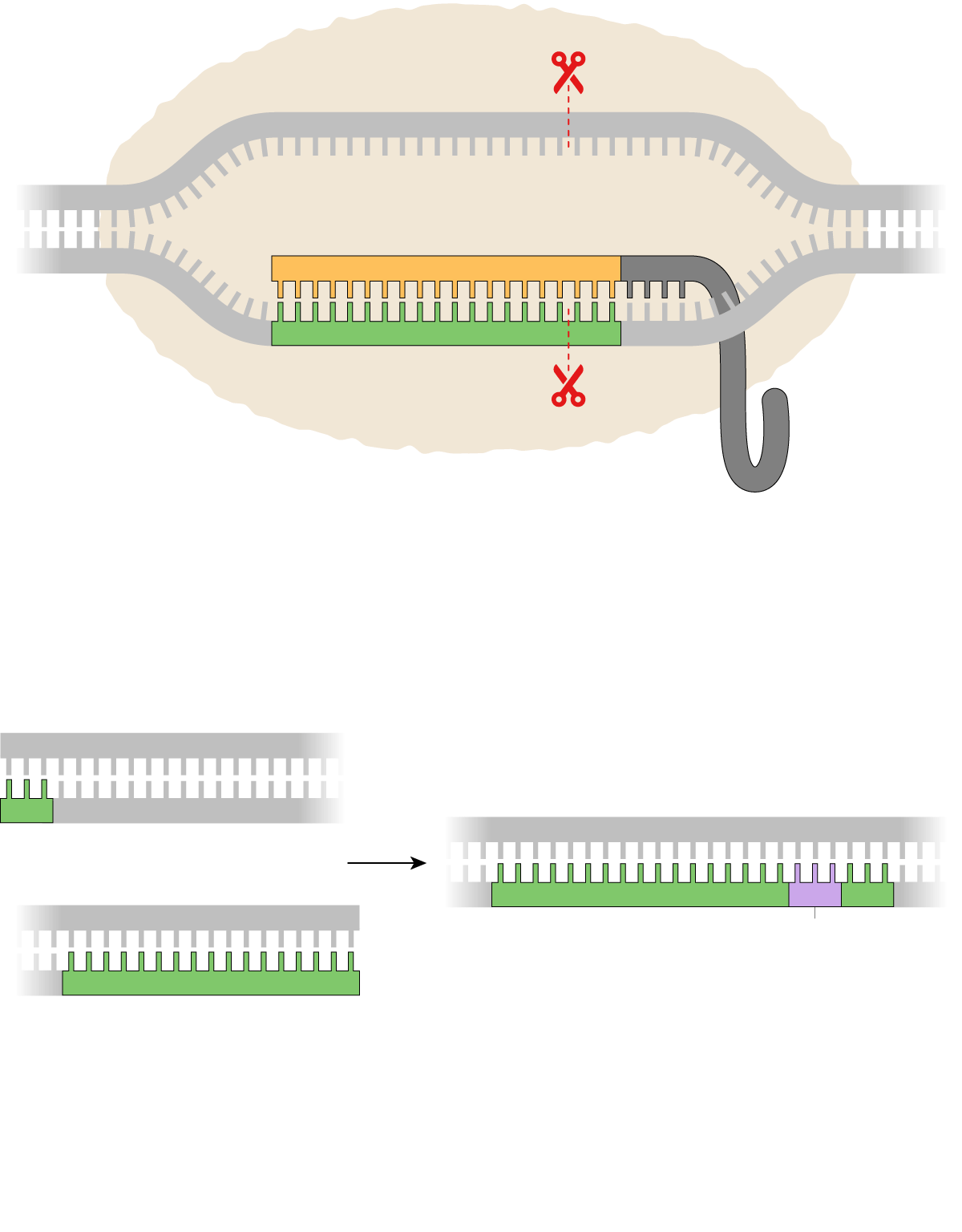
Ten years ago this week, Jennifer Doudna and her colleagues published the results of a test-tube experiment on bacterial genes. When the study came out in the journal Science on June 28, 2012, it did not make headline news. In fact, over the next few weeks, it did not make any news at all.Looking back, Dr. Doudna wondered if the oversight had something to do with the wonky title she and her colleagues had chosen for the study: “A Programmable Dual RNA-Guided DNA Endonuclease in Adaptive Bacterial Immunity.”“I suppose if I were writing the paper today, I would have chosen a different title,” Dr. Doudna, a biochemist at the University of California, Berkeley, said in an interview.Far from an esoteric finding, the discovery pointed to a new method for editing DNA, one that might even make it possible to change human genes.“I remember thinking very clearly, when we publish this paper, it’s like firing the starting gun at a race,” she said.In just a decade, CRISPR has become one of the most celebrated inventions in modern biology. It is swiftly changing how medical researchers study diseases: Cancer biologists are using the method to discover hidden vulnerabilities of tumor cells. Doctors are using CRISPR to edit genes that cause hereditary diseases.Editing the genome with CRISPR

Read more →

Antibiotic use and sepsis make recent improvements

Hospitals and policymakers alike have banded together to improve recognition and treatment of sepsis, a life-threatening reaction to infection, which by some estimates, accounts for 30-50% of all deaths in hospitalized patients. Lessening sepsis’ deadly effects means clinicians need to move quickly to recognize the signs and symptoms, and initiate treatment with antibiotics.
However, a parallel movement within healthcare to limit the unnecessary use of antibiotics has caused some experts to wonder whether efforts to treat sepsis faster, and the use of time-to-treatment thresholds as a hospital performance measure, could lead to overuse of antibiotics and the emergence of antimicrobial resistance.
New research from the University of Michigan Medical School, the VA Ann Arbor and Kaiser Permanente helps puts these fears to rest.
Led by Hallie Prescott, M.D. of the U-M Health Division of Pulmonary and Critical Care and Vincent Liu, M.D., of Kaiser Permanente Division of Research, the study looked at data from more than 1.5 million patients from 152 hospitals nationwide from 2013 through 2018. Patients included came to the emergency department with signs of systemic inflammatory response syndrome (SIRS), which includes increased heart rate, abnormal body temperature, among other signs.
The research team analyzed the use of antibiotics in these patients, including how many received antibiotics, when their treatment started, how long they were on the medications and the broadness of spectrum of the antibiotics: in other words, how many different bacteria species the antibiotics would kill.
“We showed in the overall cohort, that antibiotic use decreased. There was a slight decrease in the proportion treated within 48 hours, a more impressive decrease in the average number of days of antibiotic treatment, and also a decrease in the use of broad-spectrum antibiotics,” said Prescott.
The findings are published in JAMA Internal Medicine.
About half of the people who met the criteria for SIRS received antibiotics within 12 to 48 hours after admission, a practice that decreased slightly over time. At the same time, 30-day mortality (how many people died within 30 days of their hospital stay), length of hospitalization, and the development of multi-drug resistant bacteria also decreased.
“This study adds to our national conversation about how to combat sepsis most effectively. It also confirms that we now need to look for new opportunities to mitigate sepsis by finding patients at high risk before they arrive at the hospital, identifying hospitalized patients most likely to benefit from specific treatments, and enhancing their recovery after they survive sepsis,” said Liu.
Prescott agrees.
“The pushback has been [time-to-treatment for sepsis] should not be a performance measure because it’s going to cause more harm than good, and I think our data shows it probably does more good than harm,” Prescott said. “We have shown that 152 hospitals have been able to make improvements in stewardship and sepsis treatment at the same time, contrary to popular belief.”
This work was supported by grant RO1 HSO26725 (to Drs. Prescott and Liu) from the US Agency for Healthcare Research and Quality and grant IIR 20-313 (to Dr. Prescott) from the US Department of Veterans Affairs, Health Services Research and Development Service.
Story Source:
Materials provided by Michigan Medicine – University of Michigan. Original written by Kelly Malcom. Note: Content may be edited for style and length.

Read more →

Deadly fungus can multiply by having sex, which could produce more drug-resistant, virulent strains

Researchers at McMaster University have unlocked an evolutionary mystery of a deadly pathogen responsible for fueling the superbug crisis: itcan reproduce by having sex.
And while such fraternizing is infrequent, scientists report it could be producing more drug-resistant and more virulent strains of Candida auris, capable of spreading faster.
C. auris is a fungus that can cause severe infections and sometimes death, often striking immunocompromised hospital patients.
Unlike animals and plants, microorganisms of this nature usually divide and reproduce asexually, so one produces two, two produce four and so on, all genetically identical to each other, through a process of very simple division and without the exchange of genetic material.
“One of the really complex and puzzling questions about this fungal pathogen is its origin and how it reproduces in nature,” says Jianping Xu, a professor in McMaster’s Department of Biology and researcher with Canada’s Global Nexus for Pandemics and Biological Threats.
For the study, recently published online in Computation and Structural Biotechnology Journal, researchers analyzed nearly 1,300 strains available on a public database of C. auris genome sequences. They searched for and confirmed recombination events, or sexual activity.

Read more →

Study shows link between cyberbullying and suicidality in early adolescence

Young adolescents who are targets of cyberbullying are more likely to report suicidal thoughts and attempts, an association that goes above and beyond the link between suicidality and traditional offline bullying, according to new research from the Lifespan Brain Institute (LiBI) of Children’s Hospital of Philadelphia (CHOP) and the University of Pennsylvania. The findings were published today in JAMA Network Open.
“At a time when young adolescents are spending more time online than ever before, this study underscores the negative impact that bullying in the virtual space can have on its targets,” said senior author Ran Barzilay, MD, PhD, an assistant professor at LiBI. “Given these results, it may be prudent for primary care providers to screen for cyberbullying routinely in the same way that they might screen for other suicide risk factors like depression. Educators and parents should also be aware of the substantial stress bullying in the cyberworld places on young adolescents.”
Suicide rates among children have been steadily rising. According to the CDC, suicide was the second leading cause of death among individuals between the ages of 10 and 24 in 2018. The factors contributing to suicidality in children and adolescents are not fully understood, but research has shown that environmental stressors play a role. Traditional bullying and peer victimization are well established suicide risk factors among youth.
In modern times, and particularly since the COVID-19 pandemic, a substantial proportion of peer interaction, including bullying, occurs online, through text messages or social media platforms. However, prior to this study, it was not clear whether being a target of cyberbullying is an independent risk factor for suicidality.
To better understand whether cyberbullying is unique in its association with suicidality in early adolescence, the researchers collaborated with Anat Brunstein Klomek, PhD at Baruch Ivcher School of Psychology at Reichman University in Israel. Together, the researchers analyzed data collected between July 2018 and January 2021 from the Adolescent Brain Cognitive Development study (ABCD Study), a diverse sample of over 10,000 US children between the ages of 10 and 13.
As part of the ABCD Study, participants filled out a cyberbullying questionnaire, which asked whether they had ever been a target or perpetrator of cyberbullying, defined as “purposefully trying to harm another person or be mean to them online, in texts or group texts, or on social media (like Instagram or Snapchat).” Traditional offline bullying was surveyed through a separate questionnaire, which broke down behavior into three categories: overt aggression, such as threatening or hitting; relational aggression, such as not inviting or leaving someone out; and reputational aggression, such as spreading rumors or gossiping.
To determine suicidality, the researchers examined whether participants reported past or current suicidal thoughts or acts.
Of the 10,414 ABCD Study participants included in the study, 7.6% responded that they had experienced suicidal thoughts or acts, 8.9% reported being targets of cyberbullying, and 0.9% reported cyberbullying others. The authors found that being a target of cyberbullying was associated with suicidality, whereas being a perpetrator of cyberbullying was not. That finding was distinct from traditional offline bullying, where being either a target or perpetrator of bullying is linked with suicidality.
Additionally, the researchers found that being bullied online only partly overlaps with being bullied offline, supporting the notion that cyberbullying is a distinct phenomenon, independent of offline experiences of bullying. This may suggest that adolescents affected by cyberbullying are different from those affected by offline bullying.
“Our findings suggest being a target of cyberbullying is an independent risk factor for youth suicidality,” Dr. Barzilay said. “For policy makers wishing to optimize youth suicide prevention efforts, this study should further encourage interventions for those who are being bullied online.”
Story Source:
Materials provided by Children’s Hospital of Philadelphia. Note: Content may be edited for style and length.

Read more →

Smoking cessation through a healthcare system model is shown more effective than telephone 'quitlines' in the short term

A healthcare system model that offered tobacco cessation treatment to smokers being discharged from a hospital produced a higher rate of tobacco abstinence during the three- month program than referral to a state-based telephone quitline, but the advantage disappeared at six months when both treatments produced comparable quit rates, researchers from Massachusetts General Hospital (MGH) have found. In a study in JAMA Internal Medicine, the team suggested that extension beyond three months of the health system approach, which includes cessation medication and telephone-based behavioral support, could potentially keep individuals tobacco-free.
“Hospital admission is a great opportunity for smokers to begin tobacco cessation treatment,” says lead author Nancy Rigotti, MD, director of the MGH Tobacco Research and Treatment Center. “That treatment is only effective, however, if it continues after discharge. How to best meet that ongoing need is the question our study was designed to address.”
Cigarette smoking is responsible for around 480,000 deaths annually, the leading cause of preventable deaths in the United States. In 2020, an estimated 13 percent of adults were regular smokers, and more than 3.2 million of them were admitted to a hospital. Researchers from MGH, Vanderbilt University Medical Center, and University of Pittsburgh Medical Center conducted a study of 1,409 adults who smoked and received brief in-hospital tobacco treatment at one of these medical centers to determine the most effective post-discharge model for cessation.
Participants were randomized to one of two models, each offering tobacco cessation medication and counseling by telephone for up to three months. One model used the national system of state-based telephone quitlines, which provides cessation counseling and free samples of nicotine patches, gum, or lozenges. The medical center model provided people who smoked with nicotine medications at hospital discharge and continued counseling with a series of automated and live phone calls.
The study found that three months after discharge, more participants in the healthcare system model were tobacco-free for at least seven days (which was biochemically verified), compared to the quitline model. But at six months — three months after healthcare system intervention ended — the difference in smoking abstinence rates between the two models had narrowed and was not statistically significant.
“Our findings prompt the question of whether continuing smoking cessation past three months — the standard duration of treatment — would sustain the superior results of the healthcare system model,” says senior author Hilary Tindle, MD, MPH, founding director of the Vanderbilt Center for Tobacco Addiction and Lifestyle. “It’s possible that more counseling or medication, or both, could generate more engagement with the program and thus better results over time.”
That thought was echoed by co-author Esa Davis, MD, MPH, director of the University of Pittsburgh Medical Center Tobacco Treatment Service. “We found that keeping smoking cessation within the healthcare system where it could be managed like other chronic medical conditions could be an effective form of treatment,” she remarked. “This approach presents a challenge for healthcare systems, however, which future research needs to address.”
Rigotti is professor of Medicine, Harvard Medical School (HMS) and associate chief for academic advancement, Division of General Internal Medicine, MGH. Tindle is associate professor of Medicine, HMS. Davis is associate professor of Medicine, University of Pittsburgh Medical Center.
The study was funded by the National Heart, Lung, and Blood Institute.
Story Source:
Materials provided by Massachusetts General Hospital. Note: Content may be edited for style and length.

Read more →

Microbial link between Western-style diet and incidence of colorectal cancer uncovered

New research builds the case that a Western-style diet — rich in red and processed meat, sugar and refined grains/carbohydrates — is tied to higher risk of colorectal cancer through the intestinal microbiota.
Investigators from Brigham and Women’s Hospital with collaborators looked at data from more than 134,000 participants from two U.S.-wide prospective cohort studies. The team analyzed dietary patterns as well as DNA from Escherichia coli strains found in more than 1,000 colorectal tumors.
The team looked for bacterial strains carrying a distinct genetic island known as polyketide synthase (pks). Pks encodes an enzyme that has been shown to cause mutations in human cells. Overall, the team found that Western diet was associated with colorectal tumors containing high amounts of pks+ E. coli but not with tumors containing little to no amount of pks+ E. coli.
“These findings support our hypothesis that Western-style diets increase colorectal cancer risk through its effect on pks+ E. coli,” said corresponding author Shuji Ogino, MD, PhD, MS, of the Program in Molecular Pathological Epidemiology in the Department of Pathology at the Brigham. “This is the first study to link Western diet with specific pathogenic bacteria in cancer. Our next question is which component of western-style diet and lifestyle relates to colorectal cancer containing this bacterial species.”
Story Source:
Materials provided by Brigham and Women’s Hospital. Note: Content may be edited for style and length.

Read more →

Researchers determine 1st crystal structure of LAG3

Immune checkpoint inhibitors have revolutionized cancer care. The therapy works by preventing tumors from shutting down the immune response, which in turn allows T cells to kill cancer cells. Established checkpoint inhibitors target the proteins PD-1 and CTLA-4 and are used to treat a variety of solid tumor types, including melanoma and lung cancer. However, the U.S. Food and Drug Administration recently approved a new immune checkpoint inhibitor targeting the protein LAG3. This anti-LAG3 antibody, called relatlimab, was administered in combination with the anti-PD-1 antibody nivolumab to treat advanced melanoma.
Despite this therapeutic breakthrough, little has been known about the structure of the LAG3 protein. In the absence of a three-dimensional structure, LAG3-based drugs must be designed “in the dark” using inefficient screening methods. A team of Moffitt Cancer Center researchers has become the first in the world to visualize the molecular structure of the LAG3 protein. In a new article published in Nature Immunology, they describe the crystal structure of LAG3 and how it interacts with molecules produced by cancer cells.
“When I started my lab at Moffitt, I noticed a growing interest in LAG3 as an immunotherapy target. I was surprised at how little we knew about the LAG3 structure and its molecular mechanism, despite about 30 years of literature highlighting its role in the immune system,” said Vince Luca, Ph.D., assistant member of the Drug Discovery Department.
Luca and his team used X-ray crystallography to “see” the structure of the LAG3 protein at nearly atomic resolution. The researchers also mapped out the regions of LAG3 that bound to signaling molecules called FGL1 and MHCII and two different anti-LAG3 antibodies. From this information, they were able to determine which antibody binding sites were ideal to inhibit LAG3 activity.
Through their investigations, the researchers discovered how structural interactions of LAG3 and FGL1 inhibit T cell function. They found that binding of the two molecules causes LAG3 to cluster on the surface of T cells, which they hypothesize may contribute to the inhibitory activity of LAG3 by blocking the T cells from properly recognizing tumor cells.
These combined data reveal several important insights into the three-dimensional structure of LAG3 and how it interacts with other molecules, which may lead to better targeted therapeutic approaches in the future.
“Collectively, our structural, epitope mapping and functional studies provide an improved framework for understanding LAG3 molecular function. In the future, additional structures of LAG3 bound to ligands and antibodies will refine our knowledge of the LAG3 signaling axis to illuminate how extracellular binding events fine-tune LAG3-mediated changes in T cell activity. In turn, such structural insights should guide the development of maximally effective LAG3-based immunotherapies,” said Luca.
This study was supported by the National Institutes of Health (R35GM133482, P01AI120943, R01CA230610, P30CA076292), the V Foundation for Cancer Research and the Rita Allen Foundation Scholars Program.
Story Source:
Materials provided by H. Lee Moffitt Cancer Center & Research Institute. Note: Content may be edited for style and length.

Read more →