What is the Marburg virus and how can it be avoided?

Published18 minutes agoSharecloseShare pageCopy linkAbout sharingImage source, Getty ImagesTwo people in Ghana have died from the Marburg virus – and 98 been quarantined – raising fears of a mass outbreak of this highly infectious disease, which causes fever, muscle pains, diarrhoea, vomiting and, in many cases, death through extreme blood loss. What is the Marburg virus?A cousin of the equally deadly Ebola virus, according to the World Health Organization (WHO), the Marburg virus, was first identified after 31 people were infected and seven died in simultaneous outbreaks in 1967 in:Marburg and Frankfurt, GermanyBelgrade, SerbiaImage source, Getty ImagesThe outbreak was traced to African green monkeys imported from Uganda. But the virus has been linked to other animals since then. And among humans, it is spread mostly by people who have spent long periods in caves and mines populated by bats. This is Ghana’s first outbreak – but a number of African countries have had them, including:the Democratic Republic of the CongoKenyaSouth AfricaUgandaZimbabweA 2005 outbreak in Angola killed more than 300 people.But in Europe, only one person has died in the past 40 years – and one in the US, after returning from expeditions to caves in Uganda.Major outbreaks:2017, Uganda: three cases, three died2012, Uganda: 15 cases, four died2005, Angola: 374 cases, 329 died1998-2000, DR Congo: 154 cases, 128 died1967, Germany: 29 cases, seven diedSource: WHOWhat illness does it cause?The virus begins abruptly with:a feversevere headachemuscle painsThis is often followed, three days later, by:watery diarrhoeastomach painnausea vomitingThe WHO says: “The appearance of patients at this phase has been described as showing ‘ghost-like’ drawn features, deep-set eyes, expressionless faces and extreme lethargy.”Many people go on to bleed from various parts of the body and die eight to nine days after first falling ill, because of extreme loss of blood and shock.On average, the virus kills half those infected, the WHO says, but the most harmful strains have killed up to 88%.Image source, Getty ImagesHow is it spread?The Egyptian rousette fruit bat often harbours the virus.African green monkeys and pigs can also carry it.Among humans, it spreads through bodily fluids and bedding contaminated with them.And even if people recover, their blood or semen, for example, can infect others for many months afterwards. How can it be treated?There is no specific vaccine or treatments for the virus. But a range of blood products, drug and immune therapies are being developed, the WHO says.And doctors may be able to alleviate the symptoms by giving hospital patients plenty of fluids and replacing lost blood.How can it be contained? People in Africa should avoid eating or handling bushmeat, Gavi, an international organisation promoting vaccine access, says.People should also avoid contact with pigs in areas with an outbreak, the WHO says.Men who have had the virus should use condoms for a year after the onset of symptoms or until their semen twice tests negative.And those burying people who have died from the virus should avoid touching the body.

Read more →

What Pregnancy and Childbirth Do to the Bodies of Young Girls

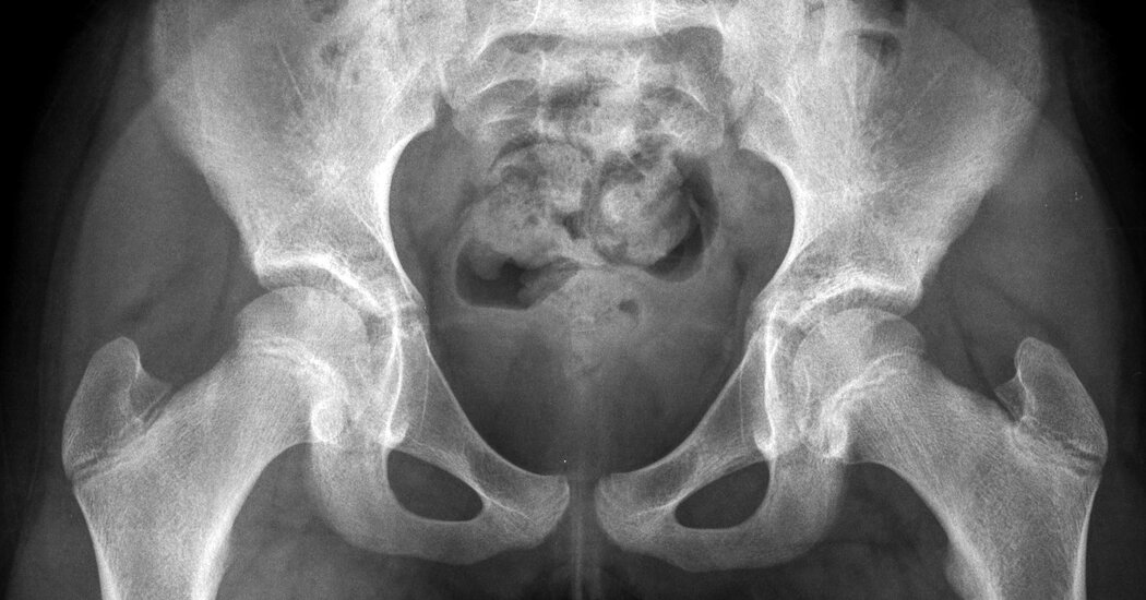
An Ohio 10-year-old’s recent abortion has generated fierce debate. Doctors in countries where pregnancy is common in adolescents say the toll of childbirth on young bodies is brutal.After the account a 10-year-old Ohio girl crossing state lines to get an abortion drew national attention last week, some prominent abortion opponents suggested the child should have carried her pregnancy to term.But midwives and doctors who work in countries where pregnancy is common in young adolescent girls say those pushing for very young girls to carry pregnancies to term may not understand the brutal toll of pregnancy and delivery on the body of a child.“Their bodies are not ready for childbirth and it’s very traumatic,” said Marie Bass Gomez, a midwife and the senior nursing officer at the reproductive and child health clinic at Bundung Maternal and Child Health Hospital in Gambia.The critical issue is that the pelvis of a child is too small to allow passage of even a small fetus, said Dr. Ashok Dyalchand, who has worked with pregnant adolescent girls in low-income communities in India for more than 40 years.“They have long labor, obstructed labor, the fetus bears down on the bladder and on the urethra,” sometimes causing pelvic inflammatory disease and the rupture of tissue between the vagina and the bladder and rectum, said Dr. Dyalchand, who heads an organization called the Institute of Health Management Pachod, a public health organization serving marginalized communities in central India.“It is a pathetic state particularly for girls who are less than 15 years of age,” he added. “The complications, the morbidity and the mortality are much higher in girls under 15 than girls 16 to 19 although 16 to 19 has a mortality twice as high as women 20 and above.”The phenomenon of young girls having babies is relatively rare in the United States. In 2017, the last year for which data was available, there were 4,460 pregnancies among girls under 15, with just under half ending in abortion, according to the Guttmacher Institute, which supports abortion rights and surveys clinics regularly.But globally, complications relating to pregnancy and childbirth are the leading cause of death for girls aged 15-19, according to the World Health Organization.Young maternal age is associated with an increased risk of maternal anemia, infections, eclampsia and pre-eclampsia, emergency cesarean delivery and postpartum depression, according to a 2014 evaluation published in the Journal of Neonatal-Perinatal Medicine.Babies born to girls are more often premature and have low birth weight, said Dr. Willibald Zeck, the maternal and newborn health coordinator for the United Nations Population Fund, who frequently delivered babies for young mothers while working as a gynecologist in Tanzania and later oversaw maternal health programs in Nepal and the Philippines.While a pregnant 10-year-old in Ohio might have access to prenatal care and a cesarean section that would blunt the effects of obstructed labor, the experience of pregnancy in a young girl is the same in India as it is in the United States, Dr. Dyalchand said. “The girls would go through more or less exactly the same kind of complication: The only difference is because of access to better health care they may not have the same kind of terrible outcomes. But that doesn’t mean that the girl’s body and her life doesn’t get scarred.”Dr. Shershah Syed, a gynecologist and expert on maternal mortality in Pakistan, regularly provides care to pregnant girls as young as 11. He said good prenatal care can avert the development of a hole between the wall of the bladder or rectum and the vagina — called a fistula — which causes leaked urine or feces that is not only painful (the leaked urine causes burning sores) but also a source of enormous shame and humiliation.But even good prenatal care cannot prevent the hypertension or urinary tract infections that are common in very young mothers, he said.“In normal physiology a 10-year-old child is not supposed to be pregnant. The point is, she’s a child and the child cannot deliver a child, she’s not ready,” Dr. Syed said, adding: “And the mental torture she will go through, that is not measurable.” In the cases he has seen, early pregnancy arrests the very young mother’s physical growth, and also often her mental development because many girls leave school and lose normal social interaction with peers, he said. But while an anemic mother struggles to carry the pregnancy, fetuses appropriate nutrients and continue to grow, until they have well surpassed what a young mother’s pelvis can deliver.“They go to labor for three days, four days, five days, and after that labor, usually the baby is dead. And then when the head is collapsed, then the baby is delivered,” said Dr. Syed, who is one of South Asia’s pre-eminent experts on the repair of obstetric fistula, a common outcome of obstructed labor in pregnant girls.In nearly all these cases, the girl has developed vesicovaginal fistula, a hole between the wall of the bladder and the vagina. In a quarter of cases, the prolonged labor will also cause fistula of the rectum, so that the girl constantly leaks both urine and feces.If fistula sufferers learn that treatment is available and make their way to his clinic, Dr. Syed said he can repair the condition. But the process requires a long recovery: fistula of the bladder takes about five weeks to heal, while a rectal fistula needs four or five months.In 1978, Dr. Dyalchand began his career in public health at a small district hospital in rural Maharashtra, on the western coast of India. In his first week, two young pregnant girls bled to death — one while in labor, the other at the entrance to the hospital, before she ever made it inside. It started him on a long career of working with communities to convince them to delay the age of marriage and first conception in girls.That intervention has shown considerable success, and, Dr. Dyalchand noted, India has also been steadily expanding abortion access. The procedure is legal up to 24 weeks of pregnancy.In Gambia, Ms. Bass Gomez said that her clinic is able to offer good prenatal care to pregnant girls, but that does little to blunt the larger trauma of the experience. Her clinic is designed to serve adults, she said. “But when you have a child walk in equally pregnant it’s really traumatizing for the child,” she said. “It’s not comfortable, that environment, it’s not set for them. You can tell they are struggling. There’s a lot of shame and disgrace.”

Read more →

Discovery advancing epilepsy research

A team of Florida State University College of Medicine researchers has found a link between a specific protein in the brain and increased vulnerability to neurodegeneration for individuals with temporal lobe epilepsy (TLE).
Their findings are published in the Journal of Neurophysiology.
TLE is the most common form of epilepsy in adults and is often resistant to medication. Professor of Biomedical Sciences Sanjay Kumar, who led the study, said the team used a novel technique that made it possible to study small amounts of tissue from hard-to-reach regions within the brain. Kumar, FSU researcher Stephen Beesley and former doctoral student Thomas Sullenberger focused on a chemical messenger called glutamate and one of its receptors, N-methyl-D-aspartate (NMDA).
Glutamate plays a major role in learning and memory, and it must be present in the right concentration at the right time for the brain to function properly. It is also the body’s most abundant amino acid, a building block of protein.
The team discovered that although two proteins commonly associated with NMDA — GluN1 and GluN2 — were evenly distributed in a critical hippocampal region of the brain, a third one — GluN3 — was distributed on a gradient. A pattern of neuron loss in the hippocampal and para-hippocampal regions of the brain is a hallmark feature of TLE.
“The relationship between GluN3 and cell loss was not known until this research,” Kumar said. “This advance in cellular biology is an important step for developing therapies to help patients.”
Because GluN3 makes neurons more susceptible to calcium-induced cellular damage, the discovery helps researchers narrow the focus to identify exactly where neurons are dying and in how large an area.
Kumar has applied to patent the novel technique, known as area-specific tissue analysis (ASTA), that he developed. ASTA’s added precision created an improved method of testing for both the presence and volume of specific proteins linked to TLE.
Ultimately, Kumar said, discovering the gradient distribution of GluN3 will allow researchers to access more relevant tissue samples.
“This research shows how area-specific tissue analysis can be a useful tool,” he said. “I’m excited to explore what further research with this technique can uncover.”
The Kumar Lab focuses on deciphering the basic mechanisms underlying TLE, as well as identifying and isolating vulnerable cells and circuits within the hippocampal region to promote the discovery of more effective therapies and interventions.
The work is supported in part by a grant from the National Institute of Neurological Disorders and Stroke, a division of the National Institutes of Health.
Story Source:
Materials provided by Florida State University. Original written by Audrey Post. Note: Content may be edited for style and length.

Read more →

Discovery of molecular signatures of immature neurons in human brain throughout life provide insights into brain plasticity

A team led by researchers at the Perelman School of Medicine at the University of Pennsylvania has used advanced techniques to show that, in a key memory region of the brain called the hippocampus, immature, plastic neurons are present in significant numbers throughout the human lifespan. The findings, published this month in Nature, hope to resolve a long-running controversy over the existence of “adult neurogenesis” — the production of new immature neurons in the mature human brain. The discovery also paves the way for the deeper study of adult neurogenesis and its roles in memory, mood, behavior, and brain disorders.
“Many mammals generate new neurons in their brains throughout their lifespans which play a critical role in the brain’s plasticity, or ability to change and adapt over time. This ability to repair itself is especially important when the brain is damaged, which is what happens during a stroke or brain injury,” said senior author Hongjun Song, PhD, a Perelman Professor of Neuroscience at Penn. “This plasticity is also important for understanding diseases like Alzheimer’s, which affect a patient’s memory, among other functions.”
The existence of adult neurogenesis in humans has been debated for decades. For almost a century, neuroscientists assumed that, once the mammalian brain matured, no new neurons were produced in it — the existing ones had to last throughout adulthood. Eventually, studies began to provide evidence of newly produced, immature neurons in the adult brain, in mice, humans, and other mammals, especially in the olfactory (smell) region, and in the hippocampus. Immature neurons in the hippocampus were and continue to be of particular interest, since this brain region play major roles in learning, memory and mood regulation, and are reduced in the brains of people with Alzheimer’s disease.
However, in the past few years, other studies have found no evidence of significant adult neurogenesis in the human hippocampus. It has been difficult for neuroscientists to settle the debate, because they haven’t had an easy, sensitive, and specific method for identifying newly produced, immature neurons in samples of adult human brain tissue.
Song, Ming and their team overcame this challenge with the help of two powerful and relatively new tools. The first is single-nucleus RNA sequencing, which records essentially all gene activity in any individual cell. The second is machine learning, a type of artificial intelligence that, in this case, the researchers used to sift through enormous gene-activity datasets — for mice and humans — to learn the subtle differences between mature and immature hippocampal neurons.
Using these methods, the researchers confirmed the presence of immature hippocampal neurons, chiefly of a type called granule cells, across a wide range of human brain samples from infancy to age 92. The immature granule cells represented usually at least a few percent of the granule cell population, even in elderly brains. The researchers did not find significant numbers of immature neurons in other regions in adult human brains.
The analysis uncovered a broad pattern of gene activity characteristic of immature granule cells, and showed how that pattern shifts during normal aging, differs between humans and mice, and is altered in Alzheimer’s disease. Consistent with prior studies, the researchers found that the proportion of immature granule cells among all granule cells was greatly reduced in Alzheimer’s brains — by more than half compared to age-matched non-Alzheimer’s brains.
Further hinting at the power of this type of analysis for exploring disease origins, the researchers looked at the expression, across the lifespan in immature granule cells, of known risk genes for brain disorders including Alzheimer’s and autism spectrum disorders. They found that several of these risk genes began to be expressed in immature granule cells at ages when the linked disorder is thought to arise.
The study included the identification of the hippocampal progenitor cells that derive from neural stem cells and, in turn, give birth to new granule cells. These experiments indicated that the progenitors are relatively scarce, but serve as steady sources of new granule cells, which mature very slowly, over time spans of a year or more.
“In the future, we hope this type of research can help to further understand the causes of brain diseases like psychiatric disorders and Alzheimer’s, which can inform the possibility of treating these diseases,” said senior author Guo-li Ming, MD, PhD, a Perelman Professor of Neuroscience.
The research was made possible with close collaborations with neurosurgery team at Penn and CHOP as well as contributions from tissue banks across the country. The research was supported by the National Institutes of Health (R35NS097370, R35NS116843), the Lieber Institute for Brain Development, and the Dr. Miriam and Sheldon G. Adelson Medical Research Foundation.

Read more →

Loss of 'youth' protein may drive aging in the eye

Loss of the protein pigment epithelium-derived factor (PEDF), which protects retinal support cells, may drive age-related changes in the retina, according to a new study in mice from the National Eye Institute (NEI). The retina is the light-sensitive tissue at the back of the eye, and aging-associated diseases of the retina, like age-related macular degeneration (AMD), can lead to blindness. This new finding could lead to therapies to prevent AMD and other aging conditions of the retina. The study was published in the International Journal of Molecular Sciences. NEI is part of the National Institutes of Health.
“People have called PEDF the ‘youth’ protein, because it is abundant in young retinas, but it declines during aging,” said Patricia Becerra, Ph.D., chief of NEI’s Section of Protein Structure and Function and senior author of the study. “This study showed for the first time that just removing PEDF leads to a host of gene changes that mimic aging in the retina.”
The retina is composed of layers of cells that function together to detect and process light signals, which the brain uses to generate vision. The retina’s light-sensing photoreceptors sit above the retinal pigment epithelium (RPE), a layer of support cells. The RPE nourishes photoreceptors and recycles pieces of the photoreceptor cells called “outer segments,” which get used up and their tips shed each time photoreceptors detect light. If the RPE cannot provide recycled components of older outer segment tips back to photoreceptors, these cells lose their ability to make new segments, and eventually become unable to sense light. And without nutrients supplied by the RPE, photoreceptors die. In people with AMD or certain types of retinal dystrophies, senescence (aging) or death of RPE cells in the retina leads to vision loss.
Previous work from Becerra’s lab and others has shown that PEDF protects retinal cells, preventing both damage to the cells and abnormal growth of blood vessels in the retina. RPE cells produce and secrete the PEDF protein. The protein then binds to its receptor, PEDF-R, which is also expressed by RPE cells. Binding by PEDF stimulates PEDF-R to break down lipid molecules, key components of the cell membranes that enclose photoreceptor outer segments and other cellular compartments. This breakdown step is a key part of the outer segment recycling process. And while researchers have known that PEDF levels drop in the retina during the aging process, it was not clear whether this loss of PEDF was causing, or merely correlated with, age-related changes in the retina.
To examine the retinal role of PEDF, Becerra and colleagues studied a mouse model that lacks the PEDF gene (Serpin1). The researchers examined the cellular structure of the retina in the mouse model, finding that the RPE cell nuclei were enlarged, which may indicate changes in how the cells’ DNA is packed. The RPE cells also had turned on four genes associated with aging and cellular senescence, and levels of the PEDF receptor were significantly below normal. Finally, unprocessed lipids and other photoreceptor outer segment components had accumulated in the RPE layer of the retina. Similar changes in gene expression and defects in RPE metabolism are found in the aging retina.
“One of the most striking things was this reduction in the PEDF receptor on the surface of the RPE cells in the mouse lacking the PEDF protein,” said the study’s lead author, Ivan Rebustini, Ph.D., a staff scientist in Becerra’s lab. “It seems there’s some sort of feedback-loop involving PEDF that maintains the levels of PEDF-R and lipid metabolism in the RPE.”
While at first glance, the retinas of these PEDF-negative mice appear normal, these new findings suggest that PEDF is playing a protective role that helps the retina weather trauma and aging-related wear and tear.
“We always wondered if loss of PEDF was driven by aging, or was driving aging,” said Becerra. “This study, especially with the clear link to altered lipid metabolism and gene expression, indicates the loss of PEDF is a driver of aging-related changes in the retina.”
The study was supported by the NEI intramural program.
Story Source:
Materials provided by NIH/National Eye Institute. Note: Content may be edited for style and length.

Read more →

Covid Rises Across U.S. Amid Muted Warnings and Murky Data

CHICAGO — Covid-19 is surging around the United States again in what experts consider the most transmissible variant of the pandemic yet.But something is different this time: The public health authorities are holding back.In Chicago, where the county’s Covid warning level was raised to “high” last week, the city’s top doctor said there was no reason for residents to let the virus control their lives. The state health director in Louisiana likened a new rise in Covid cases there to a downpour — “a surge within a surge” — but characterized the situation as concerning but not alarming.And the public health officer in King County, Wash., Dr. Jeffrey Duchin, said on Thursday that officials were discussing reissuing a mask mandate but would prefer that the public mask up voluntarily. “We’re not going to be able to have infinite series of mandates forcing people to do this, that and the other,” he said.The latest surge, driven by a spike of BA.5 subvariant cases in this country since May, has sent infections rising in at least 40 states, particularly in the Great Plains, West and South. Hospitalizations have climbed by 20 percent in the past two weeks, leaving more than 40,000 people in American hospitals with the coronavirus on an average day.More than two years after the pandemic began, though, public health officials are sounding only quiet warnings amid a picture that they hope has been changed by vaccines, treatments and rising immunity. Deaths are rising, but only modestly so far in this new wave. And state and local public health officials say they also must now factor in a reality that is obvious along the streets from Seattle to New York City: Most Americans are meeting a new Covid wave with a collective shrug, shunning masks, joining crowds indoors and moving on from the endless barrage of virus warnings of months past.“I feel strongly that you can’t just kind of cry wolf all the time,” said Dr. Allison Arwady, the commissioner of the Chicago health department, who said she would wait to see whether hospitals become strained before considering another citywide mask mandate. “I want to save the requirements around masks or updating vaccine requirements for when there’s a significant change.”A bar in the River North neighborhood of Chicago last week. Most Americans are meeting a new Covid wave with a collective shrug.Jamie Kelter Davis for The New York TimesComplicating the country’s understanding of this BA.5 wave is a dearth of data. Not since the earliest months of the pandemic has there been so little precise information about the number of actual infections in the United States. As public testing sites have closed and at-home testing — if people test at all — has grown common, the publicly reported data has become scarce and spotty.Still, experts say, the outlines of a new wave are undeniable.“You don’t have to count every raindrop to know it’s raining,” said Dr. Joseph Kanter, Louisiana’s state health officer and medical director. “And it’s pouring right now.”In that state, the health department analyzes a wide range of data to track the spread of the virus, including case counts, samples from a growing network of wastewater testing sites, test positivity rate and hospitalization metrics.Coronavirus cases in the United States by regionThis chart shows how reported cases per capita have changed in different parts of the country.

Read more →

Researcher uses graphene for same-time, same-position biomolecule isolation and sensing

New research led by University of Massachusetts Amherst assistant professor Jinglei Ping has overcome a major challenge to isolating and detecting molecules at the same time and at the same location in a microdevice. The work, recently published in ACSNano, demonstrates an important advance in using graphene for electrokinetic biosample processing and analysis and could allow lab-on-a-chip devices to become smaller and achieve results faster.
The process of detecting biomolecules has been complicated and time consuming. “We usually first have to isolate them in a complex medium in a device and then send them to another device or another spot in the same device for detection,” says Ping, who is in the College of Engineering’s Mechanical and Industrial Engineering Department and is also affiliated with the university’s Institute of Applied Life Sciences. “Now we can isolate them and detect them at the same microscale spot in a microfluidic device at the same time — no one has ever demonstrated this before.”
His lab achieved this advance by using graphene, a one-atom-thick honeycomb lattice of carbon atoms, as microelectrodes in a microfluidic device.
“We found that, compared to typical inert-metal microelectrodes, the electrolysis stability for graphene microelectrodes is more than 1,000 times improved, making them ideal for high-performance electrokinetic analysis,” he says.
Also, Ping added, since monolayer graphene is transparent, “we developed a three-dimensional multi-stream microfluidic strategy to microscopically detect the isolated molecules and calibrate the detection at the same time from a direction normal to the graphene microelectrodes.”
The new approach developed in the work paves the way to the creation of lab-on-a-chip devices of maximal time and size efficiencies, Ping says. Also, the approach is not limited to analyzing biomolecules and can potentially be used to separate, detect and stimulate microorganisms such as cells and bacteria.
Story Source:
Materials provided by University of Massachusetts Amherst. Note: Content may be edited for style and length.

Read more →

Brains of children with autism may not always 'see' body language, study finds

Noticing and understanding what it means when a person leans into a conversation or takes a step back and crosses their arms is a vital part of human communication. Researchers at the Del Monte Institute for Neuroscience at the University of Rochester have found that children with autism spectrum disorder may not always process body movements effectively, especially if they are distracted by something else.
“Being able to read and respond to someone’s body language is important in our daily interactions with others,” said Emily Knight, M.D., Ph.D., clinical and postdoctoral fellow in Pediatrics and Neuroscience, is the first author of the study recently published in Molecular Autism. “Our findings suggest that when children with autism are distracted by something else, their brains process the movements of another person differently than their peers.”
Key differences in brain processes
Using electroencephalogram (EEG), researchers recorded the brain waves of children with and without autism as they watched videos of moving dots that were arranged to look like a person. In these videos the dots moved to represent actions such as running, kicking, or jumping, and at times were turned in different directions or jumbled to no longer move like a person. The six- to 16-year-olds were asked to either focus on the color of the dots or to focus on whether the dots moved like a person. Researchers found the brainwaves of children with autism did not process when the dots moved like a person if they were focused on the dot color.
“If their brain is processing body movements less they might have a harder time understanding other people, and need to pay extra attention to body language in order to see it,” said Knight. “Knowing this can help guide new ways to support people with autism.”
“This is more evidence of how the brain of someone with autism is processing the world around them,” said John Foxe, Ph.D., lead author of the study. “This research is a vital step in creating a more inclusive space for people with autism by giving a glimpse of how their brain processes an unspoken part of communication.”
Additional authors include Ed Freedman, Ph.D., from the University of Rochester Medical Center, John Butler, Ph.D., Aaron Krakowski, and Sophie Molholm, Ph.D., of Einstein College of Medicine. This research was supported by the National Institute of Mental Health and the University of Rochester Intellectual and Developmental Disability Research Center (UR-IDDRC) and the Rose F. Kennedy Intellectual and Developmental Disabilities Research Center (RFK-IDDRC).
Story Source:
Materials provided by University of Rochester Medical Center. Original written by Kelsie Smith Hayduk. Note: Content may be edited for style and length.

Read more →

Houston residents' chemical exposure increased post-Hurricane Harvey, study finds

Researchers at Oregon State University used silicone wristbands to measure Houston residents’ increased exposure to hazardous chemicals in the wake of Hurricane Harvey in 2017.
The wristbands recorded exposures to 162 different chemicals, including pesticides, flame retardants, industrial compounds, phthalates and polycyclic aromatic hydrocarbons.
Researchers followed up with study participants a year after Harvey to approximate a baseline so they could parse out which exposures were caused by the storm. On average, 75% of the chemicals detected across both timepoints were found in higher concentrations immediately after the hurricane, but people’s baseline exposure was already high.
“Houston is one of our most industrialized cities,” said co-author Kim Anderson, head of OSU’s Department of Environmental and Molecular Toxicology and the inventor of the study’s wristbands. “When we look a year after the storm, we see that several neighborhoods that are closer to industrial zones — socioeconomically disadvantaged neighborhoods — had higher concentrations of chemicals right from the get-go, and that was only exacerbated when the hurricane came in.”
The silicone wristbands absorb chemicals from the air and from skin contact, making them a useful screening tool. Anderson has used them in similar studies in Africa, Europe and South America.
Many of the chemicals recorded in the Houston study have not yet been thoroughly tested to determine their potential health effects, researchers said. But some heavier polycyclic aromatic hydrocarbons have been found to be carcinogenic, and phthalate exposure can have adverse effects on reproductive health.

Read more →

Tissue nanotransfection technology proves useful in non-viral topical gene editing to close complex cutaneous wounds

The Indiana Center for Regenerative Medicine and Engineering (ICRME) at Indiana University School of Medicine is home to tissue nanotransfection (TNT) regenerative medicine technology that achieves functional tissue reprogramming in the live body. Last year, ICRME researchers published on how to manufacture the TNT 2.0 silicon chip hardware in Nature Protocol. Now, their research demonstrates for the first time that TNT can serve as a non-viral, topical, gene-editing delivery device.
TNT is a minimally invasive device that can reprogram tissue function in the live body by applying pulses of harmless, electric sparks to deliver specific genes of interest to the skin.
“TNT-based delivery can achieve cell-specific gene editing,” said corresponding author Chandan K. Sen, PhD, the J. Stanley Battersby Chair and distinguished professor of surgery, director of the ICRME at IU School of Medicine and executive director of the Indiana University Health Comprehensive Wound Care Center. “Your skin has thousands of genes and in chronic wounds many key genes are silenced by DNA methylation. TNT-based gene editing technology can remove that barrier.”
In this study, genome-wide methylation was observed in the chronic wound tissue of patients. This was reproduced in an experimental murine model. TNT-based, cell-specific gene editing rescued wound healing. Results were published recently in the Journal of Clinical Investigation.
Previous TNT application studies reported on the rescue of injured legs, diabetic neuropathy, crushed nerve and the stroke-affected brain. This is the first time promoter methylation of genes is recognized as a critical barrier to wound healing. In this study, ICRME investigators found that P53 methylation and gene silencing as a critical barrier to cutaneous wound epithelial-to-mesenchymal transition (EMT), a mechanism that is necessary to close skin wounds. TNT based non-viral keratinocyte-specific demethylation of P53 gene rescued EMT and achieved wound closure.
Chronic wounds can result in serious and sometimes life-threatening complications from an abundance of dying and necrotic tissue, such as cellulitis, lower-extremity amputation and sepsis. Treating chronic wounds is estimated to cost the United States health care system $28 billion annually, which amplifies the need to test novel treatments to prevent amputation, save lives and lower health care costs.
“Inspired by observations in chronic wound patients, this work has achieved an important milestone highlighting the need to de-silence genes at the wound-site,” said first author Kanhaiya Singh, PhD, assistant professor of surgery and an investigator at the ICRME.
This study was supported by the National Institute of Diabetes and Digestive and Kidney Diseases, the U.S. Department of Defense and the Lilly Endowment INCITE program.
Story Source:
Materials provided by Indiana University School of Medicine. Original written by Christina Griffiths. Note: Content may be edited for style and length.

Read more →