Drug side effects across pediatric development stages identified

Side effects from pediatric drug treatment are responsible for nearly 10 percent of childhood hospitalizations, with nearly half of those being life-threatening. Despite the need to know more about these drugs and the adverse events they can have on children, little evidence is currently available.
Clinical trials remain the gold standard for identifying adverse drug events (ADEs) for adults, but these have both ethical and methodological concerns for the pediatric population. The rapidly changing biologic and physiologic developments only enhance the challenges of understanding the potential impacts of different drug treatments at various stages of childhood.
Researchers at the Columbia University Irving Medical Center developed a novel algorithm that identified nearly 20,000 ADEs signals (information on a new or known side effect that may be caused by a particular drug) across the seven pediatric development stages and made them freely available. This process is strengthened by a novel approach that allows neighboring development stages to enhance the signal detection power, which helps it overcome limited data within individual stages.
This use of predictive modeling on real-world data can help address a critical gap in healthcare research around the understudied pediatric community.
DBMI associate professor Nicholas Tatonetti and Nick Giangreco, a recent Systems Biology PhD graduate at Columbia University, shared these findings in the study A database of pediatric drug effects to evaluate ontogenic mechanisms from child growth and development, which was recently published in Med.
“For many reasons, children have historically not been included in clinical trials,” Tatonetti said. “There are many ethical issues around including children in trials, and there are several limitations when children are included that make it difficult to assess the effectiveness and safety of drugs.”
Because of these factors, few drugs are specifically approved for use in children, though once drugs are approved for adults, physicians can prescribe them “off-label” to children.
Read more →

A.I. Predicts the Shape of Nearly Every Protein Known to Science

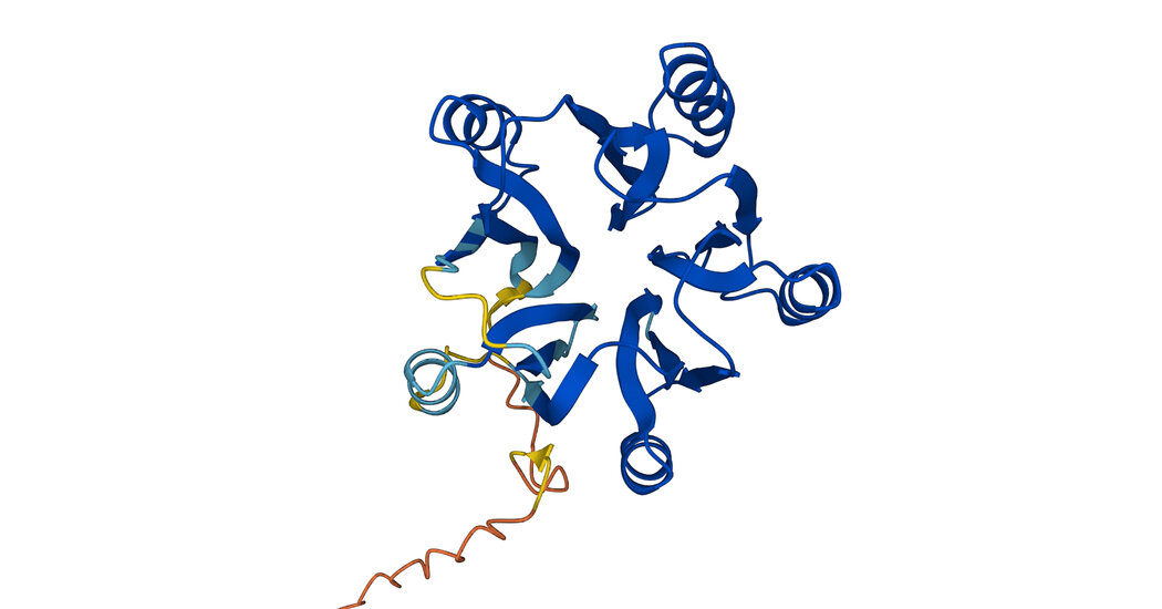
DeepMind has expanded its database of microscopic biological mechanisms, hoping to accelerate research into all living things.In 2020, an artificial intelligence lab called DeepMind unveiled technology that could predict the shape of proteins — the microscopic mechanisms that drive the behavior of the human body and all other living things.A year later, the lab shared the tool, called AlphaFold, with scientists and released predicted shapes for more than 350,000 proteins, including all proteins expressed by the human genome. It immediately shifted the course of biological research. If scientists can identify the shapes of proteins, they can accelerate the ability to understand diseases, create new medicines and otherwise probe the mysteries of life on Earth.Now, DeepMind has released predictions for nearly every protein known to science. On Thursday, the London-based lab, owned by the same parent company as Google, said it had added more than 200 million predictions to an online database freely available to scientists across the globe.With this new release, the scientists behind DeepMind hope to speed up research into more obscure organisms and spark a new field called metaproteomics.“Scientists can now explore this entire database and look for patterns — correlations between species and evolutionary patterns that might not have been evident until now,” Demis Hassabis, the chief executive of DeepMind, said in a phone interview.Proteins begin as strings of chemical compounds, then twist and fold into three-dimensional shapes that define how these molecules bind to others. If scientists can pinpoint the shape of a particular protein, they can decipher how it operates.This knowledge is often a vital part of the fight against illness and disease. For instance, bacteria resist antibiotics by expressing certain proteins. If scientists can understand how these proteins operate, they can begin to counter antibiotic resistance.Previously, pinpointing the shape of a protein required extensive experimentation involving X-rays, microscopes and other tools on a lab bench. Now, given the string of chemical compounds that make up a protein, AlphaFold can predict its shape.The technology is not perfect. But it can predict the shape of a protein with an accuracy that rivals physical experiments about 63 percent of the time, according to independent benchmark tests. With a prediction in hand, scientistic can verify its accuracy relatively quickly.Kliment Verba, a researcher at the University of California, San Francisco, who uses the technology to understand the coronavirus and to prepare for similar pandemics, said the technology had “supercharged” this work, often saving months of experimentation time. Others have used the tool as they struggle to fight gastroenteritis, malaria and Parkinson’s disease.The technology has also accelerated research beyond the human body, including an effort improve the health of honeybees. DeepMind’s expanded database can help an even larger community of scientists reap similar benefits.Like Dr. Hassabis, Dr. Verba believes the database will provide new ways of understanding how proteins behave across species. He also sees it as way of educating a new generation of scientists. Not all researchers are versed in this kind of structural biology; a database of all known proteins lowers the bar to entry. “It can bring structural biology to the masses,” Dr. Verba said.

Read more →

Sophisticated mechanism that bacteria use to resist antibiotics

Researchers have discovered a significant and previously unknown mechanism that many bacteria use to resist antibiotics.
Using a combination of computation and physical observation in the laboratory, the researchers have unraveled a sophisticated process that some commonly occurring bacteria use to save themselves from the rifamycin class of antibiotics, which occur naturally and are also manufactured to treat infectious diseases.
Rifamycins work by binding to RNA polymerase, a protein essential for bacterial life.
The resistant bacteria, which occur widely in the environment and in some human pathogens, have developed a protein that can eject the antibiotic from RNA polymerase. Once the rifamycin is dislodged, they use specially adapted proteins to attack and destroy it.
“What we’ve discovered is a brand-new trick up the sleeves of bacteria to evade this class of antibiotics,” explains researcher Gerry Wright, who leads the McMaster-based Global Nexus for Pandemics and Biological Threats. “It’s like a one-two punch. It’s fascinating and it’s so crafty.”
The discovery shows that the mechanisms of antimicrobial resistance (AMR) are more complex and highly evolved than scientists had previously recognized.

Read more →

Coming wave of opioid overdoses 'will be worse than it's ever been before'

Over the past 21 years of opioid overdose deaths — from prescription drugs to heroin to synthetic and semisynthetic opioids such as fentanyl — geography has played a role in where opioid-involved overdose deaths have occurred, reports a new Northwestern Medicine study.
But the coming wave will not discriminate between rural and urban areas, the study findings suggest. Every type of county — from the most rural to the most urban — is predicted to see dramatic increases in deaths from opioid-involved overdoses. The reason opioid overdoses have reached historical highs comes from combining synthetic opioids with stimulants such as cocaine and methamphetamines, a lethal cocktail that is hard to reverse during an overdose, the study authors said.
“I’m sounding the alarm because, for the first time, there is a convergence and escalation of acceleration rates for every type of rural and urban county,” said corresponding author Lori Post, director of the Buehler Center for Health Policy and Economics at Northwestern University Feinberg School of Medicine. “Not only is the death rate from an opioid at an all-time high, but the acceleration of that death rate signals explosive exponential growth that is even larger than an already historic high.”
The study will be published July 28 in JAMA Network Open.
The study examined geographic trends in opioid-involved overdose deaths between 1999 and 2020 to determine if geography played a role in the three waves and the theorized fourth wave of America’s opioid crisis. The authors used data recorded in the Centers for Disease Control and Prevention’s WONDER database for 3,147 counties and county equivalents categorized on a six-point urbanicity scale (most urban to most rural).
First study to look at acceleration rates systematically by geography
While some researchers have looked at an acceleration rate from one year to the next, the study authors said, to their knowledge, no one has examined acceleration rates of opioid-involved overdose death rates systematically by geography for every year.

Read more →

Organoid production breakthrough to help accelerate disease and drug development research

As the idea of growing tiny human organs in lab dishes has moved in recent years from futuristic science fiction to actual bioscientific reality, the usefulness of organoids as a research tool for studying the digestive system swiftly encountered a bottleneck — these valuable tissues are quite difficult to make.
Even with highly trained teams using all the best ingredients and equipment, one batch of starting material can generate lots of tiny spheroids of organ precursor cells that can then be grown into specific organoid types. But the next batch may produce few spheroids or none at all.
As a result, lab teams often experience delays making the organoids they need for pre-clinical experiments that seek to test the safety or potency of potential medications or for basic research to delve deeper into the genetic and molecular activities that cause disease.
Now, in a paper published June 28, 2022, in Stem Cell Reports, a team of experts at Cincinnati Children’s reports developing a method that overcomes this production bottleneck. The new method already is being used to advance organoid studies within the medical center. But since the materials involved can be frozen and thawed and still produce high-quality organoids, this discovery makes it possible to ship starter materials to other labs anywhere in the world — which could spark dramatically accelerated use of human gastrointestinal organoids throughout medical research.
“This method can make organoids a more accessible tool,” says first author Amy Pitstick, MS, manager of the Pluripotent Stem Cell Facility at Cincinnati Children’s. “We show that the aggregation approach consistently produces high yields and we have proven that precursor cells can be thawed from cyrogenic storage to produce organoids of the small intestine.”
“Using this approach will make it possible for many research labs to use organoids in their experiments without the time and expense of learning how to grow induced pluripotent stem cells (iPSCs),” says corresponding author Chris Mayhew, PhD, director of the Pluripotent Stem Cell Facility. “The ability to freeze the precursor cells also will allow labs to easily make organoids without having to start each new experiment with complicated and highly variable iPSC differentiation.”
About the new process

Read more →

Novel drug promotes nervous system repair in animal models of stroke

A new groundbreaking study from the University of Cincinnati shows promise that a new drug may help repair damage caused by strokes.
Researchers from UC and Case Western Reserve University published the pioneering preclinical study in the journal Cell Reports July 26.
Currently, there are no FDA approved drugs to repair the damage caused by a stroke. The study found a drug called NVG-291-R enables nervous system repair and significant functional recovery in an animal model of severe ischemic stroke. Genetic deletion of the molecular target of the drug also shows similar effect on neural stem cells.
“We are very excited about the data showing significant improvement in motor function, sensory function, spatial learning and memory,” said Agnes (Yu) Luo, PhD, associate professor in the Department of Molecular Genetics and Biochemistry in UC’s College of Medicine and the study’s senior author.
Luo said the drug would be a “substantial breakthrough” if the early results translate into clinical settings. Further study and validation of results from independent groups will be needed to determine if the drug is similarly effective to repair the damage of ischemic strokes in human patients. Additional studies will be needed to research if NVG-291-R effectively repairs damage caused by hemorrhagic strokes in both animal models and human patients.
“Most therapies being researched today primarily focus on reducing the early damage from stroke,” Luo said. “However, our group has focused on neurorepair as an alternative and now has shown that treatment with NVG-291-R not only results in neuroprotection to reduce neuronal death but also robust neuroreparative effects.”
The study also found the drug was effective even when treatment began as late as seven days after the stroke’s onset.
“The only current FDA-approved drug for treatment of stroke does not repair damage and must be administered within 4.5 hours of stroke onset.” Luo said. “Most therapies being researched need to be applied within 24-48 hours of a stroke’s onset. A product that works to repair damage from stroke even a week after symptom onset would change the paradigm for stroke treatment.”
Jerry Silver, PhD, co-author of the study and professor of neurosciences at CWRU’s School of Medicine, said the study showed the drug repaired damage through at least two avenues: creating new neuronal connections and enhancing migration of newly born neurons derived from neuronal stem cells to the site of the damage.
“NVG-291-R’s ability to enhance plasticity was demonstrated by using staining techniques that clearly showed an increase in axonal sprouting to the damaged part of the brain,” Silver said. “This enhanced plasticity is an excellent validation of the same powerful mechanisms that we and other researchers were able to demonstrate using NVG-291-R in spinal cord injury.”
NervGen Pharma Corp. holds the exclusive worldwide rights to NVG-291, and the drug is also currently being tested in a Phase 1 clinical trial in healthy human subjects. NervGen plans to initiate patient safety and efficacy trials in spinal cord injury, Alzheimer’s disease and multiple sclerosis in 2022 and 2023.
The research was supported by a National Institute of Neurological Disorders and Stroke grant (grant no. R01NS107365). The study authors have inventorship in a patent application that has been submitted by CWRU based partially on these results. Silver is an advisor to NervGen, a startup pharmaceutical company that has licensed from CWRU an issued patent (#9937242) covering the ISP peptide.
Story Source:
Materials provided by University of Cincinnati. Original written by Tim Tedeschi. Note: Content may be edited for style and length.

Read more →

New needle-free nasal vaccine shows promise for COVID-19

New research shows that a needle-free mucosal bacteriophage (phage) T4-based COVID-19 vaccine is effective against SARS-CoV-2 infection. The findings were published in mBio, an open access journal of the American Society for Microbiology.
In recent years, the Food and Drug Administration authorized mRNA- and adenovirus-based SARS-CoV-2 vaccines. These vaccines are intramuscularly injected in 2 or more doses and are effective in preventing COVID-19, but they do not induce efficient mucosal immunity or prevent viral transmission.
In the new study, senior study authors Venigalla B. Rao, Ph.D., from the Bacteriophage Medical Research Center, Department of Biology, The Catholic University of America, Washington, D.C., and Ashok K. Chopra, Ph.D., CSc, Department of Microbiology & Immunology, The University of Texas Medical Branch, Galveston, Texas, and their colleagues report the first non-infectious, bacteriophage T4-based, multicomponent, needle and adjuvant-free mucosal vaccine. Both of the senior authors are elected fellows of the American Academy of Microbiology.
In experiments conducted in mice, intranasal administration of 2 doses of the phage T4-COVID-19 vaccine 21-days apart induced robust mucosal immunity, in addition to strong systemic humoral and cellular immune responses. The intranasal vaccine induced broad virus neutralization antibody titers against multiple variants and triggered Th1-biased cytokine responses, strong CD4+ and CD8+ T cell immunity, and high secretory IgA titers in sera and bronchoalveolar lavage of vaccinated mice. All these responses were much stronger in intranasally vaccinated mice than that induced by the injected vaccine. Furthermore, the nasal vaccine provided complete protection and sterilizing immunity against the mouse-adapted SARS-CoV-2 MA10 strain, the ancestral WA-1/2020 strain, and the most lethal Delta variant in mouse models.
Additionally, the T4-COVID-19 vaccine elicited broad virus-neutralizing antibodies against SARS-CoV-2 variants in sera and bronchoalveolar lavage, did not affect the gut microbiota, exhibited minimal lung lesions in vaccinated and challenged mice and is stable at ambient temperature.
“This intranasally administered vaccine generates superior mucosal immunity in mice in addition to inducing robust humoral and cell-mediated immune responses, and provides complete protection and sterilizing immunity against SARS-CoV-2 variants. The vaccine is stable, adjuvant-free and cost-effectively manufactured and distributed, making it a strategically important next-generation COVID-19 vaccine for ending this pandemic,” said Drs. Rao and Chopra. “This modular, needle-free, phage T4 mucosal vaccine delivery platform is an excellent candidate to design efficacious mucosal vaccines against other respiratory infections and for emergency preparedness against emerging epidemic and pandemic pathogens.”
Story Source:
Materials provided by American Society for Microbiology. Note: Content may be edited for style and length.

Read more →

High-tech imaging reveals details about rare eye disorder

Using a new imaging technique, researchers from the National Eye Institute have determined that retinal lesions from vitelliform macular dystrophy (VMD) vary by gene mutation. Addressing these differences may be key in designing effective treatments for this and other rare diseases. NEI is part of the National Institutes of Health.
“The NEI’s long-term investment in imaging technology is changing our understanding of eye diseases,” said NEI Director Michael F. Chiang, M.D. “This study is just one example of how improved imaging can reveal subtle details about pathology in a rare eye disease that can inform the development of therapeutics.”
VMD is an inherited genetic disease that causes progressive vision loss through degeneration of the light-sensing retina. Genes implicated in VMD include BEST1, PRPH2, IMPG1, and IMPG2. Depending on the gene and mutation, age of onset and severity vary widely. All forms of the disease have in common a lesion in the central retina (macula) that looks like an egg yolk and is a build-up of toxic fatty material called lipofuscin. VMD affects about 1 in 5,500 Americans and there is currently no treatment for this condition.
Johnny Tam, Ph.D., head of the NEI Clinical and Translational Imaging Unit, used multimodal imaging to evaluate the retinas of patients with VMD at the NIH Clinical Center. Tam’s multimodal imaging uses adaptive optics — a technique that employs deformable mirrors to improve resolution — to view live cells in the retina, including the light-sensing photoreceptors, retinal pigment epithelial (RPE) cells, and blood vessels in unprecedented detail.
Tam and his team collaborated with clinicians at the NEI Eye Clinic to characterize 11 participants using genetic testing and other clinical assessments, and then evaluated their retinas using multimodal imaging. Assessment of cell densities (photoreceptors and RPE cells) near VMD lesions revealed differences in cell density according to the various mutations. IMPG1 and IMPG2 mutations had a greater effect on photoreceptor cell density than RPE cell density. The opposite was true with PRPH2 and BEST1 mutations. In participants with only one affected eye, the researchers noted similar effects on cell density in the unaffected eye, despite lacking lesions.
Tam is using multimodal imaging on a variety of other rare retinal diseases and more common ones including age-related macular degeneration.
Story Source:
Materials provided by NIH/National Eye Institute. Note: Content may be edited for style and length.

Read more →

People with poor sleep behaviors may be at risk for fatty liver disease

People with sedentary lifestyles and unhealthy sleep behaviors could develop fatty liver disease, according to new research published in the Endocrine Society’s Journal of Clinical Endocrinology & Metabolism.
Fatty liver disease is the leading chronic liver disease worldwide, affecting about a quarter of the adult population. This type of liver disease is fueled by metabolic disorders such as obesity and type 2 diabetes. Fatty liver disease may progress to end-stage liver disease, posing a major health and economic burden to society.
“People with poor nighttime sleep and prolonged daytime napping have the highest risk for developing fatty liver disease,” said Yan Liu, Ph.D., of the Guangdong Provincial Key Laboratory of Food, Nutrition and Health and Sun Yat-sen University in Guangzhou, China. “Our study found a moderate improvement in sleep quality was related to a 29% reduction in the risk for fatty liver disease.”
The researchers analyzed self-reported sleep behaviors from 5,011 Chinese adults with fatty liver disease and found late bedtime, snoring and daytime napping for over 30 minutes were significantly associated with an increased risk of fatty liver disease. A moderate improvement in sleep quality led to a 29% reduction in fatty liver disease risk. People with a sedentary lifestyle and central obesity experienced more prominent adverse effects from poor sleep quality than others.
“Our study provides evidence that even a moderate improvement in sleep quality is sufficient to reduce the risk for fatty liver disease, especially in those with unhealthy lifestyles,” Liu said. “Given that large proportions of subjects suffering from poor sleep quality are underdiagnosed and undertreated, our study calls for more research into this field and strategies to improve sleep quality.”
Other authors of this study include: Jialu Yang, Shiyun Luo, Rui Li, Jingmeng Ju, Zhuoyu Zhang, Jiahua Fan and Min Xia of the Guangdong Provincial Key Laboratory of Food, Nutrition and Health and Sun Yat-sen University; and Jichuan Shen, Minying Sun and Wei Zhu of the Guangzhou Center for Disease Control and Prevention in Guangzhou, China.
The study received funding from the National Key R&D Program of China, Sun Yat-sen University, the Key Project of Medicine Discipline of Guangzhou, the Basic Research Project of the Key Laboratory of Guangzhou and the Natural Science Foundation of the Guangdong Province.
Story Source:
Materials provided by The Endocrine Society. Note: Content may be edited for style and length.

Read more →

Put down devices, let your mind wander, study suggests

People consistently underestimate how much they would enjoy spending time alone with their own thoughts, without anything to distract them, according to research published by the American Psychological Association.
“Humans have a striking ability to immerse themselves in their own thinking,” said study lead author Aya Hatano, PhD, of Kyoto University in Japan. “Our research suggests that individuals have difficulty appreciating just how engaging thinking can be. That could explain why people prefer keeping themselves busy with devices and other distractions, rather than taking a moment for reflection and imagination in daily life.”
The research was published in the Journal of Experimental Psychology: General.
In a series of six experiments with a total of 259 participants, the researchers compared people’s predictions of how much they would enjoy simply sitting and thinking with their actual experience of doing so. In the first experiment, they asked people to predict how much they would enjoy sitting alone with their thoughts for 20 minutes, without being allowed to do anything distracting such as reading, walking around or looking at a smartphone. Afterward, participants reported how much they had enjoyed it.
The researchers found that people enjoyed spending time with their thoughts significantly more than they had predicted. This held true across variations of the experiment in which participants sat in a bare conference room or in a small, dark tented area with no visual stimulation; variations in which the thinking period lasted for three minutes or for 20 minutes; and one variation in which the researchers asked people to report on their enjoyment midway through the task instead of after it was over. In every case, participants enjoyed thinking more than they had expected to.
In another experiment, the researchers compared one group of participants’ predictions of how much they would enjoy thinking with another group’s predictions of how much they would enjoy checking the news on the internet. Again, the researchers found that people underestimated their enjoyment of thinking. The thinking group expected to enjoy the task significantly less than the news-checking group, but afterward, the two groups reported similar enjoyment levels.
These results are especially important in our modern era of information overload and constant access to distractions, according to study co-author Kou Murayama, PhD, of the University of Tübingen in Germany. “It’s now extremely easy to ‘kill time.’ On the bus on your way to work, you can check your phone rather than immerse yourself in your internal free-floating thinking, because you predict thinking will be boring,” he said. “However, if that prediction is inaccurate, you are missing an opportunity to positively engage yourself without relying on such stimulation.”
That missed opportunity comes at a cost because previous studies have shown that spending time letting your mind wander has some benefits, according to the researchers. It can help people solve problems, enhance their creativity and even help them find meaning in life. “By actively avoiding thinking activities, people may miss these important benefits,” Murayama said.
It is important to note that participants did not rate thinking as an extremely enjoyable task, but simply as more enjoyable than they thought it would be, according to Murayama. On average, participants’ enjoyment level was around 3 to 4 on a 7-point scale. Future research should delve into which types of thinking are most enjoyable and motivating, according to Murayama. “Not all thinking is intrinsically rewarding, and in fact some people are prone to vicious cycles of negative thinking,” he said.
Future research should also explore the reasons why people underestimate how much they will enjoy thinking, according to the researchers. The results also need to be replicated in more diverse populations than the current study, in which all participants were college students in Japan or the U.K.

Read more →