Mechanism of bacterial toxins in deadly attacks

Only one thousandth of a milligram of the bacterial botulinum toxin is necessary to kill a living organism. The toxin unfolds its lethal effect by preventing the release of neurotransmitters at the point where nerve cells attach to muscles, thereby paralyzing them. As simple as it may seem, this process is in fact a sophisticated and multi-staged procedure. No less complex and in fact very effective is the intoxication process of toxin complexes (Tc), virulence factors of many bacteria, including insect and human pathogens.
A bacterial syringe delivers a deadly enzyme
The mechanism of action of Tc toxins has only recently been uncovered to a greater extent by the work of Stefan Raunser’s team in structural biology at the MPI Dortmund. “Unraveling the structure of the Tc toxin subunits and their assembly by cryo electron microscopy (cryo-EM) enabled us to understand the key steps of toxin activation and membrane penetration,” says Raunser. The scientists showed that the subunits of the Tc toxin complex work together like a syringe: once the subunits are assembled, structural changes in the complex trigger the opening of a cocoon which contains a toxic enzyme, which is then secreted in a unique injection mechanism via a channel into the host cell. There, it unfolds its deadly effect by disturbing the regulation of the cell’s cytoskeleton, which consists of a network of polymerized actin (F-actin) filaments involved in many essential cellular processes.
Setting up the opponent by reducing the striking distance
“For a long time, we struggled with obtaining a complete picture of the intoxication process, since we lacked the structural data of the secreted enzymes, one of which is TccC3,” reflects Raunser. Until recently, it was only known that TccC3 transfers an ADP-ribose group to actin promoting its aberrant polymerization, which leads to actin filament clumping. “TccC3 is what we call a “difficult” system for structural investigations due to its size and high flexibility,” says Hartmut Oschkinat. “Only by applying solution NMR, could we overcome this challenge and visualize the protein’s 3D structure for the first time.” By fusing two further cryo-EM snapshots of TccC3 bound to F-actin and of the modified F-actin alone, the scientists have uncovered the enzyme’s unique mechanism of action. “TccC3 acts like a boxer who sets up his opponent to make him vulnerable to the attack” says Stefan Raunser. In the first step, the enzyme binds to a region between two consecutive actin subunits of F-actin. TccC3 then opens a gate, which brings the molecule NAD+ that contains the ADP-ribose group within striking distance to a reactive site on actin. Once the bulky ADP-ribose group is transferred to F-actin, it is no longer accessible for its depolymerizing factors, whereby F-actin can no longer be broken down and thus clumps.
In addition to this discovery, the scientists’ findings have helped formulate an explanation for the strikingly high efficiency of the enzyme. When the enzyme detaches from F-actin, its gate mechanism prevents a futile rebinding to the already modified actin as preparation for the next attack. “It is amazing how all these mechanisms evolved to increase the toxins potency to the max. And nature did quite a good job since botulinum toxins, ricin and other biotoxins are still considered the most toxic substances known,” Raunser concludes.
Story Source:
Materials provided by Max Planck Institute of Molecular Physiology. Note: Content may be edited for style and length.

Read more →

Stickers and a smartphone for easy nitrite detection on foods

Nitrates and nitrites give processed meats their characteristic pink color and robust flavor. Although many consumers want to limit consumption of these preservatives because the substances can form potentially cancer-causing compounds, knowing how much is in a food has been difficult to determine. Now, researchers reporting in ACS Applied Materials & Interfaces have developed a color-changing film that consumers can stick onto foods and easily analyze nitrite levels by snapping a picture with a smartphone.
Cured and processed meats, such as salami and bacon, are often treated with nitrite or nitrate salts to keep them looking and tasting fresh. Though nitrate is relatively stable, it can be converted to the more reactive nitrite ion in the body. When in the acidic environment of the stomach or under the high heat of a frying pan, nitrite can undergo a reaction to form nitrosamines, which have been linked to the development of various cancers. Some methods to determine nitrite levels in foods already exist, but they are not very consumer-friendly and often require expensive and laborious techniques and instruments. To help consumers make more informed decisions, Saúl Vallejos, José M. García and colleagues wanted to develop an easy-to-use nitrite quantification system.
To accomplish this, the researchers developed a film they called “POLYSEN,” which stands for “polymeric sensor,” made of four monomers and hydrochloric acid. Disks punched from the material were placed on meat samples for 15 minutes, allowing the monomer units and acid in the film to react with nitrite in a four-step azo coupling reaction. The disks were then removed and dipped in a sodium hydroxide solution for one minute to develop the color. When nitrite was present, the film’s yellowish hue deepened with higher nitrite levels in the food. To quantitate the color change, the researchers created a smartphone app that self-calibrates when a chart of reference disks is photographed in the same image as the sample disks.
The team tested the film on meats they prepared and treated with nitrite, in addition to store-bought meats, and found that the POLYSEN-based method produced results similar to those obtained with a traditional and more complex nitrite detection method. In addition, POLYSEN complied with a European regulation for migration of substances from the film to the food. The researchers say the new approach could be a user-friendly and inexpensive way for consumers to determine nitrite levels in foods.
The authors acknowledge funding from La Caixa Foundation and the Spanish Agencia Estatal de Investigación. They also acknowledge the support and collaboration of the company Inforapps.
Story Source:
Materials provided by American Chemical Society. Note: Content may be edited for style and length.

Read more →

Rapid, at-home prototype saliva test that's as good as RT-PCR

At-home COVID-19 tests have become an easy way to self-diagnose. But current tests have drawbacks, such as the length of time it takes to get an answer, or how accurately the test can identify a positive case. And most of them require the uncomfortable procedure of sticking a swab up one’s nose. Now, researchers reporting in ACS Sensors have developed a SARS-CoV-2 saliva assay and prototype device that combine speed and ease with high sensitivity.
The two main options for at-home COVID-19 testing today are rapid antigen tests and those based on reverse transcription polymerase chain reaction (RT-PCR). Rapid antigen testing delivers a result directly to the user in about 15 minutes, but it’s not very sensitive and can provide a false-negative signal, meaning someone could unknowingly infect others. Though RT-PCR is known as the “gold-standard” assay because of its high sensitivity and specificity for SARS-CoV-2, it requires the sample to be sent away to a lab and be analyzed by specialized personnel. The analysis itself can take up to an hour, but the total time from swab to answer can take days. Plus, both methods require the uncomfortable process of swabbing the back of your nose.
Non-invasive saliva-based tests exist, but they also rely on the slow and specialized RT-PCR approach. A similar method called reverse transcription loop-mediated isothermal amplification (RT-LAMP) can also detect viral SARS-CoV-2 RNA at a level of specificity and sensitivity consistent with RT-PCR, but it’s quicker, cheaper and easier to use. Therefore, Weihua Guan and colleagues wanted to see if they could use RT-LAMP to create a fast and sensitive COVID-19 test that only requires a saliva sample; a palm-sized, portable device; and a smartphone.
The researchers integrated several steps into one compact machine, which they call the saliva-based SARS-CoV-2 self-testing with RT-LAMP in a mobile device (SLIDE). Its five distinct modules conduct all of the steps needed for RT-LAMP: heating the sample, mixing it with RT-LAMP reagents, carrying out the reaction, detecting how much viral RNA is present and communicating that result to a smartphone. To use SLIDE, a person simply spits into a vial on a cartridge that they insert into the device, and results are sent to a smartphone within 45 minutes. In lab tests, SLIDE successfully detected and quantitated a mock saliva sample spiked with inactivated SARS-CoV-2 virus particles, as well as a real saliva sample from someone known to be positive for COVID-19. In both cases, the results were consistent with those from RT-PCR, which suggests that the SLIDE device could be a quick, easy and sensitive way to tell whether someone has COVID-19, say the researchers.
The authors acknowledge funding from the National Institutes of Health, National Science Foundation and Penn State Coronavirus Research Seed Fund.
Story Source:
Materials provided by American Chemical Society. Note: Content may be edited for style and length.

Read more →

'RNA fishing' reveals new driver of melanoma malignancy and metastasis

Researchers at the Centre for Genomic Regulation (CRG) have identified several proteins involved in the progression of melanoma, the deadliest form of skin cancer. One of the proteins — PDIA6 — was found to be particularly important for driving malignancy. Experiments with mice showed that melanoma cells with reduced levels of PDIA6 had an impaired ability to metastasize to the lung.
The researchers found that PDIA6 promoted melanoma malignancy by binding to RNA molecules inside the tumour’s cell. The authors of the study, published in the journal Nucleic Acids Research with the support of “La Caixa” Foundation identified the region on the surface of PDIA6 that binds to RNA molecules. With further research, this information can help design new therapeutic compounds that prevent the spread of melanoma from one part of the body to another.
PDIA6 has been previously linked to the progression of lymphoma, breast and lung cancer. If further research finds that PDIA6 also binds to RNA in these other cancer types, it could lead to a therapeutic strategy that targets the same mechanism of action across different tumours.
The finding was made possible thanks to an ‘RNA fishing’ technique specifically designed to identify RNA-binding proteins. These are a class of proteins that carry out a variety of biological functions, an in cancer, confer resilience to cells by helping them adapt to changing environments and quickly respond to external threats, promoting cancer malignancy and resistance to therapy.
The technique, also known as RNA interactome capture, involves fishing out all messenger RNAs in a cell. Researchers then see which proteins are attached to them, which informs on the level and diversity of RNA-binding activities taking place inside the cells.
“RNA-binding proteins are of great therapeutic interest. The technique in this study can help us measure their activity, something not possible at such global scale with conventional methods. When used to compare tumour and non-tumour cells, the approach helps us identify which proteins might play important roles in cancer progression, and carrying out further experiments tells us how they work. We used this blueprint to uncover new vulnerabilities in melanoma, and we hope this can be repeated to find new therapeutic targets in other types of cancers too,” explains Dr. Fátima Gebauer, researcher at the CRG and senior author of the study.
Out of the hundreds of RNA-binding proteins identified by the technique, the researchers selected 24 to carry out further analysis using varied criteria, for example because their RNA-binding activity had not been described or they were not previously linked to cancer progression. The researchers singled-out PDIA6 to further characterize its function, including transplantation experiments in mouse models. Future studies could delve into the roles of the other 23 proteins found.
“We are just scratching the surface of the potential of RNA-binding proteins as therapeutic targets. Identifying RNA-binding activities through functional methods, followed by detailed molecular understanding of their capacities, will pave the way for the design of inhibitors that may greatly improve personalized therapy for cancer,” concludes Dr. Gebauer.
The work was carried out in collaboration with the University of Halle in Germany and the University of Oxford in the United Kingdom, with the support of the “la Caixa” foundation via a CaixaResearch Health grant. The project started in 2014 funded by “La Marató de TV3.”
Story Source:
Materials provided by Center for Genomic Regulation. Note: Content may be edited for style and length.

Read more →

New research could lead to simple blood test for brain tumors

University of Bristol research could lead to better detection of the most common type of malignant brain cancer.
The development of a simple blood test for glioblastomas (GBMs) could mean earlier diagnosis and more effective and personalised treatment options.
Bristol-led research, published in the journal The Royal Society Interface, involved the development of mathematical models to assess the current use of biomarkers in the detection of GBMs and how such biomarker-based strategies can be improved.
This research is part of a wider University of Bristol-led CRUK project to develop an affordable, point of care blood test to diagnose brain tumours. This cross-disciplinary project combines biomarker discovery, development of fluorescent nanoparticle and new testing techniques with computational modelling.
In this recent study mathematical models were developed and paired with experimental data. The researchers found that for the prospective GBM biomarker Glial fibrillary acidic protein (GFAP) lowering the current biomarker threshold could lead to earlier detection of GBMs. The team also used computational modelling to explore the impact of tumour characteristics and patient differences on detection and strategies for improvements.
Dr Johanna Blee, lead author and Research Associate in the University of Bristol’s Department of Engineering Mathematics, said:
“Our findings provide the basis for further clinical data on the impact of lowering the current detection threshold for the known biomarker, GFAP, to allow earlier detection of GBMs using blood tests. With further experimental data, it may also be possible to quantify tumour and patient heterogeneities and incorporate errors into our models and predictions for blood levels for different tumours. We have also demonstrated how our models can be combined with other diagnostics such as scans to enhance clinical insight with a view to developing more personalised and effective treatments.
“These mathematical models could be used to examine and compare new biomarkers and tests for brain tumours as they emerge. We are hopeful this research will ultimately aid the development of a simple blood test for brain tumours, enabling earlier and more detailed diagnoses.”
Story Source:
Materials provided by University of Bristol. Note: Content may be edited for style and length.

Read more →

Children with rare genetic disorders more likely to be diagnosed with developmental, behavioral and mental health problems, study finds

A major study of children with intellectual disabilities has highlighted the additional challenges that they often face, including a much-increased likelihood of being diagnosed as autistic, as well as Attention Deficit Hyperactivity Disorder (ADHD) and other mental health difficulties.
With the advent of rapid whole genome sequencing, children presenting with an intellectual disability or developmental delay are recommended to have their DNA sequenced to identify the underlying genetic cause.
To capitalise on this recent NHS development, researchers at the University of Cambridge, University College London and Cardiff University established IMAGINE ID (https://imagine-id.org/), a national UK cohort study that aims to discover how genetic changes affect children and young people’s behaviour, in order to inform better care of families and children now and in the future.
Writing in The Lancet Psychiatry today, the researchers have published the results of an analysis of data from almost 2,800 young people with rare genomic variants — changes to their DNA — that are associated with intellectual disability.
Professor Lucy Raymond from the University of Cambridge, the study’s senior author, said: “Thanks to all the families that have taken part in our research, we’ve been able to conduct the largest study to date of the impact of rare genetic variants associated with intellectual disability. What we’ve found from parents is that these children are extremely likely to develop other neurodevelopmental or mental health conditions, which can present additional challenges both to the children and their families.”
All the participants were aged between four and 19 years. Just under three-quarters (74%) had an intellectual disability caused by a duplication or deletion of sections of DNA — a so-called copy number variant (CNV). The remaining young people had a disability caused by a single ‘spelling error’ in their DNA — a change in the A, C, G or T nucleotides — referred to as a single nucleotide variant (SNV).

Read more →

Biden to Issue Second Executive Order on Abortion

The order is short on specifics, directing federal officials to consider helping those traveling out of state for abortions.President Biden intends to sign an executive order on Wednesday aimed at helping Americans cross state lines for abortions, the White House said. It would be his second order intended to preserve abortion access after the Supreme Court struck down Roe v. Wade in June.Both orders, however, are short on specifics, instead directing the Department of Health and Human Services to sort out how the policies would work. Last month, the president signed an order intended to ensure access to abortion medication and emergency contraception.Wednesday’s order asks the department’s secretary, Xavier Becerra, to “consider action to advance access” to abortion, including through Medicaid, for those who travel out of state, the White House said in a news release. It also calls for Mr. Becerra to “consider all appropriate actions” to ensure health care providers comply with federal nondiscrimination laws, and promote research on maternal health.The order comes after voters in Kansas on Tuesday overwhelmingly rejected an amendment that would have erased abortion rights from its state constitution. Also on Tuesday, the Biden administration sued Idaho over its strict new law that the Justice Department said would inhibit emergency room doctors from performing abortions that are necessary for women facing medical emergencies.

Read more →

Over 100,000 monkeypox vaccines procured, government says

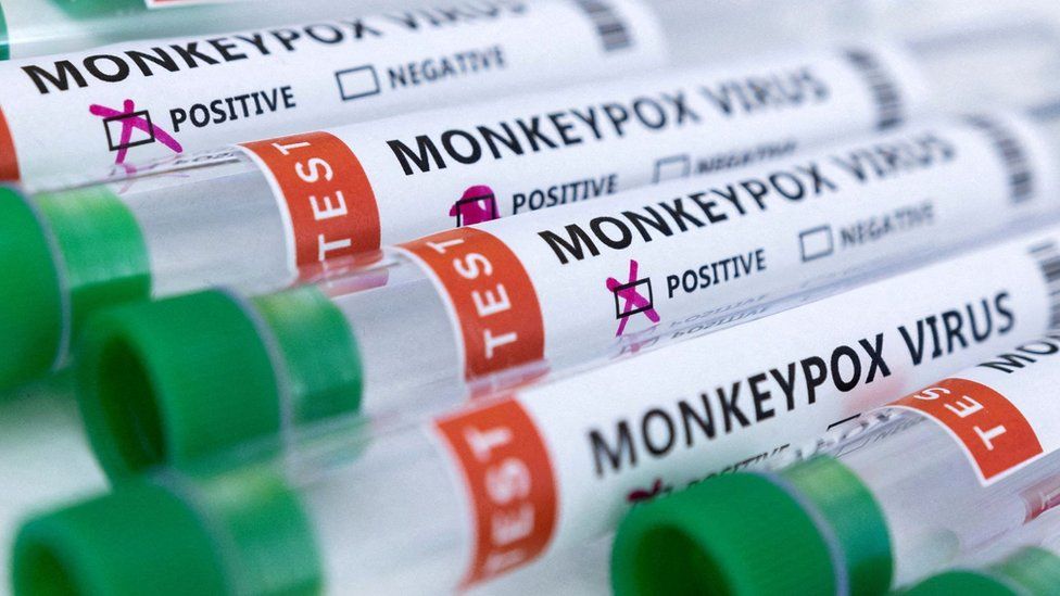
Published1 day agoSharecloseShare pageCopy linkAbout sharingImage source, ReutersMore than 100,000 doses of the monkeypox vaccine have been acquired in order to combat the spread of the virus, the government has said.Last month the NHS stepped up its monkeypox vaccination programme in England as infections rose.Vaccines minister Maggie Throup said the majority of vaccines were being made available in London, with about 75% of confirmed cases in the capital.But she urged people to wait to be invited to receive their jabs.While anyone can get monkeypox, the majority of those with the virus are gay, bisexual and other men who have sex with men.The latest figures show that nationally there have been 2,436 confirmed cases, with 1,778 of those in London.What is monkeypox and how do you catch it?Monkeypox jab to be offered to at-risk menMonkeypox outbreak mostly in young men in LondonAcross the capital, there are more than 18 clinics offering vaccinations, including the Dean Street sexual health clinic in Soho, Chelsea and Westminster Hospital, Guy’s Hospital in Southwark, Mortimer Market Centre in Camden and Barking Hospital Outpatient Centre East.During a visit to a clinic in Hammersmith, Ms Throup told BBC London: “We have procured over 100,000 doses and that is the most of any other EU country. So we are definitely ahead of the game on that.”Last week, the European Commission’s Health Emergency Preparedness and Response Authority (HERA) said 109,090 doses had been procured since the outbreak began.Spain received 5,300 doses while Portugal, Germany and Belgium were next in line to receive vaccinations, according to HERA.WHO declares highest alert over monkeypoxMs Throup added: “We are still at a bit of an unknown with this and we have reached out to the sexual health clinics asking them what numbers they have in their clinics of those needing to be jabbed. “We have got to get the first doses in people’s arms – that is our priority. We need to make sure we have the right cohorts of people coming forward.”More than 16,000 cases of monkeypox have been reported in 75 countries, according to WHO director general Dr Tedros Adhanon Ghebreyesus.He said there had been five deaths as a result of the outbreak.Image source, ReutersAnalysisBy BBC London’s political editor Tim DonovanOne issue is how best to organise this vaccination programme.Perhaps in the absence of clear public messaging – but through an effective grapevine – people have been queuing round the block for a weekend drop-in centre at Guy’s Hospital. It may have given the impression of panic or a system creaking under pressure.But at Chelsea and Westminster Hospital’s four clinics they’ve been successfully harnessing the logistical lessons learned from Covid.They’ve been poring over their patient database, inviting by text those deemed most at risk, and processing dozens of people by appointment every day.There are some who think monkeypox needs a “tsar” to be appointed to oversee the handling of the response to the outbreak.Maggie Throup says for the moment she’s content with the way vaccines are being distributed and that people with symptoms are being well advised.But it’s a two-dose treatment and for the moment, although more doses are being manufactured, the minister isn’t saying when they will be available.Follow BBC London on Facebook, Twitter and Instagram. Send your story ideas to hellobbclondon@bbc.co.ukMore on this storyMonkeypox vaccinations to be stepped up in London22 JulyWhat is monkeypox and how do you catch it?1 day agoMonkeypox jab to be offered to at-risk men21 JuneMonkeypox outbreak mostly in young men in London1 JuneRelated Internet LinksUK Health Security AgencyThe BBC is not responsible for the content of external sites.

Read more →

Dynamic travel restrictions can prevent rapid dispersion of new COVID-19 variants

A study of COVID-19 variant transmission into and across Canada shows that international travel restrictions were a key intervention for reducing or slowing spread, according to a report published today in eLife.
The results suggest that reducing the number of virus importations that can spark domestic outbreaks within a country through dynamic travel bans allows governments more time to prepare for a new variant — by ramping up testing, contact tracing and vaccination programmes.
The COVID-19 pandemic has highlighted the importance of genomic epidemiology — that is, genetic sequencing of SARS-CoV-2 samples from different regions and times — to understand the origin and movement of virus variants internationally, especially variants of concern or interest. These methods have been used widely in the UK, US, Brazil, New Zealand and Europe, and have illustrated the variation in epidemic dynamics between countries that took different public health approaches to containing the virus.
“Large-scale SARS-CoV-2 genomic epidemiology analyses in Canada have so far been limited to a study on the early epidemic within Quebec,” says lead author Angela McLaughlin, Research Assistant at the British Columbia Centre for Excellence in HIV/AIDS, and a PhD candidate in Bioinformatics, University of British Columbia, Canada. “We wanted to elaborate on this research with a national-scale analysis for the first and second COVID-19 waves. We also wanted to evaluate the impact of international travel restrictions in March 2020 on international importations of the virus and to understand why the virus persisted into 2021.”
The team used available sequence data from Canadian COVID-19 cases and data on the prevalence of circulating variants in other countries to estimate the viruses’ geographical origins. From this, they identified more than 2,260 introductions of new variants into Canada, including 680 sublineages — viruses introduced from other countries that went on to circulate within the Canadian population. They also identified 1,582 singletons — viruses introduced that did not appear to spread within the Canadian population.
Just as travel restrictions were introduced in April 2020, the importation rate reached its maximum (58.5 sublineages per week), including 31.8 from the US and 31.2 introduced solely into Quebec. Two weeks after travel restrictions took effect, the overall sublineage importation rate had dropped 3.4-fold and within four weeks had dropped 10.3-fold.
Despite these reductions, however, new virus variants continued to be introduced at a low level until August 2020 when there was a small spike in cases leading into the second wave. This suggests that wildtype sublineages introduced in the summer when prevalence and immunity were low contributed the highest proportion of COVID-19 cases in the second wave. In turn, this implies that even a low level of ongoing virus importations of similarly transmissible variants can contribute to viral persistence. By mid-October, travel restrictions were relaxed further, and importation rates rebounded quickly and contributed to the second wave.
By categorising transmission sources as within-province, between province, the US and other international sources, the team could see where the new virus importations were originating. They found that most first-wave virus introductions (January to July 2020) came from the US, followed by Russia, Italy, India, Spain and the UK, and were primarily imported into Quebec and Ontario. In the second wave (August 2020 to end February 2021), the origin of new sublineages was still dominated by the US, with increased relative contributions from India, the UK, Asia, Europe and Africa.
That the US was a large contributor of COVID-19 cases in 2020 was not unanticipated by the authors, given its high COVID-19 prevalence throughout 2020 and the long land border shared between the two countries. Even when international arrivals into Canada declined by 77.8% from 2019 to 2020, the number of truck drivers and crew members (air, ship and train) only declined by 24.8%, and accounted for almost half of all international arrivals after April 2020. Although essential key workers supporting the supply chain, these arrivals may have inadvertently facilitated additional importations from the US — suggesting this is one area where better public health measures, such as contact tracing and rapid testing, could have helped prevent the movement of new variants.
“These analyses shed light on the natural epidemiological history of SARS-CoV-2 in the context of public health interventions and show how sublineage-based genomic surveillance can be used to identify gaps in a country’s epidemic response,” concludes senior author Jeffrey Joy, Research Scientist at the British Columbia Centre for Excellence in HIV/AIDS and Assistant Professor at the Department of Medicine, University of British Columbia. “Broad and longstanding restrictions against non-essential international travel is not necessarily an advisable policy in light of economic impacts. However, our analysis suggests that swift and stringent travel bans towards localities harbouring a high frequency of a new variant of concern, or an outbreak of an entirely new virus, not yet identified domestically, should be seriously considered to reduce the probability of seeding multiple, simultaneous outbreaks and overwhelming healthcare systems.”
Story Source:
Materials provided by eLife. Note: Content may be edited for style and length.

Read more →

The bacteria powering a truly green revolution in personal electronics

AMHERST, Mass. – Researchers at the University of Massachusetts Amherst recently announced that they have figured out how to engineer a biofilm that harvests the energy in evaporation and converts it to electricity. This biofilm, which was announced in Nature Communications, has the potential to revolutionize the world of wearable electronics, powering everything from personal medical sensors to personal electronics.
“This is a very exciting technology,” says Xiaomeng Liu, graduate student in electrical and computer engineering in UMass Amherst’s College of Engineering and the paper’s lead author. “It is real green energy, and unlike other so-called ‘green-energy’ sources, its production is totally green.”
That’s because this biofilm—a thin sheet of bacterial cells about the thickness of a sheet of paper—is produced naturally by an engineered version of the bacteria Geobacter sulfurreducens. G. sulfurreducens is known to produce electricity and has been used previously in “microbial batteries” to power electrical devices. But such batteries require that G. sulfurreducens is properly cared for and fed a constant diet. By contrast, this new biofilm, which can supply as much, if not more, energy than a comparably sized battery, works, and works continuously, because it is dead. And because it’s dead, it doesn’t need to be fed.
“It’s much more efficient,” says Derek Lovley, Distinguished Professor of Microbiology at UMass Amherst and one of the paper’s senior authors. “We’ve simplified the process of generating electricity by radically cutting back on the amount of processing needed. We sustainably grow the cells in a biofilm, and then use that agglomeration of cells. This cuts the energy inputs, makes everything simpler and widens the potential applications.”
The secret behind this new biofilm is that it makes energy from the moisture on your skin. Though we daily read stories about solar power, at least 50% of the solar energy reaching the earth goes toward evaporating water. “This is a huge, untapped source of energy,” says Jun Yao, professor of electrical and computer engineering at UMass, and the paper’s other senior author. Since the surface of our skin is constantly moist with sweat, the biofilm can “plug-in” and convert the energy locked in evaporation into enough energy to power small devices.
“The limiting factor of wearable electronics,” says Yao, “has always been the power supply. Batteries run down and have to be changed or charged. They are also bulky, heavy, and uncomfortable.” But a clear, small, thin flexible biofilm that produces a continuous and steady supply of electricity and which can be worn, like a Band-Aid, as a patch applied directly to the skin, solves all these problems.
What makes this all work is that G. sulfurreducens grows in colonies that look like thin mats, and each of the individual microbes connects to its neighbors through a series of natural nanowires. The team then harvests these mats and uses a laser to etch small circuits into the films. Once the films are etched, they’re sandwiched between electrodes and finally sealed in a soft, sticky, breathable polymer that you can apply directly to your skin. Once this tiny battery is “plugged in” by applying it to your body, it can power small devices.
“Our next step is to increase the size of our films to power more sophisticated skin-wearable electronics,” says Yao, and Liu points out that one of the goals is to power entire electronic systems, rather than single devices.
This research was nurtured by the Institute for Applied Life Sciences (IALS) at UMass Amherst, which combines deep and interdisciplinary expertise from 29 departments to translate fundamental research into innovations that benefit human health and well-being.
Contacts: Jun Yao, juny@umass.edu
                 Daegan Miller, drmiller@umass.edu

Read more →