Monkeypox: US declares outbreak a public health emergency
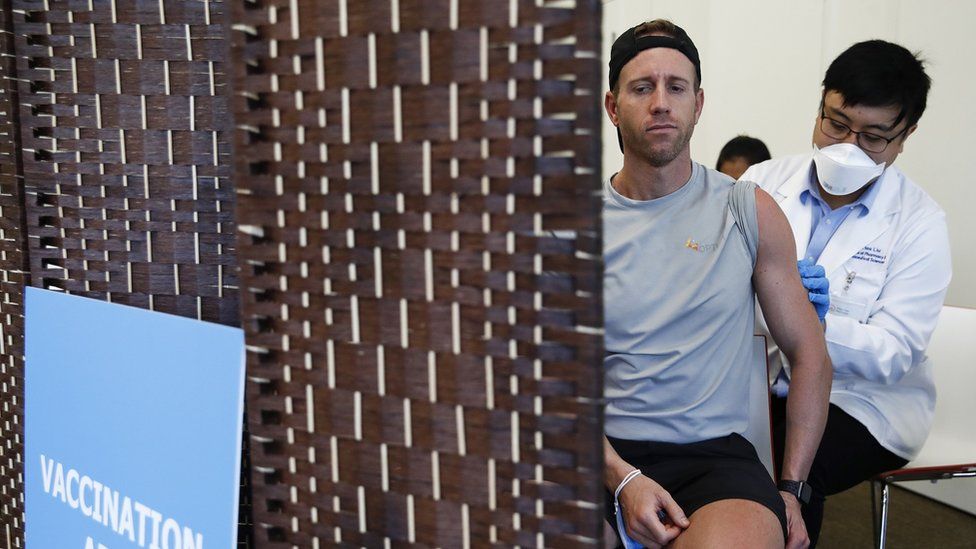
Published9 hours agoSharecloseShare pageCopy linkAbout sharingImage source, EPAThe US government has declared the monkeypox outbreak a public health emergency following a spike in cases.The decision will speed up the distribution of vaccines, treatments and federal resources to curb the spread of the virus.It comes less than a fortnight after the World Health Organization (WHO) issued its highest emergency alert following a worldwide surge in cases.Cases have topped 6,600 in the US, according to health officials.A quarter of these cases have appeared in the state of New York, which declared its own state of emergency over the disease last week.The two states with the next highest caseloads – California and Illinois – declared states of emergency earlier this week.More than 26,000 cases have been confirmed worldwide this year, data from the US Centers for Disease Control and Prevention (CDC) shows.Some public health experts have expressed concern the emergency declaration could further stigmatise the disease. Although anyone can catch monkeypox, the outbreak has been largely concentrated among men who have sex with men.But it is not purely a sexually transmitted infection (STI), and can also be passed on by close contact with an infected person.What is monkeypox and how do you catch it? WHO declares highest alert over monkeypoxMonkeypox: Handing out health advice without stigmaThe virus typically causes pimple-like rashes, which can be extremely itchy and painful, to develop and spread across the body, as well as other complications.Infections are usually mild and clear up on their own in adult patients without treatment or hospitalisation – but the WHO has warned that young children have died at higher rates from the disease.Officials in the US and elsewhere have recommended that people at the highest risk of exposure – including some gay and bisexual men, as well as some healthcare workers – should get the jab on a priority basis.San Francisco ‘in a very scary place’Pressure had been building on President Joe Biden to do more to stop the spread of monkeypox. Public health officials say the disease is particularly prevalent amongst men who have sex with men.In cities like here in San Francisco, with a large LGBT presence, politicians have claimed not enough is being done to protect the community. Unlike when Covid-19 surfaced, there are pre-existing vaccines for monkeypox. But many who have tried to get the jab have reported waiting in long lines, only to be told there aren’t enough to go round. Some have questioned whether Mr Biden should have called this nationwide state of emergency earlier – and whether this will be enough to prevent further spread. San Francisco introduced a public health state of emergency on Monday. In doing so, the mayor invoked memories of the HIV/Aids crisis in the 1980s – when she says the city was ignored. “We are in a very scary place”, she said. The declaration on Thursday comes amid reports that vaccines and treatments for monkeypox are in short supply across the country.The Department of Health and Human Services said last week that it has ordered more than five million more doses, to be delivered until next May.Earlier this week, the White House also announced the appointment of a team to co-ordinate and oversee the national monkeypox response. It includes Dr Demetre Daskalakis, a New York physician who has led successful initiatives to curb the spread of HIV in the US.This video can not be playedTo play this video you need to enable JavaScript in your browser.More on this storyWHO declares highest alert over monkeypox23 JulyMonkeypox: Handing out health advice without stigma28 May

Read more →

Plant-based 'beef' reduces CO2 emissions but threatens 1.5M ag jobs

Plant-based alternatives to beef have the potential to help reduce carbon dioxide emissions, but new economic models show their growth in popularity could disrupt the agricultural workforce, threatening more than 1.5 million industry jobs.
By embracing meat protein alternatives, U.S. food production could reduce its agricultural carbon footprint between 2.5% and 13.5%, mostly by decreasing the number of cows needed for beef production by two to 12 million, according to new research published by Cornell University, Johns Hopkins University and international partners.
Acting to reduce climate change is important, the researchers said, but technological disruption can have many consequences — both positive and negative — across the economy, such as the issue of livelihoods, working conditions, human rights, fair wages and health equity.
“A reduced carbon footprint and increased food system resource-use efficiency are reasons alternative proteins could be in a portfolio of technologies and policies to promote more-sustainable food systems,” said lead author Daniel Mason-D’Croz, a senior research associate at Cornell.
“Still, plant-based alternatives to beef are not silver bullets,” he said, “with their impact on other environmental dimensions of the food system — such as total water use — ambiguous.”
The researchers explored the potential disruption of plant-based beef alternatives by comparing the economic consequences under a range of scenarios, where plant-based beef alternatives replaced 10%, 30% or 60% of current U.S. beef demand.
“In the aggregate, food system changes would have a small, but potentially positive impact on national gross domestic product,” said Mason-D’Croz.
“But these changes would not be felt equally across the economy,” he said, “with substantial disruptions observed across the food system, particularly in the beef-value chain, which could contract substantially by as much as 45% under the 60%-replacement scenario — challenging the livelihoods of the more than 1.5 million people employed in these sectors.”
“There are good reasons for regulators and policymakers to encourage these up-and-coming technologies,” said senior author Mario Herrero, professor of sustainable food systems and global change. “Politicians must remain aware of unintended negative consequences and commit to mitigating changes that are ethically concerning, including harms to disadvantaged workers and hard-hit local communities and small producers.”
Story Source:
Materials provided by Cornell University. Original written by Blaine Friedlander, courtesy of the Cornell Chronicle. Note: Content may be edited for style and length.

Read more →

Mitochondrial DNA mutations linked to heart disease risk

Mitochondria are organelles found within most cells, best known for generating the chemical energy required to power cellular functions. Increasingly, however, researchers are discovering how mitochondrial function — and dysfunction — play critical roles in numerous diseases, and even aging.
In a new study published in the August 4, 2022 online issue of Immunity, scientists at University of California San Diego School of Medicine and Salk Institute for Biological Studies report a surprising link between mitochondria, inflammation and DNMT3A and TET2, a pair of genes that normally help regulate blood cell growth, but when mutated, are associated with an increased risk of atherosclerosis.
“We found that the genes DNMT3A and TET2, in addition to their normal job of altering chemical tags to regulate DNA, directly activate expression of a gene involved in mitochondrial inflammatory pathways, which hints as a new molecular target for atherosclerosis therapeutics,” said Gerald Shadel, PhD, co-senior study author and director of the San Diego Nathan Shock Center of Excellence in the Basic Biology of Aging at Salk Institute. “They also interact with mitochondrial inflammatory pathways, which hints at a new molecular target for atherosclerosis therapeutics.”
While studying the roles of DNMT3A and TET2 mutations in clonal hematopoiesis, which happens when stem cells begin making new blood cells with the same genetic mutation, co-senior study author Christopher Glass, MD, PhD, professor in the departments of Medicine and Cellular and Molecular Medicine at UC San Diego School of Medicine, and colleagues noted that abnormal inflammatory signaling related to DNMT3A and TET2 deficiency in blood cells played a major role in the inflammation response that promotes development of atherosclerosis.
But the question remained how DNMT3A and TET2 genes were involved in inflammation and atherosclerosis — the buildup of fatty plaques in arteries and the primary underlying cause of cardiovascular disease. It is estimated approximately half of Americans between the ages of 45 and 84 have atherosclerosis, which is the single leading cause of death in the United States and westernized nations.
“The problem was we couldn’t work out how DNMT3A and TET2 were involved because the proteins they code seemingly do opposite things regarding DNA regulation,” said Glass. “Their antagonistic activity led us to believe there may be other mechanisms at play, which prompted us to take a different approach and contact Shadel, who had uncovered the same inflammatory pathway years earlier while examining responses to mitochondrial DNA stress.”
What they found

Read more →

Study finds two protein pathways downregulated in postnatal heart

Published today in Stem Cell Reports, researchers from the University of Minnesota Medical School discovered two signaling pathways that are downregulated in human hearts after birth. These pathways, mitogen-activated protein kinase (MAPK) and phosphoinositide 3-kinase (PI3K)-AKT, lead to maturation of heart cells, allowing them to adapt functionally to their postnatal role. The researchers showed that manipulating these pathways enabled better outcomes in experiments to generate cardiac myocytes, the beating cells of the heart, in vitro from pluripotent stem cells.
Heart disease is one of the leading causes of death worldwide, partially because the adult human heart does not have any heart stem cells allowing it to regenerate after damage. Induced pluripotent stem cells, which are undifferentiated human stem cells derived by reprogramming of adult cells and typically cells from skin tissue, can be made to differentiate into cardiac myocytes in vitro, but they preferentially make immature versions of these cells. This essentially makes fetal-type cells rather than post-natal cardiomyocytes. The inability to generate mature cardiomyocytes has hindered the development of cell therapy for heart disease as well as the generation of cells for the in vitro study of cardiac physiology and toxicology.
“The last decade has seen an explosion in research to produce human cardiac myocytes from pluripotent stem cells,” said Bayardo Garay, a graduate student in the Medical Scientist Training Program. “Our study advances these efforts by providing a practical way to generate cells that are relevant to human disease.”
To discover these two pathways, the investigators mined previously published genomic data on cardiomyocytes from different stages of development. “Maturation of stem cell-derived somatic cells in a dish is difficult to achieve, but is critical for the generation of human in vitro models of health and disease,” said Brenda Ogle, PhD, Professor and Head of the Department of Biomedical Engineering and director of the Stem Cell Institute. “This work cleverly utilizes existing data to identify novel pathways that control cardiomyocyte maturation and that can be easily manipulated with small molecules to accelerate cardiomyocyte maturation.”
Researchers conclude that suppression of the MAPK and PI3K-AKT pathways on in vitro-derived human cardiomyocytes for only five days results in enhanced maturity in many domains. According to the study’s corresponding author, Rita Perlingeiro, PhD, of the Cardiology Division/Department of Medicine, “this multidisciplinary work is the fruit of the university’s investment in collaborative science, and brings the cells we can produce in the lab significantly closer to being clinically relevant to cardiac disease in humans.”
The project was supported by grants from the National Institutes of Health and the American Heart Association. The project also received seed funds from the University of Minnesota Lillehei Heart Institute and the Institute for Engineering in Medicine Group Grant. The University of Minnesota University Imaging Centers also supported the work with resources and staff.
This project was supported by NIH grants R01 AR071439 and AR078571, (R.C.R.P.), R01 HL104025 and HL106592 (S.C.D.), R01 HL155993 (J.V.B.), NHLBI R01 HL137204 (B.O.), R01 AR055685 (M.K.), T32 HL144472-01A1 (J.B.); NSF grants GFRP (S.G.), and CMMI-1553255 (P.W.A.), and predoctoral fellowship from the American Heart Association (T.R.). B.I.G. was supported by NHLBI F30 HL151138 and NIGMS T32 GM008244. This project also received seed funds from the University of Minnesota Lillehei Heart Institute (R.C.R.P.) and the Institute for Engineering in Medicine (IEM) Group Grant (B.O.). This work was also supported by the resources and staff at the University of Minnesota University Imaging Centers (UIC), SCR_020997.
Story Source:
Materials provided by University of Minnesota Medical School. Original written by Alex Smith. Note: Content may be edited for style and length.

Read more →

Hyaluranic acid, a naturally occurring compound, awakens stem cells to repair damaged muscle

A new study published in the journal Science reveals a unique form of cell communication that controls muscle repair. In damaged muscle, stem cells must work together with immune cells to complete the repair process, yet how these cells coordinate to ensure the efficient removal of dead tissue before making new muscle fibers has remained unknown. The scientists have now shown that a natural substance called hyaluronic acid, which is used in cosmetics and injections for osteoarthritis, is the key molecule that manages this fundamental interaction.
“When muscles get damaged, it is important for immune cells to quickly enter the tissue and remove the damage before stem cells begin repair,” said Dr. Jeffrey Dilworth, senior scientist at The Ottawa Hospital and professor at the University of Ottawa and senior author on the study. “Our study shows that muscle stem cells are primed to start repair right away, but the immune cells maintain the stem cells in a resting state while they finish the cleanup job. After about 40 hours, once the cleanup job is finished, an internal alarm goes off in the muscle stem cells that allows them to wake up and start repair.”
Dr. Dilworth and his team identified hyaluronic acid as the key ingredient in this internal alarm clock that tells muscle stem cells when to wake up. When muscle damage occurs, stem cells start producing and coating themselves with hyaluronic acid. Once the coating gets thick enough, it blocks the sleep signal from the immune cells and causes the muscle stem cells to wake up.
Using mouse and human tissues, Dr. Dilworth and his team also discovered how muscle stem cells control the production of hyaluronic acid using epigenetic marks on the Has2 gene.
“Interestingly, aging is associated with chronic inflammation, muscle weakness and a reduced ability of muscle stem cells to wake up and repair damage,” said lead author Dr. Kiran Nakka, a research associate with Dr. Dilworth who conducted this research as part of his postdoctoral studies. “If we could find a way to enhance hyaluronic acid production in the muscle stem cells of older people it might help with muscle repair.”
The authors note that the regenerative effect of hyaluronic acid seems to depend on it being produced by the muscle stem cells. The team is currently examining if drugs that modify the epigenetics of muscle stem cells could be used to increase their production of hyaluronic acid.
Story Source:
Materials provided by The Ottawa Hospital. Note: Content may be edited for style and length.

Read more →

How pathogenic gene variants lead to heart failure

Cardiomyopathy is not a uniform disease. Rather, individual genetic defects lead to heart failure in different ways, an international consortium reports in Science.
The molecular and cellular mechanisms that lead to heart failure in people with cardiomyopathy are determined by the specific gene variant that each patient carries, according to newly published research based on the first comprehensive single-cell analysis of cardiac cells from healthy and failing hearts.
The work, reported in the journal Science, was conducted by 53 scientists from six countries in North America, Europe, and Asia.
The study shows that cell type compositions and gene activation profiles change according to the genetic variants. The investigators say the findings can inform the design of targeted therapies that take into account each patient’s underlying gene defect responsible for their particular form of cardiomyopathy.
The team studied 880,000 single heart cells
Examining the genes activated in about 880,000 single cells from 61 failing hearts and 18 healthy donor hearts as reference was a complex endeavor which required an interdisciplinary team. The organs were procured by the Brigham and Woman’s Hospital in Boston, USA, University of Alberta in Canada, the Heart and Diabetes Center North Rhine-Westphalia in Bad Oeynhausen, Ruhr University Bochum in Germany and Imperial College London, UK.

Read more →

Single-cell approaches and deep learning to map all stages of fruit fly embryo development

Scientists have constructed the most complete and detailed single-cell map of embryo development in any animalto date, using the fruit fly as a model organism.
Published in Science, this study, co-led by Eileen Furlong at EMBL and Jay Shendure at the University of Washington, harnesses data from over one million embryonic cells spanning all stages of embryo development and represents a significant advance at multiple levels. This fundamental research also aids scientists’ ability to pursue questions like how mutations lead to different developmental defects. In addition, it provides a path to understand the vast non-coding part of our genome that contains most disease-associated mutations.
“Just capturing the entirety of embryogenesis — all stages and all cell types — to obtain a more complete view of the cell states and molecular changes that accompany development is a feat in its own right,” said Eileen Furlong, Head of EMBL’s Genome Biology unit. “But what I’m really excited about is the use of deep learning to obtain a continuous view of the molecular changes driving embryonic development — down to the minute.”
Embryonic development begins with the fertilisation of an egg, followed by a series of cell divisions and decisions that give rise to a very complex multi-cellular embryo that can move, eat, sense, and interact with its environment. Researchers have been studying this process of embryonic development for over a hundred years, but only in the last decade have new technologies enabled scientists to identify molecular changes that accompany cell transitions at a single-cell level.
These single-cell studies have raised tremendous excitement as they demonstrated the complexity of cell types in tissues, even identifying new cell types, and revealed their developmental trajectories in addition to underlying molecular changes. However, attempts to profile the entirety of embryo development at single-cell resolution have been out of reach due to many technical challenges in sampling, costs, and technologies.
In this regard, the fruit fly (Drosophila melanogaster), a pre-eminent model organism in developmental biology, gene regulation, and chromatin biology, has some key advantages when it comes to developing new approaches to address this. Fruit fly embryonic development occurs extremely rapidly; within just 20 hours after fertilisation, all tissues have formed, including the brain, gut, and heart, so the organism can crawl and eat. This, in combination with the many discoveries made in fruit flies that have propelled understanding of how genes and their products work, encouraged the Furlong lab and their collaborators to take on this challenge.

Read more →

Insight into how the intestine repairs damaged tissue

Investigators at Cedars-Sinai and the University of California, San Francisco (UCSF) have identified a component in the intestine that plays a critical role in repairing damaged tissue.
Scientists found that endothelial cells in the lymphatic vessels produce molecules that are essential for the maintenance and regulation of stem cells and tissues in the intestine. These lymphatic endothelial cells reside near specialized stem cell niches, which are microenvironments that support stem cell regeneration.
The findings were published in the peer-reviewed journal Cell Stem Cell.
“It’s important for us to understand niches and how lymphatics communicate with stem cells as part of the niche,” said Ophir Klein, MD, PhD, senior author of the study and executive director of Cedars-Sinai Guerin Children’s. “Deciphering the mechanisms that explain how the ecosystem that supports stem cells works will help to lay the foundation for future discoveries that could one day lead to therapeutic strategies to repair damaged tissue.”
The intestine undergoes continuous renewal to withstand the wear and tear that result from breakdown of foods, and from the presence of waste that can injure and kill cells. The intestine needs to replenish itself constantly with healthy cells, and fortunately, it has an exceptional capacity to regenerate cells.
The division of intestinal stem cells to make more cells is regulated by their surrounding niche, which is comprised of several cell types and is an essential source of signals. However, it is unclear which niche cells produce signals during different states of injury.

Read more →

How microglia contribute to Alzheimer's disease

One of the hallmarks of Alzheimer’s disease is a reduction in the firing of some neurons in the brain, which contributes to the cognitive decline that patients experience. A new study from MIT shows how a type of cells called microglia contribute to this slowdown of neuron activity.
The study found that microglia that express the APOE4 gene, one of the strongest genetic risk factors for Alzheimer’s disease, cannot metabolize lipids normally. This leads to a buildup of excess lipids that interferes with nearby neurons’ ability to communicate with each other.
“APOE4 is a major genetic risk factor, and many people carry it, so the hope is that by studying APOE4, that will also provide a bigger picture of the fundamental pathophysiology of Alzheimer’s disease and what fundamental cell processes have to go wrong to result in Alzheimer’s disease,” says Li-Huei Tsai, director of MIT’s Picower Institute for Learning and Memory and the senior author of the study.
The findings suggest that if researchers could find a way to restore normal lipid metabolism in microglia, that might help to treat some of the symptoms of the disease.
MIT postdoc Matheus Victor is the lead author of the paper, which appears today in Cell Stem Cell.
Lipid overload
About 14 percent of the population has the APOE4 variant, making it the most common genetic variant that has been linked to late-onset, nonfamilial Alzheimer’s disease. People who carry one copy of APOE4 have a threefold higher risk of developing Alzheimer’s, and people with two copies have a tenfold higher risk.

Read more →