Revealing roles of dementia proteins in normal memory

New research has revealed how the tau protein, a critical element in the formation of Alzheimer’s disease, is also involved in normal learning processes in the healthy brain — potentially providing a focal point for future drug therapies.
In the study, published in The EMBO Journal, Flinders University researchers have provided new insights into the tau protein, whose role has long been enigmatic, finding it may help molecular processes of memory formation.
Employing a sensitive method named proximity labelling, the team aimed to identify all proteins that tau comes in contact with within brain cells, labelling and identifying the whole collection of interacting proteins as they went.
Looking at the collection of proteins that interact with tau, and which specific functions these interactions support, the researchers found that while tau binds to proteins supporting brain cell structure, it also interacted with proteins that control vesicles and cell surface receptors for neurotransmitters, both necessary for learning and memory in the brain.
“Our new study took a snapshot of all partners tau engages with to support normal brain function,” says senior study author Dr Arne Ittner, Senior Research Fellow in Neuroscience in the Flinders Health and Medical Research Institute.
“Out of a wealth of partners, we identified one enzyme that critically controls neurotransmitter sensors. This enzyme, called NSF, is inhibited by tau, particularly in Alzheimer’s.”
Changes in the connections between brain cells, called synapses, underly the processes involved in formation and retention of memory. These changes happen at the molecular level and help us store and retrieve memories, such as places visited or of loved ones.

Read more →

Caterpillar-like bacteria crawling in our mouths

Likely in order to survive in the oral cavity, bacteria evolved to divide along their longitudinal axis without parting from one another. A research team co-led by environmental cell biologist Silvia Bulgheresi from the University of Vienna and microbial geneticist Frédéric Veyrier from the Institut national de la recherche scientifique (INRS) just published their new insights in Nature Communications. In their work, they described the division mode of these caterpillar-like bacteria and their evolution from a rod-shaped ancestor. They propose to establish Neisseriaceae oral bacteria as new model organisms that could help pinpoint new antimicrobial targets.
Although our mouth houses over 700 species of bacteria and its microbiota is, therefore, as diverse as that of our gut, not much is known about how oral bacteria grow and divide. The mouth is a tough place to live in for bacteria. The epithelial cells lining the inner surface of the oral cavity are constantly shed and, together with salivary flow, organisms that inhabit this surface will therefore struggle for attachment. It is perhaps to better stick to our mouth that bacteria of the family Neisseriaceae have evolved a new way to multiply. Whereas typical rods split transversally and then detach from each other, some commensal Neisseriaceae that live in our mouths, however, attach to the substrate with their tips and divide longitudinally – along their long axis. In addition to that, once cell division is completed, they remain attached to one another forming caterpillar-like filaments. Some cells in the resulting filament also adopt different shapes, possibly to perform specific functions to the benefit of the whole filament. The researchers explain: “Multicellularity makes cooperation between cells possible, for example in the form of division of labor, and may therefore help bacteria to survive nutritional stress.”
The team of researchers first employed electron microscopy to survey bacterial cell shape across the Neisseriaceae family that include the two standard cell shapes (rod and coccus) in addition to the caterpillar-like filaments. By comparing their cell shapes and genomes throughout the Neisseriaceae family, they could infer that the multicellular, longitudinally dividing bacteria evolved out of rod-shaped, transversally dividing bacteria. Moreover, they could pinpoint which genes were likely responsible for the unusual multiplication strategy. They then used fluorescence labelling techniques to visualize the progression of cell growth in the multicellular bacteria and finally compared the genetic make-up of these with ‘classic’, rod-shaped species. Finally, they tried to recreate that evolution by introducing the genetic changes into rod-shaped Neisseriaceae. Although they could not force rod-shaped bacteria to become multicellular, genetic manipulation resulted into longer and thinner cells. “We speculate that in the course of evolution, through a reworking of the elongation and division processes, the cell shape changed, perhaps to better thrive in the oral cavity”, Frédéric Veyrier (INRS). 
“Apart from helping us to understand how cell shape evolved, multicellular Neisseriaceae may be useful to study how bacteria learned to live attached to the surface of animals, the only place they have been found to occur so far. Half of us is carrying them in our mouths, by the way”, explains Silvia Bulgheresi from the Department of Functional and Evolutionary Ecology at the University of Vienna. However, Philipp Weber from the University of Vienna, PhD student in Bulgheresi’s team, who also worked on the study, highlights that “expanding the cell biology field to additional morphologies and symbiotic species is also crucial to increase the pool of protein targets (e.g., antibiotic targets) for biopharmaceutical applications.” Sammy Nyongesa, PhD student in Veyrier’s team from INRS, adds: “An evolutionary approach, such as that undertaken here for the Neisseriaceae, can shed light on new, unforeseen protein targets”. 
Story Source:
Materials provided by University of Vienna. Note: Content may be edited for style and length.

Read more →

Wastewater viral loads can provide advance warning of COVID-19 outbreaks

Scientists show that there is a close association between clinical cases of COVID-19 and viral loads in wastewater, with the viral loads picking up to two days before the cases were detected.
The Tokyo 2020 Olympics and Paralympics were held during July 21 and September 21, 2021, a time when the incidence and spread of COVID-19 was prevalent. Thus, a rigorous and multi-pronged testing approach was enacted in order to limit the spread of the virus while allowing the Games to proceed.
Following from previous research, a team lead by Associate Professor Masaaki Kitajima at Hokkaido University has shown the association between SARS-CoV-2 viral loads in wastewater and cases reported at the Olympic and Paralympic Village. Their findings were published in the journal JAMA Network Open.
The athletes and support staff at the Olympic and Paralympic Village were tested daily; in addition, wastewater in the sewage system was also sampled and tested (wastewater-based epidemiology, WBE) daily to determine viral loads. The results were reported to the Tokyo 2020 Organizing Committee.
In this study, the authors wanted to examine the association between clinically reported cases and viral loads in wastewater. They correlated the results of 360 samples collected from manholes in 7 distinct areas of the Village with confirmed COVID-19 cases obtained from the Organizing Committee, and with data of close contacts tests from a previous report.
The researchers found that SARS-CoV-2 was present in 151 wastewater samples — 53 from the Olympics and 98 from the Paralympics. The number of confirmed cases was also higher in the Paralympics. The strongest correlation between SARS-CoV-2 RNA load in wastewater and the presence of clinical positive areas was found in areas that had maximum viral loads in wastewater in a three-day span (two days before to the day of clinical positive area).
The study suggests that WBE and clinical tests are complementary, and that the testing strategy played a role in preventing COVID-19 clusters in the Village. This study of one of the world’s largest mass gatherings provides novel evidence on the implementation and use of WBE in communities where all members undergo daily testing, and could be used to trace and control COVID-19 clusters in the future.
Story Source:
Materials provided by Hokkaido University. Note: Content may be edited for style and length.

Read more →

Researchers show how mutations in 'dark genome' cause pancreatic malformations

Researchers at the Centre for Genomic Regulation (CRG) have identified a DNA sequence that is crucial for pancreatic differentiation and function — and for the first time — describe how it works.
Patients with mutations in a DNA sequence — which they coin EnhP — develop pancreas malformations. It is the most clear-cut example to date of an inherited disease that is caused by mutations that do not disrupt the DNA sequence of a gene.
Disorders?caused by mutations in a single DNA sequence, for example Huntington disease or sickle cell anaemia, are known as monogenic diseases. In the vast majority of cases, such mutations disrupt a protein-coding gene. In this case, the mutations in EnhP disrupt a single “enhancer” instead of a single gene.
Our genomes contain hundreds of thousands of DNA elements that are thought to act as enhancers. These enhancer DNA sequences act as switches to turn on the transcription of their target genes in the right tissues.
According to the authors of the study, published today in Developmental Cell, EnhP is by no means the only enhancer defect to cause disease. Mutations in enhancers may be the cause of a monogenic disease in many patients in which laboratory tests have failed to disclose causal gene mutations.
Understanding the role of enhancers in disease could change how we practice medicine. “Clinical genetics is shifting from a focus on sequencing protein-coding genes to sequencing whole genomes. It is now theoretically possible to discover disease-causing mutations that lie outside of traditional areas of the genome, although it is still challenging to discern which parts of the genome are truly vulnerable to mutations,” explains Dr. Jorge Ferrer, senior author of the study, Coordinator of the Medical Genomics Transversal Programme at the CRG and Group Leader at CIBERDEM.

Read more →

Study offers insights into how pancreatic cancer develops

Pancreatic cancer has few treatment options and limited survival, with only 9% of patients still living five years after diagnosis.
But a detailed analysis of pancreatic cancer by researchers at Washington University School of Medicine in St. Louis has revealed details of two key transition points in the development of these tumors — the shift from normal cells to precancerous cells, and the change from precancerous to cancerous cells. Understanding these transitions will help lead to the development of novel therapies. The study also provides insights into treatment resistance and how immunotherapy could be harnessed to treat this aggressive tumor type.
The study, published Aug. 22 in the journal Nature Genetics, is part of the Human Tumor Atlas Network, funded by the National Cancer Institute’s Cancer Moonshot program, all part of the National Institutes of Health (NIH).
Also, as part of an ongoing phase 1 immunotherapy clinical trial at Siteman Cancer Center — based at Barnes-Jewish Hospital and Washington University School of Medicine — the researchers are conducting the same detailed analyses performed in the current study to see how tumors from patients respond to two investigational drugs that prime the immune system to attack the cancer.
“Pancreatic cancer is so difficult to treat, and to develop better treatments we need to understand how normal, healthy cells in the pancreas transition to becoming cancerous,” said co-senior author and computational biologist Li Ding, PhD, the David English Smith Distinguished Professor of Medicine and a professor of genetics. “This marks the first time these transitions have been mapped out in such detail in human tumors. Our findings are jumping off points for the future development of new treatment strategies for this deadly cancer.”
The researchers conducted a deep analysis of the genetics and protein manufacturing of 83 pancreatic tumor samples donated by 31 patients who participated in the study. They noted how the tumors differed across the volume of the tumor and at various times as the patients underwent treatment.

Read more →

Untapped potential of stem cells could aid repair of spinal cord damage

Scientists at the Francis Crick Institute have identified a group of latent stem cells that respond to injury in the central nervous system of mice. If a similar type of cell exists in humans, they could offer a new therapeutic approach to treat brain and spinal cord injuries.
After disease or injury, stem cells help repair the damage by replacing cells that have died. In some organs, like the skin and intestine, these stem cells are constantly active, while in others, so called ‘latent stem cells’ lie waiting for harm to occur before being triggered into action.
In their study published in Developmental Cell today (Monday 22 August), the researchers identified a group of latent stem cells in the central nervous system of mice. These are part of the ependymal cells that line the walls of compartments in the brain and spinal cord that hold cerebrospinal fluid.
The cells were identified by chance when the team used a fluorescence tool to look for immune cells called dendritic cells in the brain. The ependymal cells that the tool identified were found to arise from embryonic progenitor cells that shared a same protein as dendritic cells on their surface, which revealed them to the scientists.
Working with neuroscientist colleagues at the Francis Crick Institute and developmental biologists at the Institute of Molecular Medicine in Lisbon, they found that in healthy mice, these cells stay still and waft small hairs on their surface to help the flow of cerebrospinal fluid.
However, in injured mouse spinal cords, these cells responded by dividing, migrating towards the damaged area and differentiating into astrocytes, one of the major cell types of the nervous system.
The team also looked at these cells in detail in the lab and found they demonstrated key hallmarks of stem cell behaviour. They divided continuously over a long period of time, and were also able to differentiate into all three main cell types of the central nervous system — neurons, astrocytes and oligodendrocytes.
Bruno Frederico, co-corresponding author and postdoctoral training fellow in the Immunobiology laboratory at the Crick says, “While we don’t know if these cells exist in humans, if they do, it would be interesting to see if they also default to becoming astrocytes rather than neurons in response to damage. This might help explain why the mammalian central nervous system does not have a strong ability to repair itself after injury.
“If we could find a way to overcome the barriers that are stopping the differentiation into neurons and oligodendrocytes after spinal cord injury, it could present a new avenue of therapies to treat spinal cord injuries.”
The researchers suggest that unlocking the potential of these cells could help the body produce new neurons, which are responsible for receiving and sending key signals for movement, after spinal injury.
Caetano Reis e Sousa, co-corresponding author and principal group leader at the Crick, says: “There was uncertainty over whether ependymal cells can have neural stem cell capabilities, but this study underscores their potential.
“We hope that studying these cells will help build a more complete picture of the role different types of stem cells play in repairing damage, which could have important implications for regenerative medicine.”
Story Source:
Materials provided by The Francis Crick Institute. Note: Content may be edited for style and length.

Read more →

The locked library: Disease causes cells to reorder their DNA incorrectly

Imagine you’re trying to do a job and all of the information you need to do it is in a few books at the library. Except, those books are randomly arranged along with all the other books on shelves across the whole building. Without that vital information from the books you were looking for, you wouldn’t perform your job very well.
This is the situation that researchers at the Perelman School of Medicine at the University of Pennsylvania found when they studied the nucleus of cells inside connective tissues deteriorating as a result of tendinosis. Disease-related disruptions in the environments that cells exist in caused the re-organization of the genome — which is the sum of an organism’s DNA sequences — inside the cell’s nucleus, changing the way cells functioned and making them unable to reorder their DNA information in the right way again. These findings, published today in Nature Biomedical Engineering, point to the possibility of new treatments — such as small-molecule therapies — to bring in a sort of librarian that could restore order to the affected cells.
“This is really important because the research tells us, for the first time, that diseased connective tissue cells change the physical structure of their genomes and stop responding to normal physical cues from their environment,” said the study’s lead author, Su Chin Heo, PhD,an assistant professor of Orthopaedic Surgery. “If we can figure out exactly why this happens, we might be able to ‘unlock’ the diseased state of these cells, and bring them back toward a healthy state.”
“Microscale” changes in the environments that cells exist in have macro-level effects because of the way they change cell behaviors and how a body functions. But this dynamic is not well understood. So Heo and colleagues set out to examine how cells in degenerating connective tissue respond to changes in their physical environment and, particularly, how the spatial organization of chromatin — the material that DNA is made out of, which has been shown to differ based on cell type — might be affected by changes brought on by disease.
To do this, the team used the latest super-resolution imaging techniques to observe human cell models, specifically tenocytes (tendon cells involved in maintaining the tissue’s structure) and mesenchymal stromal cells (similar to stem cells, they can become a variety of cells needed to build or maintain tissue).
In these models, the researchers observed that chemical and mechanical changes within environments mimicking degenerating tendons resulted in tenocytes improperly re-ordering their chromatin. And even when the researchers presented these cells with the proper mechanical environment, they saw that the cells had lost their ability to properly re-organize their genome back to a normal state — the cells could no longer respond correctly. Cells that were healthy responded well to the same chemical and mechanical prompts, so it seems that the diseased cells forgot what they were doing, or couldn’t access the right information in their crisis response.
“While we discovered that cells in diseased microenvironments lose their epigenetic memory, these results also suggest that epigenetic treatments — like small molecule medications — could restore healthy genome organization and may prove effective treatments in conditions affecting dense tissues,” said the study’s senior author, Melike Lakadamyali, PhD, an associate professor of Physiology. “That’s something that we plan to follow up on and test.”
The researchers already have secured grants studying whether cartilage cells and meniscus cells are affected similarly by disease-disrupted genomes. They’re also studying whether the aging process has a similar effect.
“Once we understand these and the specific cellular processes that makes them happen — what locks the library door — we can use small molecule drugs as skeleton keys to either try to stop it from happening or reverse the process,” said study co-senior-author Robert Mauck, PhD, a professor of Orthopaedic Surgery and director of Penn’s McKay Orthopaedic Research Laboratory.
This research was funded National Institutes of Health (K01 AR07787), the Penn Center for Musculoskeletal Disorders (P30 AR069619), the Department of Veterans Affairs (IK6 RX003416), and the NSF Science and Technology Center for Engineering Mechanobiology (CMMI-1548571).

Read more →

Dr Anthony Fauci to step down from government in December

Published6 hours agoSharecloseShare pageCopy linkAbout sharingImage source, Getty ImagesAnthony Fauci will step down as head of the National Institute of Allergy and Infectious Diseases (NIAID) and chief medical adviser to President Joe Biden. Dr Fauci, who served as director of the NIAID for 38 years, said he would leave both positions in December to “pursue the next chapter” of his career. “It has been the honour of a lifetime to have led the NIAID,” Dr Fauci, 81, said in a statement. He became the face of the nation’s Covid-19 response during the pandemic.On Monday, Mr Biden thanked him for his “spirit, energy, and scientific integrity”.”The United States of America is stronger, more resilient, and healthier because of him,” the president wrote in a statement.In July, Dr Fauci said he would retire before the end of Mr Biden’s current term. The face of America’s fight against Covid-19Dr Fauci first joined the National Institutes of Health in 1968, when Lyndon Johnson was president. He was appointed to director of the NIAID, the infectious national disease branch, in 1984, while the AIDS epidemic raged. He has served under seven presidents since – from Republican Ronald Reagan to Democrat Joe Biden. It wasn’t until 2020, with the onset of the coronavirus pandemic, that he became the most famous doctor in America. Dr Fauci became a frequent media presence in the US and abroad as he emerged as the face of America’s fight against coronavirus. He also became polarising figure during that time. This video can not be playedTo play this video you need to enable JavaScript in your browser.While he gained fans – a petition to name him People magazine’s “Sexiest Man Alive” in 2020 gathered more than 28,000 signatures – he also angered some on the right who saw him as the public face of lockdowns and mask mandates.And he occasionally clashed with former president Donald Trump over the pandemic response. Though Dr Fauci is leaving government, he made clear on Monday that he was not retiring from medicine altogether. “I plan to pursue the next phase of my career while I still have so much energy and passion for my field,” he said. Dr Fauci, who will turn 82 on 24 December, did not set an exact date for his departure. More on this storyKey moments: Fauci and the pandemic. Video, 00:02:45Key moments: Fauci and the pandemic16 January2:45Why are people talking about Dr Fauci’s emails?2 June 2021The face of America’s fight against Covid-1913 July 2020

Read more →

Brain stimulation boosts memory for a month

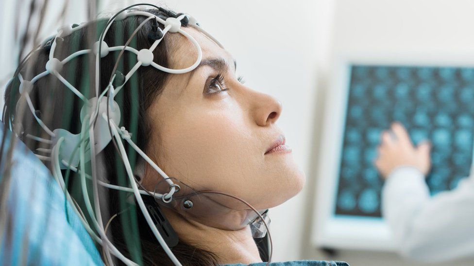
Published10 minutes agoSharecloseShare pageCopy linkAbout sharingImage source, Getty ImagesScientists have discovered they can boost people’s memory for a least a month by harmlessly stimulating parts of the brain with electricity. Volunteers performed better at word memorisation games, which tested both their immediate “working” memory and their long-term memory, experts found.Exactly what the results mean for day-to-day life is still unclear. But ideas range from helping old people cope with memory decline, to treating disease and aiding exam preparation.Dr Robert Reinhart, from Boston University, described the stimulation technique as “an entirely different approach to isolating and augmenting parts of the brain” which offered “an entirely new realm of potential treatment options”.People on the trial wore a cap filled with electrodes. A controlled electrical current, which feels similar to an itch or a tingle, was then used to precisely alter brainwaves in targeted regions of the brain. The volunteers underwent 20 minutes of stimulation daily for four days in a row. Throughout the study they had to memorise lists of words, which they were again asked to recall one month later. Dr Reinhart said the treatment “could cause selective memory improvement that lasts for at least one month”. The results, published in the journal Nature Neuroscience, showed those volunteers who were struggling with the memory games at the beginning of the experiment were those whose memory improved the most. How memory worksThe electrical signals changed the rhythm of brain activity – brainwaves – in the areas targeted. Scientists think the four rounds of stimulation reinforced those patterns and led to lingering improvements as the brain adapted and rewired itself – known as neuroplasticity. “It’s sort of connecting to the so-called language of the brain, that speaks to itself and communicates with itself through electrical impulses,” said Dr Reinhart.However, it takes different types of stimulation to boost the different types of memory: Working memory is for the here and now. It’s how you retain information in your mind – like taking notes in a class – and is vital in problem-solving and decision-making.Boosting it required low-frequency stimulation of the prefrontal cortex, at the front of the brainLong-term memory is where we bank information; it’s how we can remember our first day a school or a weddingBoosting it required high-frequency stimulation of the parietal cortex, at the back of the brainIn the word games – recalling those given at the start tests long-term memory, while recall after a month tests working memoryAll 150 people who took part in the trial were healthy, with no cognitive impairment, and were aged between 65 and 88. Becoming more forgetful is often a sign of age, but whether this form of stimulation could aid the aging brain in the real world, beyond word games, is still unknown. Dementia, including Alzheimer’s disease, is caused by a diseased brain with dying brain cells – leading to memory problems.The researchers are investigating whether the technology can be used in Alzheimer’s disease to stimulate the surviving brain cells, as well as in schizophrenia and obsessive compulsive disorder. Dr Susan Kohlhaas, the director of research at Alzheimer’s Research UK, said: “We don’t know if brain stimulation techniques have potential to help people with dementia but there is research under way in this area.”At present, the stimulation method used – transcranial alternating current stimulation – is only possible in research laboratories.So while you may be thinking of using cognitive enhancement to get through an exams – or just a pub quiz – the scientists say those sort of homegrown applications are for the distant future.Thinking caps and superbrainsHowever, researcher Shrey Grover said he can ultimately see them being used alongside more traditional methods which people use to keep their mind sharp, such as crosswords and Sudoku. “Any efforts to remain cognitively engaged are always welcome, this kind of approach is perhaps something that could be added on to things that people are already doing.”Follow James on Twitter.

Read more →

Monkeypox vaccine rationed by giving mini doses

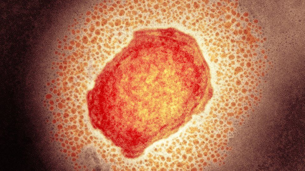
Published27 minutes agoSharecloseShare pageCopy linkAbout sharingImage source, Science Photo LibraryThree NHS sites are set to begin a pilot offering eligible patients smaller doses of monkeypox vaccine, amid global shortages of the jab. Experts say the reduced shots are just as effective and will mean more people are protected. The fractional dosing approach has been authorised in the US and the EU, as well as the UK. More than 3,000 people have been diagnosed with monkeypox in the UK since the epidemic began in May. Most cases of the virus have been among gay and bisexual men.However, anyone who comes into close contact with someone who has monkeypox could potentially contract the virus.Vaccination can help prevent new cases.UK officials say more than 33,000 regular dose shots have been given to some of those at the greatest risk of contracting the virus.But many more, who could benefit from being vaccinated, have not yet been immunised – and stocks of the vaccine are running low. Monkeypox: Can we still stop the outbreak?’I’m a different person after having monkeypox’What is monkeypox and how do you catch it? Fractional dosing could maximise the number of doses that can be administered currently, without compromising protection, says the UK Health Security Agency (UKHSA). Clinics in Chelsea and Westminster NHS Trust, Central and North West London NHS Foundation Trust and Locala Health and Wellbeing in Greater Manchester are to begin offering people the lower dose option. Although the dose is a fifth of the volume of the regular one – 0.1ml rather than 0.5ml – studies suggest it still gives good protection. Dr Mary Ramsay, Head of Immunisation at UKHSA, said: “Adopting this tried-and-tested technique will help to maximise the reach of our remaining stock, including the 100,000 doses due to arrive in the country next month – potentially enabling us to offer protection for many more thousands of people.”We will continue to remain agile in our response to the monkeypox outbreak and will adapt our approach as new science and advice becomes available.”Dr Claire Dewsnap, President of the British Association for Sexual Health & HIV, said: “We absolutely support the UKHSA-led fractional dosing pilots assessing feasibility in UK sexual health clinics. If acceptable, this would offer us the opportunity to roll out vaccine to those eligible much faster, and would address the issues of short supply of vaccine across the world.”More on this storyWhat is monkeypox and how do you catch it?5 AugustMonkeypox cases declining – particularly in London2 days ago

Read more →