Analyzing the potential of AlphaFold in drug discovery

Over the past few decades, very few new antibiotics have been developed, largely because current methods for screening potential drugs are prohibitively expensive and time-consuming. One promising new strategy is to use computational models, which offer a potentially faster and cheaper way to identify new drugs.
A new study from MIT reveals the potential and limitations of one such computational approach. Using protein structures generated by an artificial intelligence program called AlphaFold, the researchers explored whether existing models could accurately predict the interactions between bacterial proteins and antibacterial compounds. If so, then researchers could begin to use this type of modeling to do large-scale screens for new compounds that target previously untargeted proteins. This would enable the development of antibiotics with unprecedented mechanisms of action, a task essential to addressing the antibiotic resistance crisis.
However, the researchers, led by James Collins, the Termeer Professor of Medical Engineering and Science in MIT’s Institute for Medical Engineering and Science (IMES) and Department of Biological Engineering, found that these existing models did not perform well for this purpose. In fact, their predictions performed little better than chance.
“Breakthroughs such as AlphaFold are expanding the possibilities for in silico drug discovery efforts, but these developments need to be coupled with additional advances in other aspects of modeling that are part of drug discovery efforts,” Collins says. “Our study speaks to both the current abilities and the current limitations of computational platforms for drug discovery.”
In their new study, the researchers were able to improve the performance of these types of models, known as molecular docking simulations, by applying machine-learning techniques to refine the results. However, more improvement will be necessary to fully take advantage of the protein structures provided by AlphaFold, the researchers say.
Collins is the senior author of the study, which appears today in the journal Molecular Systems Biology. MIT postdocs Felix Wong and Aarti Krishnan are the lead authors of the paper.

Read more →

Data from Israel: myocarditis after COVID-19 vaccines remain rare, highest risk in young males

A new study from Israel found that the risk of developing myocarditis among males ages 16 to 19 years was about 1 in 15,000 after third dose of the Pfizer-BioNTech COVID-19 vaccine, and the cases were rare and mild, according to new research published today in the American Heart Association’s flagship journal Circulation.
Several previous studies and reports from public health agencies around the world including the U.S. Centers for Disease Control and Prevention have highlighted a possible connection and potentially increased risk of myocarditis after receiving an mRNA COVID-19 vaccine, generating considerable scientific, policy and public interest .
Typically thought to be triggered by a viral infection, myocarditis is the inflammation of the middle layer of the wall of the heart muscle, the myocardium. This condition is uncommon and may temporarily or permanently weaken the heart muscle and the heart’s electrical system, which keeps the heart beating normally. An episode of myocarditis may resolve on its own or with treatment, or may result in lasting damage to the heart. In the general population not during a global pandemic, it is estimated that approximately 10 to 20 people per 100,000 are diagnosed with myocarditis each year, according to the American Heart Association’s 2021 scientific statement on myocarditis.
Research detailing post-vaccination myocarditis in Israel after the first and second dose of the Pfizer-BioNTech COVID-19 vaccine were recently published by the country’s Ministry of Health. The incidence rate of myocarditis was low, however, it was primarily in young males after a second COVID-19 vaccination, suggesting a potential relationship between the vaccine and myocarditis. The results raised concerns about the potential for increased myocarditis after a booster dose, therefore, this new analysis was focused on the risk of myocarditis after a booster dose.
“It is important to understand the connections between this rare heart condition and COVID-19 vaccines, so we can monitor the prevalence of myocarditis and pay extra attention to those who are most at risk,” said lead study author Dror Mevorach, M.D., a professor of medicine and head of Immunology-Rheumatology Institution at Hadassah Ein Karem Medical Center and chairman of the Israeli Ministry of Health Committee for Identifying Myocarditis as an Adverse Effect of mRNA Vaccines in Jerusalem, Israel.
From July 31, 2021, to November 5, 2021, nearly 4 million (3.94 million) adults in Israel received a booster dose of the Pfizer-BioNTech vaccine, about half (48.7%) of whom were males. Health data for all reported cases of myocarditis after receiving the Pfizer-BioNTech COVID-19 vaccine were evaluated. A cardiologist and a rheumatologist reviewed and classified the data based on the Brighton Collaboration Myocarditis Case Definition.
The analysis found that after a 30-day follow up: Fewer than 100 cases (91) of myocarditis were reported, including 35 cases that occurred within the first 30 days of receiving a COVID-19 booster (a 3rd dose) of the Pfizer-BioNTech COVID-19 vaccine. 28 cases of myocarditis were probable or confirmed, and 18 occurred within the first seven days after receiving the booster dose of the Pfizer-BioNTech vaccine. All 28 cases of myocarditis were clinically defined as mild, and individuals recovered within an average of 3.5 days in the hospital. Among all age groups, the risk rates of developing myocarditis were nearly nine times higher in males than in females (1.42 vs. 0.16). Males ages 16-19 were at the highest risk, with 6 in 100,000 individuals developing myocarditis, followed by males ages 20-24 (5.21 cases per 100,000), 30-39 (1.81 cases per 100,000), and 25-29 (0.79 cases per 100,000).The risk differences declined significantly between the second and third vaccine doses across both genders and across all age groups. Authors believe there are two potential explanations for the changes. “The first is that individuals who developed myocarditis after the second COVID-19 vaccine dose did not receive a third shot, which was a medical precaution in Israel. The second potential explanation is the interval of time between doses: first and second doses are administered approximately three weeks apart, however, the time between a second dose and a booster was about 20 to 24 weeks, ” added Mevorach.
Researchers believe further study is required to better explain what may predispose young males to develop myocarditis after a COVID-19 vaccine and the pathophysiological mechanisms involved.
Co-authors are Emilia Anis, M.D., M.P.H.; Noa Cedar, M.P.H.; Tal Hasin, M.D.; Michal Bromberg, M.D., M.P.H.; Lital Goldberg, M.D., M.P.H.; Nir Levi, M.D.; Ofer Perzon, M.D.; Nur Magadle, M.D.; Barhoum Barhoum, M.D.; Elchana Parnassa, M.D.; Rita Dichtiar, M.P.H.; Yael Hershkovitz, M.Sc.; Manfred S. Green, M.B., Ch.B., Ph.D.; Nachman Ash, M.D.; Lital Keinan-Boker, M.D., Ph.D.; and Sharon Alroy-Preis, M.D., M.P.H. Authors’ disclosures are listed in the manuscript.
The study authors reported no funding sources for this research.

Read more →

Stem cell-gene therapy shows promise in ALS safety trial

Cedars-Sinai investigators have developed an investigational therapy using support cells and a protective protein that can be delivered past the blood-brain barrier. This combined stem cell and gene therapy can potentially protect diseased motor neurons in the spinal cord of patients with amyotrophic lateral sclerosis, a fatal neurological disorder known as ALS or Lou Gehrig’s disease.
In the first trial of its kind, the Cedars-Sinai team showed that delivery of this combined treatment is safe in humans.
The findings were reported today in the peer-reviewed journal Nature Medicine.
“Using stem cells is a powerful way to deliver important proteins to the brain or spinal cord that can’t otherwise get through the blood-brain barrier,” said senior and corresponding author Clive Svendsen, PhD, professor of Biomedical Sciences and Medicine and executive director of the Cedars-Sinai Board of Governors Regenerative Medicine Institute. “We were able to show that the engineered stem cell product can be safely transplanted in the human spinal cord. And after a one-time treatment, these cells can survive and produce an important protein for over three years that is known to protect motor neurons that die in ALS.”
Aimed at preserving leg function in patients with ALS, the engineered cells are potentially a powerful therapeutic option for this disease that causes progressive muscle paralysis, robbing people of their ability to move, speak and breathe.
None of the 18 patients treated with the therapy — developed by Cedars-Sinai scientists — had serious side effects after the transplantation, according to the data.

Read more →

Scientists develop new method to faster — and more accurately — find antigens that trigger specific immune cells

A cell’s secrets can be divulged by its surface, decorated with tens to hundreds of thousands of molecules that help immune cells determine friend from foe. Some of those protruding molecules are antigens that trigger the immune system to attack, but it can be difficult for scientists to identify those antigens, which often vary across individuals, in the molecular forest.
A team of Stanford scientists led by Polly Fordyce, an Institute Scholar at Sarafan ChEM-H, has developed a new method to faster and more accurately predict which antigens will lead to a strong immune response. Their approach, which was reported in Nature Methods on Sept. 5, could help scientists develop more effective cancer immunotherapies.
T cells, a class of immune cells, crawl along and squish past other cells as they patrol the body, using T cell receptors to molecularly read peptides, or short pieces of proteins — which are cradled within larger proteins called major histocompatibility complexes (pMHCs) that project from cell surfaces. Healthy host cells display an array of pMHCs that do not trigger an immune response, but once T cells recognize disease-indicating peptides, they become activated to find and kill cells bearing these foreign signatures. Understanding how T cells sensitively distinguish these antigenic peptides from host peptides to avoid mistakenly killing host cells has long been a mystery.
“A T cell can detect a single antigenic peptide amongst a sea of 10,000 or 100,000 non-antigenic peptides being displayed on cell surfaces,” said Fordyce, assistant professor of bioengineering and of genetics.
The key to selectivity is in the T cell crawl. T cells’ sliding puts stress on the bonds between receptors and peptides, and in most cases, that extra stress is enough to break that bond. But sometimes, it has the opposite effect. Chris Garcia, co-author of the study and professor of molecular and cellular physiology and of structural biology, and others had already shown that the most antigenic peptides are those whose interactions with T cell receptors grow stronger in response to sliding.
“It’s kind of like a Chinese finger trap,” said Fordyce. “When you pull a bit at the receptor-antigen interaction, the binding actually lasts longer.”
Cellular mimicry

Read more →

Bronchodilators don't improve smoking-related respiratory symptoms in people without COPD

Researchers supported by the National Institutes of Health have found that dual bronchodilators — long-lasting inhalers that relax the airways and make it easier to breathe — do little to help people who do not have chronic obstructive pulmonary disease (COPD), but who do have respiratory symptoms and a history of smoking.
COPD, a lung disease that obstructs the airways and leads to coughing, wheezing, and shortness of breath, affects about 15 million Americans. However, millions of others who smoke or used to smoke and have some symptoms of COPD have also been prescribed bronchodilators.
“We’ve assumed these medications worked in patients who don’t meet lung function criteria for COPD, but we never checked,” said MeiLan K. Han, M.D., a principal investigator and first author of the study. “We now know these existing medications don’t work for these patients.”
The findings of the study, which was funded by the National Heart, Lung, and Blood Institute (NHLBI), were published in the New England Journal of Medicine and simultaneously presented at the European Respiratory Society International Congress.
According to scientists, the implications are significant. First, they show the importance of diagnosing lung conditions through spirometry, a lung function test Han noted is underutilized in clinical practice. Second, they show the need for new, effective therapies for patients without COPD.
Inhalers have long been the primary go-to treatment for these patients, she explained, because doctors either assume a patient has COPD, or if they don’t, that their smoking-related symptoms could be helped by the inhalers. But while tobacco smoking causes a large spectrum of lung damage, the study showed bronchodilator therapy only helps patients with enough lung damage that would result in abnormal spirometry readings.

Read more →

DNA in Viking feces sheds new light on 55,000-year-old relationship between gut companions

Using stool samples from Viking latrines, researchers at the University of Copenhagen have genetically mapped one of the oldest human parasites — the whipworm. The mapping reflects the parasite’s global spread and its interaction with human beings, a delicate relationship that can make us healthier and ill.
Using fossilized eggs in up to 2500-year-old feces from Viking settlements in Denmark and other countries, researchers at the University of Copenhagen’s Department of Plant and Environmental Sciences and the Wellcome Sanger Institute (UK) have made the largest and most in-depth genetic analysis of one of the oldest parasites found in humans — the whipworm.
The study, published in Nature Communications, presents completely new knowledge about the parasite’s development and prehistoric dispersal. This knowledge can be applied in efforts to prevent the parasite’s drug resistance and its future spread.
The study suggests that human and parasite have developed a delicate interaction over thousands of years, whereby the parasite tries to stay “under the radar” not to be repelled, which allows it more time to infect new people. From other studies, it is known that the whipworm stimulates the human immune system and the gut microbiome, to the mutual benefit of both host and parasite.
While whipworm (Trichuris trichiura) is now rare in industrialized countries, and most often only causes minor problems among healthy individuals, the parasite is estimated to affect 500 million people in developing countries.
“In people who are malnourished or have impaired immune systems, whipworm can lead to serious illness. Our mapping of the whipworm and its genetic development makes it easier to design more effective anti-worm drugs that can be used to prevent the spread of this parasite in the world’s poorest regions,” says Professor Christian Kapel of UCPH’s Department of Plant and Environmental Sciences.

Read more →

China approves inhaled Covid vaccine

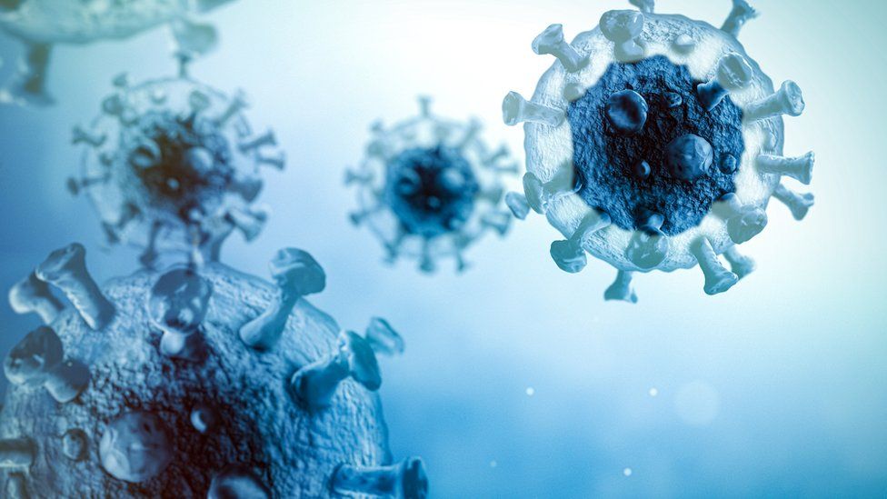
Published14 minutes agoSharecloseShare pageCopy linkAbout sharingImage source, Getty ImagesChina has become the first country to approve an inhaled Covid vaccine.Made by CanSino, it has similar ingredients to its injected vaccine, using a harmless adenovirus as a carrier for the genetic code that teaches the body how to fight Covid. Inhaled as a fine mist, Convidecia Air can provide good protection after just one breath, the company says.Other researchers, including teams in the UK and the US, have been investigating nasal spray vaccines. Scientists say these may give added immunity in the lining of the nose and upper airways, where Covid typically enters the body. The National Medical Products Administration of China granted CanSino approval for its inhaled vaccine to be used as a booster dose. It can top up protection in those who have previously had a jab, trials suggest.Who will get a Covid booster this autumn? Who can get a free flu jab?Meanwhile, the latest Covid vaccine booster programme has begun in England, Wales and Scotland.Infections are falling around the UK – but health bosses predict a resurgence of Covid and flu this autumn and winter.They are urging those eligible to protect themselves from serious illness by being vaccinated against both.More on this storyMillions invited for Covid booster jabs from Monday1 day agoChinese city of 21m locked down over Covid cases3 days agoRelated Internet LinksCanSinoThe BBC is not responsible for the content of external sites.

Read more →

Legionnaire's suspected cause of Argentina pneumonia deaths

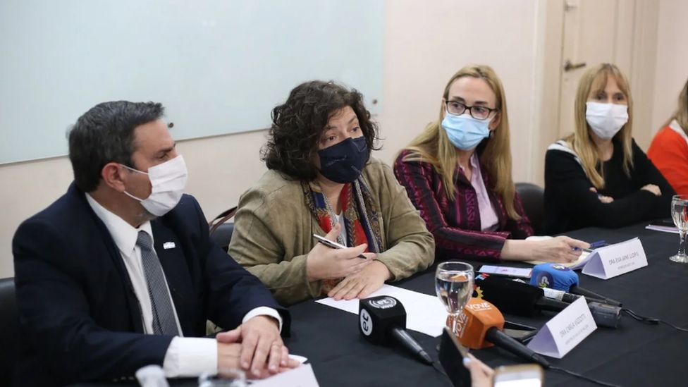
Published21 hours agoSharecloseShare pageCopy linkAbout sharingImage source, Argentinian Ministry of HealthHealth officials in Argentina have said that an infectious pneumonia outbreak which killed four people may have been caused by Legionnaire’s disease.Seven other cases have been found, most at a clinic in the northern province of Tucuman where the deaths occurred. The relatively rare lung disease is commonly linked to contaminated water or unclean air-conditioning systems. The World Health Organisation was alerted earlier this week to the cluster of infections. Doctors trying to determine the cause of the flu-like symptoms ruled out Covid-19, flu and the hantavirus – a severe respiratory disease carried by rodents – after testing the patients in the city of San Miguel de Tucuman.The symptoms included high fevers, body aches and breathing difficulties. Officials said the symptoms first appeared in six cases related to the facility which developed between 18 and 23 August. Legionella bacteria – which causes the illness – is commonly found in water sources such as rivers and lakes which sometimes find their way into artificial water systems.An estimated 10% of people who contract the disease die from complications arising from the infection.Argentinian Health Minister Carla Vizzotti said on Saturday that the authorities were working to ensure the clinic was safe for all.More on this storyArgentina investigates mystery pneumonia deaths2 days ago

Read more →

The Curious Hole in My Head

Born without my left temporal lobe, a brain region thought to be critical for language, I’ve been a research subject for much of my life.I barreled into the world — a precipitous birth, the doctors called it — at a New York City hospital in the dead of night.In my first few hours of life, after six bouts of halted breathing, the doctors rushed me to the neonatal intensive care unit. A medical intern stuck his pinky into my mouth to test the newborn reflex to suck. I didn’t suck hard enough. So they rolled my pink, 7-pound-11-ounce body into a brain scanner.Lo and behold, there was a huge hole on the left side, just above my ear. I was missing the left temporal lobe, a region of the brain involved in a wide variety of behaviors, from memory to the recognition of emotions, and considered especially crucial for language.My mother, exhausted from the labor, remembers waking up after sunrise to a neurologist, pediatrician and midwife standing at the foot of her bed. They explained that my brain had bled in her uterus, a condition called a perinatal stroke.They told her I would never speak and would need to be institutionalized. The neurologist brought her arms up to her chest and contorted her wrists to illustrate the physical disability I would be likely to develop.In those early days of my life, my parents wrung their hands wondering what my life, and theirs, would look like. Eager to find answers, they enrolled me in a research project at New York University tracking the developmental effects of perinatal strokes.But month after month, I surprised the experts, meeting all of the typical milestones of children my age. I enrolled in regular schools, excelled in sports and academics. The language skills the doctors were most worried about at my birth — speaking, reading and writing — turned out to be my professional passions.My case is highly unusual but not unique. Scientists estimate that thousands of people are, like me, living normal lives despite missing large chunks of our brains. Our myriad networks of neurons have managed to rewire themselves over time. But how?‘The worst participant’Helen Santoro developed a passion for language, despite being born without the left temporal lobe, a region of the brain considered especially crucial for language.Kayana Szymczak for The New York TimesMy childhood memories are filled with researchers following me around with pens and clipboards. My brain was scanned several times a year, and I was tasked with various puzzles, word searches and picture-recognition tests. At the end of each day of testing, the researchers would give me a sticker, which I would keep in a tin container next to my bed.When I was around 9 years old, researchers wanted to see how my brain would act when I was exhausted. I would sometimes stay up all night with my mom, eating Chinese food and watching Katharine Hepburn and Spencer Tracy movies. The next day I would stumble into the clinic half-awake, and scientists would stick electrodes on my scalp. As long wires fell from my head like Medusa’s snakes, I was finally allowed to fall asleep, blissfully unaware that the researchers were searching for abnormalities in my brain waves.Over the years, the scientists realized that I wasn’t like the other children in the study: I didn’t have any deficits to track over time. When I was around 15, my dad and I met in the cluttered Manhattan office of Dr. Ruth Nass, the pediatric neurologist leading the research. She questioned if I had actually had a perinatal stroke. In any case, she said frankly that my brain was so different from the others’ that I could no longer be in the study.I didn’t mind. I had other things going on in my life, like the beginning of high school, cross-country practice and crushes. But I had also learned enough about neuroscience to become completely consumed by the topic. When I was 17 and entering my senior year in high school, I wrote to Dr. Nass and asked if I could do an internship in her lab. She readily agreed.Hope Kean, a graduate student at M.I.T., preparing Helen Santoro for the M.R.I. scan in July.Kayana Szymczak for The New York TimesOne day in the lab, I asked if she could show me my study files. We walked into a room filled with stacks of plastic bins, each one brimming with folders and loose papers. She grabbed a folder and read it quietly. Then, peering over a piece of paper, she said, “You were the worst participant because you were perfectly fine! You threw off all of my data.”Dr. Nass, who passed away in 2019, and her colleagues would go on to publish many studies on perinatal strokes. In a 2012 paper, for example, they found that babies suffering these strokes had a higher risk of attention and behavioral problems compared with the general pediatric population. Many of these children — recruited from 1983 to 2006 from ‌‌Southern California and New York City — suffered from seizures and muscle weakness on one side of their bodies. Most also had damaged or missing areas, known as lesions, in their left hemispheres, like me. I assume that one of those data points was mine.I went to college and majored in neuroscience. After graduating in 2015, I spent two years working in a lab studying concussions. I spent hours in the magnetic resonance imaging room, watching as other peoples’ brains appeared before me on a computer screen.But I never thought much about my own brain until this spring, when I happened upon a story in Wired magazine about a woman just like me: astonishingly normal, apart from a missing temporal lobe.A critical hemisphereHelen Santoro underwent cognitive testing at M.I.T.Kayana Szymczak for The New York TimesFor more than a century, the left hemisphere of the brain has been considered the center of language production and comprehension.This idea was first proposed in 1836 by Dr. Marc Dax, a physician who observed that patients who had injuries to the left side of their brains could no longer speak properly. Twenty-five years later, Dr. Pierre Paul Broca observed a young man who had lost the ability to speak and could utter only one syllable: “Tan.” A brain biopsy following the patient’s death revealed a large lesion in the frontal part of the left hemisphere, now known as Broca’s area.In the early 1870s, Dr. Carl Wernicke, a neurologist, saw several patients who could speak fluently, but their utterances made little sense. One of these patients had a stroke in the back of her left temporal lobe, and Dr. Wernicke concluded that this section of the brain — now called Wernicke’s area — must serve as a second center for language, alongside Broca’s area.Modern brain imaging studies have further expanded our understanding of language. Much of this work has shown that two brain regions — the left sides of the temporal and frontal lobes — activate when a person is reading or hearing words. Some researchers have called this the “language network.”But other neuroscientists have argued that language processing is even broader and not confined to specific brain regions.“I believe that language in the brain is distributed throughout the entire brain,” said Jeremy Skipper, the head of the Language, Action and Brain Lab at University College London (and my former college psychology professor).Studies have shown that written words can activate the part of the brain associated with the word’s meaning. For example, the word “telephone” activates an area related to hearing, “kick” triggers a region involved in moving the legs, and “garlic” activates a part that processes smells.The areas of the brain traditionally attributed to language have lots of other functions, Dr. Skipper said. “It just depends on what other sections of the brain they are talking to and at what time and in what context.”Eight interesting brainsHelen Santoro became a participant in the Interesting Brain Project.Kayana Szymczak for The New York TimesThe Wired article described an anonymous woman from Connecticut who had no idea she lacked a left temporal lobe until undergoing an unrelated brain scan as an adult. For the past few years, the article explained, she had been part of a research project led by Evelina Fedorenko, a cognitive neuroscientist at the Massachusetts Institute of Technology.In April, I wrote Dr. Fedorenko an email telling her about my missing left temporal lobe and offering to be part of her research. She replied four and a half hours later, and soon I was booking an airplane ticket from my home in rural Colorado to Boston.There are currently eight participants, including me, in Dr. Fedorenko’s Interesting Brain Project, she told me. I haven’t met them, but four of us had presumed perinatal strokes, resulting in damage to our left hemispheres. Two participants have benign cysts in their right or left hemispheres, one had a stroke in the right hemisphere, and one had brain tissue removed from the left hemisphere because of a tumor.“The brain has incredible neuroplasticity,” said Hope Kean, a graduate student in Dr. Fedorenko’s lab who is running the Interesting Brain study as part of her dissertation.It seems that networks in the brain arrange in a particular way, but if you lose crucial brain regions as a baby — when the brain is still very plastic — these networks can reroute, Ms. Kean said.I arrived at Dr. Fedorenko’s lab in Cambridge on a hot day in July. I lay on a bed that slid into the M.R.I. machine’s narrow tube, with a cagelike device placed over my head. Ms. Kean snapped a mirror onto the headpiece so I could see a screen at the back of the scanner. As the machine started to make its banging, booming sounds, I remembered all of the times I had dozed off inside as a kid, lulled to sleep by its thundering chords.On the screen, words flashed quickly and a voice read them aloud, forming random sentences like, “Just the barest suggestion of a heel is found on teenage pumps.” Then, the words switched to a haphazard assortment of letters, creating incomprehensible sounds.After the scan was completed, the researchers and I crowded around a computer screen, where I saw a slice of my brain for the first time. I stared in disbelief, stunned that my neuronal wiring could have rerouted around this large, oblong hole where my temporal lobe should have been in the space behind my left temple and eye socket.Hope Kean, a graduate student at M.I.T., helped Helen Santoro out of the M.R.I. machine after her brain scan.Kayana Szymczak for The New York TimesIn a typical person’s brain, the sentences that I heard and read in the scanner would robustly activate the left temporal and frontal lobes, whereas the nonsense sounds would not.The researchers’ studies found that the brain of the Connecticut patient had adapted by switching sides: For her, these sentences activated the right temporal and frontal lobes, according to a case study published in the journal Neuropsychologia.My brain, however, surprised everyone, yet again.A preliminary analysis of the scans showed that, even without a left temporal lobe, I still process sentences using my left hemisphere.“I had thought that any large left hemisphere early lesion leads to the migration of the language system to the right hemisphere!” Dr. Fedorenko said. “But science is cool this way. Surprises often mean cool discoveries.”A possible reason behind this discovery, according to Dr. Fedorenko, is that my lesion is primarily in the front of my left hemisphere, leaving enough healthy tissue in the back for the language system to take root.Over the next few years, I’ll be flying back to the lab for additional scans and tests, and Dr. Fedorenko hopes to recruit even more people with unusual brains to participate in this study.I still think about the study I was in as a young child and about all of the other kids whose perinatal strokes had left many of them severely disabled. For some mysterious reason, my brain evolved around its missing lobe, whereas theirs struggled to do so. Why wasn’t I born with the developmental and cognitive problems, and they were? Why did my left side rewire to give me the syllables, words and phrases that have so enriched my life?It’s these questions that make me grateful to have been involved in this study — and to be a research participant once again.Helen Santoro, sitting at left next to her mother, watched Hope Kean, a graduate student, discuss Ms. Santoro’s brain scans with a research assistant, Niharika Jhingan, in July.Kayana Szymczak for The New York Times

Read more →

The Quiet Cost of Family Caregiving

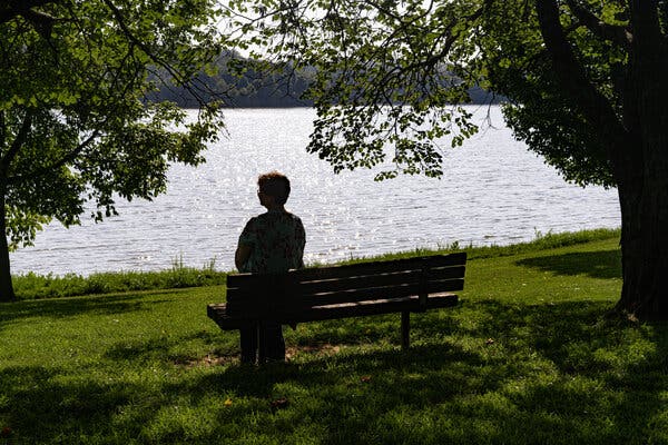
At first, Dana Guthrie thought she could help care for her parents, whose health had begun to decline, and still hold onto her job administering a busy dental practice in Plant City, Fla.“It was a great-paying job and I didn’t want to lose it,” Ms. Guthrie, 59, recalled recently. So she tried shifting to a four-day schedule, working evenings to keep up with the office demands, and she began spending a few nights a week at her parents’ home instead of her own nearby.Ultimately, though, her mother’s liver disease progressed and her father was diagnosed with dementia. The family learned that the cost of hiring home aides for two ailing 82-year-olds exceeded even a middle-class retirement income and savings. “They really needed me,” Ms. Guthrie said. In 2016, she left her job “and moved in full time.”An estimated 22 to 26 million American adults currently provide care for family members or friends, most of them older people, who need help with daily activities; more than half of those caregivers have jobs. “There’s no doubt that juggling the two can be very difficult,” said Douglas Wolf, a demographer and gerontologist at Syracuse University.Caregivers who are employed often reduce their work hours or leave the workplace altogether, research has shown. Several recent studies, however, reveal the impact of these decisions in more detail, not only on working caregivers but on employers and the general economy.Yulya Truskinovsky, an economist at Wayne State University, and her co-authors combined data from a Census Bureau survey with Social Security records to follow unusually long employment trajectories for nearly 13,000 people.Among those who became caregivers, employment dropped almost 8 percent compared to demographically similar non-caregivers, the authors found. “It happens right away, in the first year,” Dr. Truskinovsky said. “We see little evidence that they either reduce hours or switch to self-employment. They leave the labor force and remain out of it for quite a long time.”She added, “Younger caregivers are just as likely to leave the labor force as older ones.” Seven years later, the study found, those caregivers had not returned to the level of labor participation of demographically matched non-caregivers.Moreover, there were significant gender differences among those exiting the work force.Men started to reduce their workloads well before they became caregivers; then, “they leave the labor force and they don’t come back,” Dr. Truskinovsky said. The study could not provide an explanation; perhaps men take on caregiving when their work lives are already winding down.In contrast, women caregivers leave the work force more abruptly and are more likely to return — after just two years, on average — “but at lower wages or fewer hours,” Dr. Truskinovsky said.The pandemic amplified the conflict between employment and caregiving, Dr. Truskinovsky and colleagues found in another study. “Caregiving arrangements are very fragile,” she noted. While families often patch together paid and unpaid care, “it’s unstable, and if one thing falls through, your whole arrangement falls apart.”In a national sample of adults over 55, half the family caregivers reported that Covid-19 had disrupted their care schedules, forcing them to provide more care (because paid help became unavailable) or less (because of quarantines and fear of transmission). Before the pandemic, more than one-third had been employed.Caregivers facing disrupted arrangements were more likely to be furloughed or lose their jobs; they also showed far higher rates of depression, anxiety and loneliness than either non-caregivers or caregivers who did not experience disruptions.After Ms. Guthrie’s parents died, she relocated to Radcliff, Ky., where her sister lives. She is currently unemployed, and though she has been interviewing for jobs, she wonders whether she will ever be able to retire.Natosha Via for The New York TimesThe toll on working caregivers takes many forms. Susan Larson, 59, an education services specialist for the U.S. Army, has forgone promotions, even when her superiors urged her to apply. “I’m not geographically mobile,” she said.She cannot leave her home in St. Paul, Minn., where she and her husband built a handicapped-accessible addition for her mother, 83, who needs extensive assistance. The Army has been very supportive, Ms. Larson said. But she estimated that her salary would be nearly 25 percent higher if she had accepted promotions, in turn bolstering both her eventual pension and Social Security benefits.Shawn French, 51, a video game writer in Limerick, Me., and his wife welcomed her widowed father into their home three years ago. Because Mr. French works remotely, he can help his father-in-law with meals, medications and mobility; his wife handles doctors visits and other tasks.“I wouldn’t have it any other way,” Mr. French said. But the stress led him to relinquish weekend freelancing that had generated $200 to $300 a week. “We relied on it anytime things were a little tight,” he said. His wife reconfigured her work arrangement, too, which led to him being dropped from her health plan and becoming uninsured.Even when caregivers keep their jobs, another recent study indicates, almost one-quarter report either missed work (absenteeism) or reduced productivity (known as presenteeism).Presenteeism accounts for the most productivity loss, said senior author Jennifer Wolff, a gerontologist and health services researcher at Johns Hopkins University. “Absenteeism is visible, presenteeism is less so,” she said. “You show up, but you’re making doctor calls or managing insurance.”Among affected employees, work productivity dropped by one-third, on average. Based on 2015 data, the most recent available on adults 65 and older, that translates to a $49 billion loss annually.Paid family leave, although better configured for the more predictable needs of new parents, might help workers providing care for the elderly as well. When California adopted paid leave, which took effect in 2004, residency rates at nursing homes declined by about 11 percent, Dr. Wolf of Syracuse and his co-author Kanika Arora found.Although the study could not establish the reason, Dr. Wolf speculated that “the change in the law induced people to stay on the job, but they took enough time off to keep their parents out of a nursing home.” The authors’ more recent work shows that paid sick leave also helps increase family caregiving.President Biden campaigned on an ambitious caregiving plan that would have provided 12 weeks of paid family leave annually, plus tax credits to offset caregiving expenses and Social Security credits for time family caregivers spend out of the labor force. Republican opposition in the Senate has prevented its passage.Debate on how best to support family caregivers will continue, but there is little debate about their need for help. Although many workers can handle the more predictable needs of aging parents and spouses, some face intense pressures incompatible with contemporary workplaces.After Ms. Guthrie’s parents died, she relocated to Radcliff, Ky., where her sister lives. She found positions at dental practices there but has never matched the compatibility or the salary of the job she left in Florida.Currently unemployed, Ms. Guthrie has been interviewing for jobs and wondering whether she will ever be able to retire, although she doesn’t regret the sacrifices she made to care for her parents.“We were a close-knit family and I would do it again,” she said. “But I took a beating, emotionally and financially, and I haven’t really been able to recover.”

Read more →