Los Angeles County Investigates Death of Person With Monkeypox
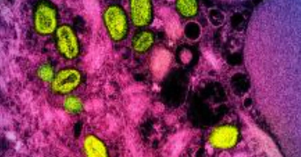
Officials said they were conducting an autopsy to see if a monkeypox infection contributed to the death of a Los Angeles County resident.Los Angeles County officials said this week that they were investigating the death of a person who had been diagnosed with monkeypox to see if the illness was a contributing cause of death.It is the second known death of a person diagnosed with the disease in the United States, which has reported more cases of the viral illness this year than anywhere in the world. Health officials are investigating what, if any, role monkeypox played in the two deaths.Dr. Rita Singhal, chief medical officer for the Los Angeles County Department of Public Health, said at a news conference on Thursday that an autopsy was being conducted on the person who died in the county and that it could take days or weeks to get the results. Dr. Singhal did not say whether the person who died had any underlying medical conditions, as was the case in the earlier fatality, in Texas.“We are early in the investigation and do not have additional details available at this time,” Dr. Singhal said. “As soon as details become available, we will share them while maintaining confidentiality and privacy.”As part of the investigation, Dr. Singhal said, county officials will work with the state of California and the Centers for Disease Control and Prevention to see if guidelines for how to treat people with monkeypox, especially those who become severely ill, need to be updated.There have been more than 56,600 confirmed cases of monkeypox in more than 100 countries, including more than 21,500 confirmed cases in the United States, according to the C.D.C.The first reported death of someone in the United States who had been diagnosed with monkeypox was in Texas last month. The Department of State Health Services there announced on Aug. 30 that an adult in Harris County who was “severely immunocompromised” and diagnosed with monkeypox had died.“The case is under investigation to determine what role monkeypox played in the death,” the department said in a statement.As of Sept. 4, the deaths of 18 people who had been diagnosed with monkeypox had been reported to the World Health Organization. The number of weekly new cases globally decreased by 25.5 percent in the week ending on Sept. 4, compared with the week before, the W.H.O. said. The outbreak continues to primarily affect men who have sex with men who have a median age of 36 years, the W.H.O. said.The spread of monkeypox appears to be declining in major U.S. cities, including New York City, Los Angeles, San Francisco and Chicago, health officials say.Dr. Rochelle P. Walensky, the C.D.C. director, cautioned two weeks ago that the decrease was not even across the country.“Week over week, our numbers are still increasing,” she said in a White House news conference. “The rate of rise is lower. But we are still seeing increases. And we are of course a very diverse country, and things are not even across the country.”

Read more →

Risk factors for heart disease and stroke largely similar in men and women globally

Women and men share most of the same risk factors for cardiovascular disease (CVD), a large international study has found — the first such study to include people not only from high income countries, but also from low- and middle-income countries where the burden of CVD is the greatest.
The study was published today in The Lancet.
The global study assessed risk factors, including metabolic (such as high blood pressure, obesity and diabetes), behavioural (smoking and diet), and psychosocial (economic status and depression) in about 156,000 people without a history of CVD between the ages of 35 and 70. Living in 21 low, middle and high-income countries on five continents, they were followed for an average of 10 years.
“Women and men have similar CVD risk factors, which emphasizes the importance of a similar strategy for the prevention of CVD in men and women,” said the paper’s first author Marjan Walli-Attaei, a research fellow at the Population Health Research Institute (PHRI) of McMaster University and Hamilton Health Sciences (HHS).
Overall, women had a lower risk of developing CVD than men, especially at younger ages.
However, diet was more strongly associated with CVD risk in woman than men — “something that’s not been previous described, and which requires independent confirmation,” said Salim Yusuf, lead investigator of the study, senior author, executive director of PHRI, professor of medicine at McMaster University, and cardiologist at HHS.
High levels of bad (LDL) cholesterol and symptoms of depression were more strongly associated with CVD risk in men than in women. The patterns of these findings were generally similar in high-income countries and upper-middle-income countries, and in low-income and lower-middle-income countries.
Funding was provided by the PHRI, Hamilton Health Sciences Research Institute, the Canadian Institutes of Health Research (including through the Strategy for Patient-Oriented Research via the Ontario SPOR Support Unit), the Ontario branch of the Heart and Stroke Foundation, and the Ontario Ministry of Health and Long-Term Care.
Story Source:
Materials provided by McMaster University. Original written by Heather Angus-Lee. Note: Content may be edited for style and length.

Read more →

Antibiotics given in infancy may have adverse impact on adult gut health

Preterm and low birth weight babies are routinely given antibiotics to prevent, not just treat, infections, which they have a high risk of developing. A new study, published in The Journal of Physiology has found that early life exposure to antibiotics in neonatal mice has long-lasting effects on their microbiota, enteric nervous system, and gut function. This could mean that babies given antibiotics may grow up to experience gastrointestinal issues.
This discovery by the research team from the Department of Anatomy and Physiology at the University of Melbourne is the first to show that antibiotics given to neonatal mice has these long-lasting effects which result in disturbed gastrointestinal function, including the speed of motility through the gut and diarrhoea-like symptoms in adulthood.
The research team gave mice an oral dose of vancomycin every day for the first ten days of their lives. They were then reared normally until they were young adults, and their gut tissue was looked at to measure its structure, function, microbiota, and nervous system. The investigators found that changes were also dependent on the sex of the mice. The females had long whole gut transit and the males had lower faecal weight than the control group. Both males and females had greater faecal water content, which is a diarrhoea-like symptom.
Mice have many similarities to humans, but they are born with more immature guts than humans and have accelerated growth due to their shorter life spans. Their gut microbiota and nervous systems are less complex than humans, so the findings cannot yet be directly associated to human children and infants. The researchers will be doing further studies on the mechanisms of antibiotics on the gut and the causes of the sex specific actions, and if early life antibiotic use has effects on metabolism and brain function.
Lead Physiologist, Dr Jaime Foong said, ‘We are very excited about the findings of our study which show that antibiotics given after birth could have prolonged effects on the enteric nervous system. This provides further evidence of the importance of microbiota on gut health and could introduce new targets to advance antibiotic treatment to very young children.’
Story Source:
Materials provided by The Physiological Society. Note: Content may be edited for style and length.

Read more →

'Jumping gene' found to be strongly linked to depression, fear, and anxiety

First characterized in Prof. Tadashi Yamamoto’s former lab in Japan in 1996, the gene Tob is well known for the role it plays in cancer. Previous research has also indicated that it has a hand in regulating the cell cycle and the body’s immune response. Now, in a multidisciplinary study that combines molecular biology with neuroscience, researchers from the Okinawa Institute of Science and Technology (OIST) have found that this gene also plays an important role in reducing depression, fear, and anxiety. Their work was published by the journal Translational Psychiatry.
“This research is about understanding stress-resilience,” explained lead author, Dr. Mohieldin Youssef, former PhD student in OIST’s Cell Signal Unit, which is led by Prof. Yamamoto. “The presence of the gene helps with stress-resilience and if it’s removed, there’s an increase in depression, fear, and anxiety.”
Tob is named for the Japanese verb “tobu,” which means to fly or to jump. This is because when the cell is exposed to a stimulus, its protein levels jump in activity. Dr. Youssef said that this has resulted in the gene being classed as an immediate-early gene, as it has such a fast response.
“The Tob gene is related to many different phenomena but working on the brain system is particularly challenging,” said Prof. Yamamoto. “Although it was previously suspected, this research is the first work that clarifies that Tob has a function in the brain against stress.”
Their conclusion that this gene is linked to anxiety, fear, and depression was drawn from several different experiments. First, the researchers exposed mice to stress and, as expected, saw the Tob protein levels increase. They then used mice which had been born without a Tob gene and found an increase in depression, fear, and anxiety. For example, when a mouse with the Tob gene was placed in a bucket of water, they would swim and try to escape. However, a mouse without the Tob gene simply floated. This lack of will to fight a difficult situation is one way that researchers determine that an animal is depressed.
What’s more, the mice without the Tob gene didn’t seem to learn. Dr. Youssef explained that when mice are put day-after-day in a place which evokes fear memory, they normally learn that it isn’t so bad and stop being as frightened. But those without the Tob gene still showed increased levels of fear observed as freezing, even after several days.
The researchers then teamed up with OIST’s former PhD student Dr. Hiroaki Hamada from the Neural Computational Unit. Through an MRI, they found that the connectivity between two key places regulating brain’s stress resilience was altered when the Tob gene was removed — the hippocampus and the pre-frontal cortex. From there, the researchers decided to look at the specific role that the gene plays within the hippocampus. They took mice without the Tob gene and injected this gene into the hippocampus, while leaving it nonexistent in other parts of the body. The level of fear and depression returned to normal, but the mice still had increased anxiety. The researchers then did the opposite — they created a mouse that had no Tob gene in the cells in the hippocampus but had it in the cells in the rest of the body. In this case, they found that the mice had normal levels of anxiety but increased fear and depression.
“We’ve concluded that the Tob gene within the hippocampus suppresses fear and depression,” explained Dr. Youssef. “But the suppression of anxiety must be regulated by another part of the brain.”
Next, researchers from OIST’s former Brain Mechanisms for Behavior Unit measured the function of the neurons within the hippocampus of the mice without the Tob gene. They found that excitation was increased, while inhibition was decreased, suggesting that the overall balance was impacted, which would impact the behavior of the mice.
Finally, the researchers conducted molecular analyses after exposing the mice to stress. Interestingly, they found that expression didn’t immediate change with stress. But, 15 minutes after exposing the mice to stress, there were changes. Other genes and proteins were impacted if the Tob gene was deleted. This suggests that the Tob gene likely has multiple direct and indirect impacts.
“Uncovering this role of the Tob gene in fear, depression, and anxiety could have vast implications for developing therapeutics for psychiatric stress,” said Dr. Youssef.

Read more →

Breaking down proteins: How starving cancer cells switch food sources

Cancer cells often grow in environments that are low in nutrients, and they cope with this challenge by switching their metabolism to using proteins as alternative “food.” Building on genetic screens, an international team of scientists could identify the protein LYSET as part of a pathway that allows cancer cells to make this switch. Their findings are now published in the journal Science.
Amino acids are the building blocks of proteins and key nutrients for cell growth and proliferation. Understanding how cells utilise amino acids in different environments is a central question in basic biology and cancer research. Tumor tissues often have a limited blood supply, and to grow under such conditions, cancer cells switch their metabolic activities. In particular, they switch from taking up nutrients delivered by blood vessels to exploiting alternative nutrients, such as breaking down surrounding proteins as a food source when facing starvation. However, the mechanisms that enable cancer cells to make this switch have remained largely elusive.
To better understand the molecular pathways that underly this nutrient switch in cancer, two groups of scientists with matching expertise teamed up: Wilhelm Palm of the German Cancer Research Center (DKFZ) in Heidelberg is a leading expert in cancer metabolism, Johannes Zuber at the Research Institute of Molecular Pathology (IMP) in Vienna brought in ample experience in functional cancer genetics. The scientists set up their study with tightly controlled nutrient conditions to mimic amino acid starvation as it occurs in many tumors. Then they used the “gene scissors” CRISPR-Cas9 to disrupt the expression of almost every gene in the genome, which allowed them to pin down several pathways involved in the nutrient switch.
Among them, the scientists spotted an uncharacterised gene that was only required for cell survival when cancer cells were feeding on extracellular proteins. This gene, which the scientists re-named “LYSET” (Lysosomal Enzyme Trafficking Factor), turned out to be critical for the function of lysosomes, small organelles that function as the stomach of cells where proteins are digested. Further experiments into the function of LYSET revealed that the gene acts as a core component of the so-called mannose-6-phosphate pathway, which is required for filling lysosomes with digestive enzymes. In the absence of LYSET, cancer cells lack enzymes in their lysosomes and are no longer able to digest proteins.
Then the scientists turned to mouse models to study the function of LYSET in real tumors. They found that the loss of LYSET strongly impaired tumor development in several types of cancer, while it was well-tolerated under normal nutrient conditions.
Wilhelm Palm, whose lab was among the first who described the ability of cancer cells to feed on extracellular proteins, says: “With LYSET, we have discovered a central component of a metabolic pathway that enables adaptations to different nutrients, a key ability of cancer cells to survive and grow in austere Tumor environments.”
“This is what made the discovery so exciting,” says Johannes Zuber. “LYSET and the mannose-6-phosphate pathway turn out to be particularly important for cancer cells and could therefore be a molecular entry point for attacking a major metabolic bottleneck in cancer.”
Story Source:
Materials provided by German Cancer Research Center (Deutsches Krebsforschungszentrum, DKFZ). Note: Content may be edited for style and length.

Read more →

SARS-CoV-2 protein caught severing critical immunity pathway

Over the past two years, scientists have studied the SARS-CoV-2 virus in great detail, laying the foundation for developing COVID-19 vaccines and antiviral treatments. Now, for the first time, scientists at the Department of Energy’s SLAC National Accelerator Laboratory have seen one of the virus’s most critical interactions, which could help researchers develop more precise treatments.
The team caught the moment when a virus protein, called Mpro, cuts a protective protein, known as NEMO, in an infected person. Without NEMO, an immune system is slower to respond to increasing viral loads or new infections. Seeing how Mpro attacks NEMO at the molecular level could inspire new therapeutic approaches.
To see how Mpro cuts NEMO, researchers funneled powerful X-rays from SLAC’s Stanford Synchrotron Radiation Lightsource (SSRL) onto crystallized samples of the protein complex. The X-rays struck the protein samples, revealing what Mpro looks like when it dismantles NEMO’s primary function of helping our immune system communicate.
“We saw that the virus protein cuts through NEMO as easily as sharp scissors through thin paper,” said co-senior author Soichi Wakatsuki, professor at SLAC and Stanford. “Imagine the bad things that happen when good proteins in our bodies start getting cut into pieces.”
The images from SSRL show the exact location of NEMO’s cut and provide the first structure of SARS-CoV-2 Mpro bound to a human protein.
“If you can block the sites where Mpro binds to NEMO, you can stop this cut from happening over and over,” SSRL lead scientist and co-author Irimpan Mathews said. “Stopping Mpro could slow down how fast the virus takes over a body. Solving the crystal structure revealed Mpro’s binding sites and was one of the first steps to stopping the protein.”
The research team from SLAC, DOE’s Oak Ridge National Laboratory, and other institutions published their results today in Nature Communications.

Read more →

Stricter blood sugar control in gestational diabetes leads to better outcomes for babies

Lowering the target blood sugar level for mothers with gestational diabetes did not reduce the risk of large babies, a new study finds, but it did reduce the risk of death or injury to the baby during birth. Caroline Crowther of the University of Auckland, New Zealand, and colleagues report these findings on September 8th in the open-access journal PLOS Medicine.
Gestational diabetes is a significant and growing health problem worldwide, which often causes the birth of especially large babies who face a high risk of obesity and type 2 diabetes later in life. Women can change their diets and take medication to control their blood sugar, but currently, it is unknown how tightly those levels should be controlled to minimize the risks to the mother and baby.
To determine if stricter control is better, researchers conducted a study of 1,100 pregnant women with gestational diabetes seen at 10 hospitals in New Zealand. During the study, each hospital switched from higher to lower blood sugar targets, and outcomes for women and babies in each group were compared. While tighter blood sugar control did not lead to babies being larger than expected, it did reduce the risk of infant death, trauma, and shoulder dystocia during birth by half. However, tighter control almost doubled the risk of serious health outcomes for the mother, such as a major postpartum hemorrhage, among other complications.
The new results can help doctors decide what blood sugar level individual patients should strive for while managing their gestational diabetes. The study is the largest randomized comparison of two blood sugar level targets reported to date in a diverse population. However, the researchers point out that there is still a need to confirm their findings through additional randomized trials and in different healthcare settings.
Crowther adds, “This unique trial allowed for the sequential implementation of the newly, recommended tighter treatment targets for women with gestational diabetes and assessed if there are true benefits, without harm, to use of tighter treatment targets.”
Story Source:
Materials provided by PLOS. Note: Content may be edited for style and length.

Read more →

New approach more than doubles stem cell editing efficiency

A Penn State-led team of interdisciplinary researchers has developed techniques to improve the efficiency of CRISPR-Cas9, the genome editing technique that earned the Nobel Prize in 2020. While CRISPR-Cas9 is faster, less expensive and more accurate than other gene-editing methods, according to project leader Xiaojun “Lance” Lian, associate professor of biomedical engineering and biology at Penn State, the technology has limitations — especially in applications to improve human health.
The researchers developed a more efficient and accessible process to apply CRISPR-Cas9 systems in human pluripotent stem cells (hPSCs), derived from federally approved stem cell lines, which Lian said could greatly advance diagnostics and treatments for genetic disorders. The approach was published Sept. 7 in Cell Reports Methods.
CRISPR-Cas9, which stands for clustered regularly interspaced short palindromic repeats and CRISPR-associated protein 9, gives scientists the ability to target precise locations of genetic code to change DNA, providing opportunities to create new diagnostic tools and potentially correct mutations to treat genetic causes of disease.
“The human genome is enormous, and CRISPR-Cas9 makes it possible for scientists to find and target a mutated gene for the purpose of studying it,” Lian said.
CRISPR uses a disc of genetic material, known as plasmid DNA, to deliver guided ribonucleic acid (RNA) that positions the Cas9 enzyme at the precise location of the target gene. When the DNA is located, Cas9 binds to it and cuts it out, allowing other DNA to repair the cut. Researchers can then see how the removal changes the gene’s expression. But there are delivery and editing efficiency problems with current DNA-based CRISPR methods, according to Lian.
“Delivery of DNA CRISPR effectors is low,” he said. “Only 20% to 30% of the targeted cells will receive gene-editing DNA when using CRISPR. Delivery of RNA into cells can be more efficient; however, when regular RNA is introduced, cells can see it as a virus. They destroy the RNA before it can make proteins — say, in a matter of a few hours — and, in doing so, destroy the gene editing attempt.”
To improve the outcome, the researchers changed the way the genome editing tools are delivered to the stem cells, using modified RNA (modRNA). The modRNA differs from plasmid DNA in that it replaces one of the base substrates found in RNA with a chemically modified version, and it is stabilized by stronger structural support.

Read more →

New advances in stem-cell derived mouse embryo model

Just two weeks after announcing the development of a mouse embryo model, complete with beating hearts and the foundations for a brain and other organs, from mouse stem cells, researchers in the laboratory of Magdalena Zernicka-Goetz, Bren Professor of Biology and Biological Engineering, have published new findings about another mouse embryo model reaching similar developmental stages, but created out of only mouse embryonic stem cells. This modification has simplified the protocol and makes the embryo model easier to be adopted in other laboratories.
The new study appears in the journal Cell Stem Cell on September 8. The research was led by graduate students Kasey Lau and Hernan Rubinstein of the University of Cambridge and the Weizmann Institute of Science, respectively.
“This discovery opens up new avenues for understanding why the great majority of human pregnancies are lost and to create knowledge that will prevent this from happening,” says Zernicka-Goetz, who is also a professor of mammalian development and stem cell biology at Cambridge University in the Department of Physiology, Development and Neuroscience. “This knowledge will also let us, with time, repair tissues and organs much more effectively than we can do now.”
“As we develop these models further, we will learn more about the signals that initiate and guide the development of organs, which will give us routes for helping to generate organs in culture that will ultimately find application in transplant surgery or in regenerative medicine,” she explains.
In a paper published in the journal Nature on August 25, the team detailed how to develop a mouse embryo model out of mouse embryonic and extraembryonic stem cells. Instead of creating mouse embryos by the natural biological method of combining egg and sperm, the team guided three populations of cultured stem cells to interact, inducing the expression of certain genes and establishing an environment for the cells to “talk” to each other. As a result, the stem cells self-organized into structures that then progressed through successive developmental stages until the mouse embryo model had beating hearts and the foundations for a brain and all other organs, in addition to the yolk sac and which facilitate gas and nutrient exchange between the embryo and the mother. This is the most advanced stage of development achieved to date in a stem cell-derived model.
Naturally, in the first few days after fertilization, three major types of tissues develop in the early mouse embryo: one will eventually become the tissues of the body, and the other two will support the embryo’s development. One of these latter two types, known as the trophectoderm, will become the placenta, which connects the fetus to the mother and provides oxygen and nutrients. The other, known as primitive endoderm, will give rise to the yolk sac, where the embryo grows and from which it receives nutrients in early development.
Three types of stem cells can be derived from each of these three tissues from the mouse embryo and cultured indefinitely in the laboratory.
Building off of the previous research, the mouse embryo model reported in the new paper are only made of a single type of cultured stem cell: the embryonic stem cells (ESCs). Untreated ESCs become the body of the embryo. Another ESC line is coaxed by researchers to become like extraembryonic endoderm stem cells, which provide one set of developmental signals. The team also drives a third ESC line to become like trophoblast stem cells, which provide a second set of developmental signals. Thus, the team is able to reconstitute the three major tissues of the developing mouse embryo by starting with only ESCs. This has simplified the protocol while still preserving the important signaling events between the three tissues, which are critical to building the body plan of the embryo.
“Of the three stem cell types, only the ESCs are pluripotent — that is to say, only the ESCs have the potential to develop into any tissue of the body,” explains Zernicka-Goetz. “But to do this, they require the other two types of extra-embryonic stem cell. ESCs can be directed to become these other two extra-embryonic cell types. In this way, we end up with three starting cell types all generated from the single ESC line.”
The paper is titled “Mouse embryo model derived exclusively from embryonic stem cells undergoes neurulation and heart development.” Lau and Rubinstein are the study’s first authors. Additional co-authors are Carlos Gantner of the University of Cambridge, Caltech postdoctoral scholar Ron Hadas, Gianluca Amadei of the University of Cambridge, Yonatan Stelzer of the Weizmann Institute of Science in Israel, and Zernicka-Goetz. Funding was provided by the National Institutes of Health, the Allen Discovery Center for Lineage Tracing, the European Research Council, the Wellcome Trust, Open Philanthropy/Silicon Valley Community Foundation, and Weston Havens Foundation.

Read more →

New study provides insights into each US state's COVID-19 vaccination rate

In the first year of its availability, 84.2% of US adults received at least one dose of COVID-19 vaccine. Progress in COVID-19 vaccination slowed after April 2021, and millions of Americans remain unvaccinated. New research in the American Journal of Preventive Medicine, published by Elsevier, finds wide geographic variance in vaccine uptake and identifies the underlying behavioral and social drivers that factor into the decision to remain unvaccinated. The findings suggest that improving COVID-19 vaccine confidence may not be solved by a “one-size-fits-all solution.”
“Many studies have explored vaccine coverage and vaccine confidence at a national level, but these are likely to vary widely by geography and sociodemographics,” said lead investigator Nina B. Masters, PhD, Epidemic Intelligence Service, and Division of Viral Diseases, National Center for Immunization and Respiratory Diseases, Centers for Disease Control and Prevention (CDC), Atlanta, GA, USA. “We decided to explore state and regional trends in COVID-19 vaccine confidence and the underlying reasons for non-vaccination.”
The study used data from the National Immunization Survey-Adult COVID Module. This is a nationally representative household cellphone survey of US adults aged 18 or older. Of the 531,798 participants surveyed (April 22 to December 31, 2021), respondents who had not received a COVID-19 vaccine were then asked how likely they were (definitely, probably, probably not, definitely will not, not sure) to get vaccinated. Additional questions were asked about the individual’s concerns about COVID-19 and confidence in vaccine safety and importance.
To understand social norms, the participants were asked how many friends and family members had received a COVID-19 vaccination, and if a healthcare provider had recommended that they receive the vaccine. For practical factors, the survey asked whether one’s work or school required COVID-19 vaccination and whether they experienced any actual or perceived difficulty in accessing a vaccination. Demographic data were also collected. The analysis examined these variables nationally and for each state for each month in the survey period, to support trends over time.
In general, rural residents were less likely to be vaccinated than urban residents, but in the Southeast and Midwest, differences in vaccination coverage by urbanicity were smaller. In most states, Black and Hispanic adults were less likely to be vaccinated than White and non-Hispanic adults. However, in the South and the Southeast, where vaccination rates were low compared to the rest of the US, vaccination coverage was similar across race and ethnicity. The Southeast and Midwest had the largest proportion of unvaccinated people who indicated that they would probably get a COVID-19 vaccine or were unsure. These regions saw similar trends in concern about COVID-19 and confidence in the importance of getting vaccinated.
Overall, individuals with low confidence in the importance of the vaccine were five times more likely to remain unvaccinated. This suggests that focused messaging about the benefits of vaccination, compared to the risk of the disease, remains a priority. Adults in rural areas — as well as those with less than a college degree, without insurance, making less than $75,000/year and under 65 — had higher prevalence of non-vaccination across all regions, while associations between vaccination and race/ethnicity varied by region.
“Our analysis supports the idea that reasons for non-vaccination are personal and individual, and there are nuances and differences in this reasoning, for example, between an unvaccinated, young individual living in a rural community in the Southeast and an unvaccinated, older individual living in the Northeast who has mostly vaccinated friends and family. It’s important to meet people where they are to build confidence in vaccines,” said Dr. Masters. “The CDC has made national, regional, and state data available on the Behavioral and Social Drivers of COVID-19 vaccination through national surveys of adults and parents. State and local health departments may be able to strengthen their ongoing efforts locally by accessing these data at the CDC COVID Data Tracker: Vaccine Confidence [https://covid.cdc.gov/covid-data-tracker/#datatracker-home].”
Story Source:
Materials provided by Elsevier. Note: Content may be edited for style and length.

Read more →